

 PDF
PDF ePub
ePub Citation
 Print
Print


Abstract
The main goals of treatment of malignant bone tumor are the prolongation of life survival and the improvement of quality of life. In growing children, however, leg length discrepancy (LLD) is one of major problem in the treatment of malignant bone tumors. Therefore, the precise understanding of growth in children is essential, and the prediction of LLD is critical in deciding the time and options of surgery. In addition, to use the adequate method of growth expectation, periodic followup and collaboration with patient's parents are needed.
REFERENCES
1. Liu XC, Fabry G, Molenaers G, Lammens J, Moens P. Kinematic and kinetic asymmetry in patients with leg-length discrepancy. J Pediatr Orthop. 1998; 18:187–189.

2. Gofton JP, Trueman GE. Studies in osteoarthritis of the hip. II. Osteoarthritis of the hip and leg-length disparity. Can Med Assoc J. 1971; 104:791–799.
3. Kujala UM, Friberg O, Aalto T, Kvist M, Osterman K. Lower limb asymmetry and patellofemoral joint incongruence in the etiology of knee exertion injuries in athletes. Int J Sports Med. 1987; 8:214–220.
4. Giles LG, Taylor JR. The effect of postural scoliosis on lumbar apophyseal joints. Scand J Rheumatol. 1984; 13:209–220.
5. Dimeglio A. Growth in pediatric orthopaedics. J Pediatr Orthop. 2001; 21:549–555.
6. Tanner JM, Davies PS. Clinical longitudinal standards for height and height velocity for North American children. J Pediatr Orthop. 1985; 107:317–329.
7. Tanner JM, Whitehouse RH. Clinical longitudinal standards for height, weight, height velocity, weight velocity, and stages of puberty. Arch Dis Child. 1976; 51:170–179.
8. Moseley RT, Weinstein SL. Lovell and Winter's Pediatirc Orthopaedics. 6th ed.Philadelphia, PA: Lippincott Williams and Wilkins;2006.
9. Greulich WW, Pyle SI. Radiographic atlas of skeletal development of the hand and wrist. 2nd ed.Stanford: Stanford University Press;1959.
10. Sauvegrain J, Nahum H, Bronstein H. Study of bone maturation of the elbow. Ann Radiol (Paris). 1962; 5:542–550.
11. Risser JC. The Iliac apophysis; an invaluable sign in the management of scoliosis. Clin Orthop Relat Res. 1958; 11:111–119.
12. Anderson M, Messner MB, Green WT. Distribution of lengths of the normal femur and tibia in children from one to eighteen years of age. J Bone Joint Surg Am. 1964; 46:1197–1202.
13. Ha JH, Choi IH, Chung CY, et al. Distribution of lengths of the normal femur and tibia in Korean children from three to sixteen years of age. J Korean Med Sci. 2003; 18:715–721.
14. Anderson M, Green WT, Messner MB. Growth and predictions of growth in the lower extremities. J Bone Joint Surg Am. 1963; 45:1–14.
15. Tupman GS. A study of bone growth in normal children and its relationship to skeletal maturation. J Bone Joint Surg Br. 1962; 44:42–67.
16. Menelaus MB. Correction of leg length discrepancy by epiphysial arrest. J Bone Joint Surg Br. 1966; 48:336–339.
17. Westh RN, Menelaus MB. A simple calculation for the timing of epiphysial arrest: a further report. J Bone Joint Surg Br. 1981; 63:117–119.
18. Moseley CF. A straight-line graph for leg-length discrepancies. J Bone Joint Surg Am. 1977; 59:174–179.
19. Moseley CF. A straight line graph for leg length discrepancies. Clin Orthop Relat Res. 1978; 136:33–40.
20. Paley D, Bhave A, Herzenberg JE, Bowen JR. Multiplier method for predicting limb-length discrepancy. J Bone Joint Surg Am. 2000; 82:1432–1446.
21. Cool WP, Carter SR, Grimer RJ, Tillman RM, Walker PS. Growth after extendible endoprosthetic replacement of the distal femur. J Bone Joint Surg Br. 1997; 79:938–942.
22. Dominkus M, Krepler P, Schwameis E, Windhager R, Kotz R. Growth prediction in extendable tumor prostheses in children. Clin Orthop Relat Res. 2001; 390:212–220.
23. Neel MD, Heck R, Britton L, Daw N, Rao BN. Use of a smooth press-fit stem preserves physeal growth after tumor resection. Clin Orthop Relat Res. 2004; 426:125–128.
24. Key JA, Ford LT. A study of experimental trauma to the distal femoral epiphysis in rabbits. II. J Bone Joint Surg Am. 1958; 40:887–896.
25. Siffert RS. The effect of staples and longitudinal wires on epiphyseal growth; an experimental study. J Bone Joint Surg Am. 1956; 38:1077–1088.
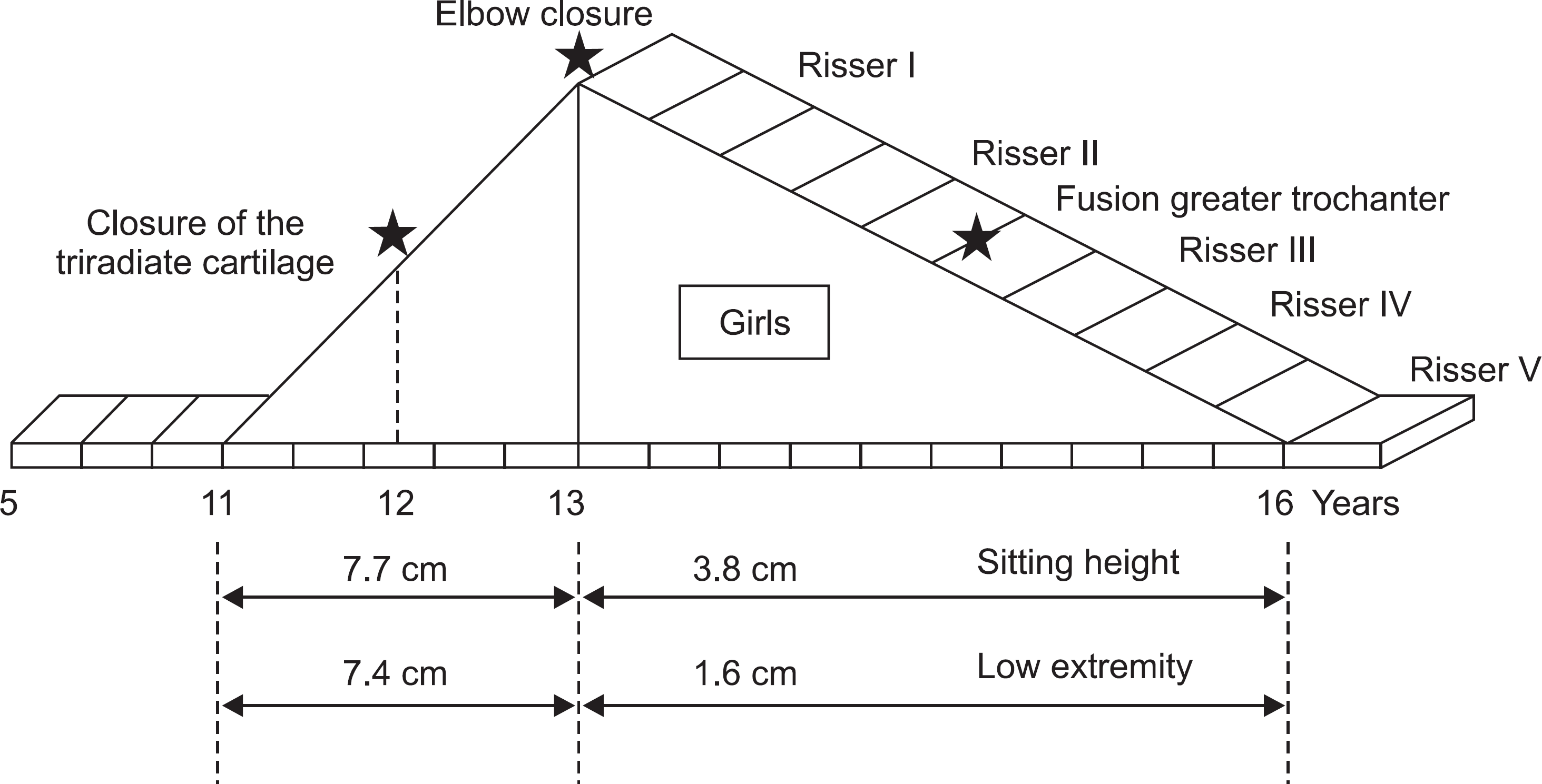
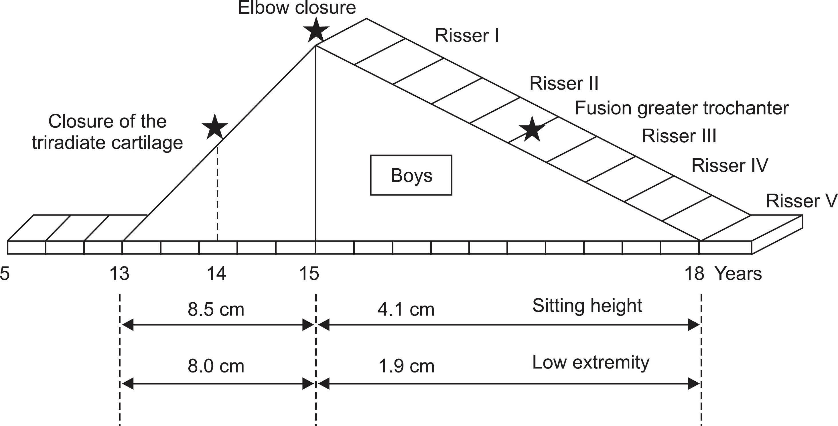
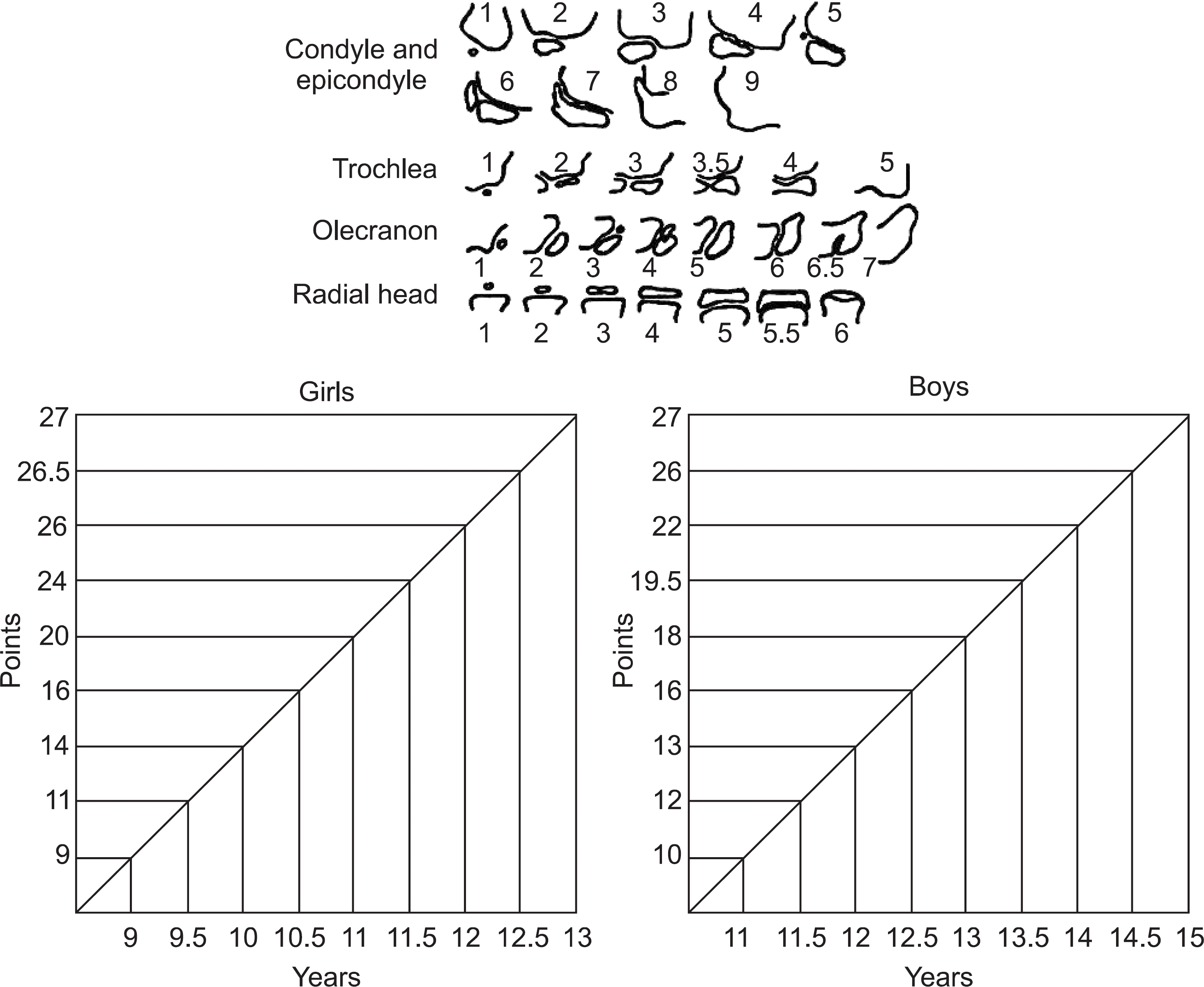
Figure 4.
This showing the stages of closure of the olecrenon in relation to the pubertal growth and the Risser sign.
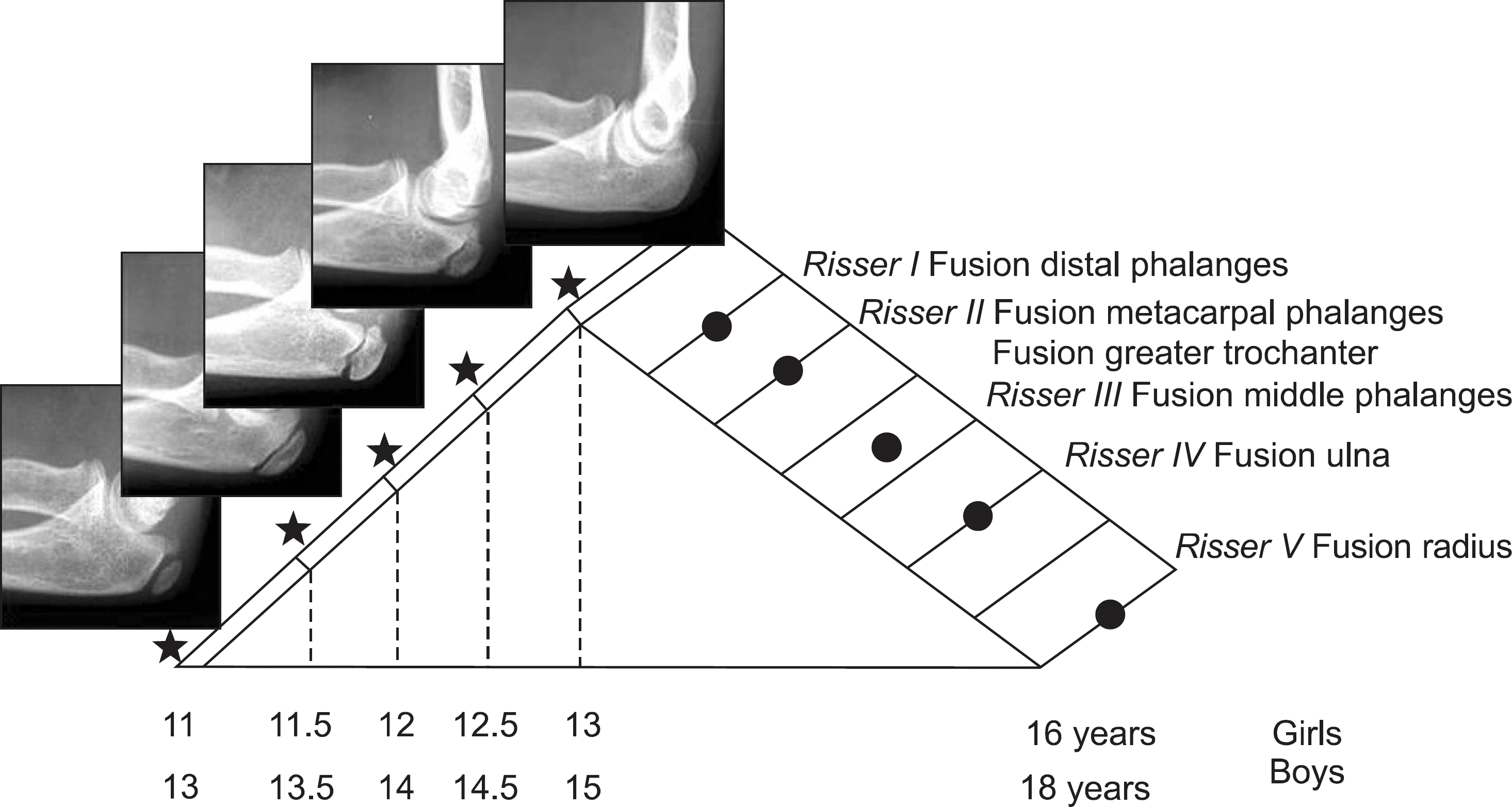
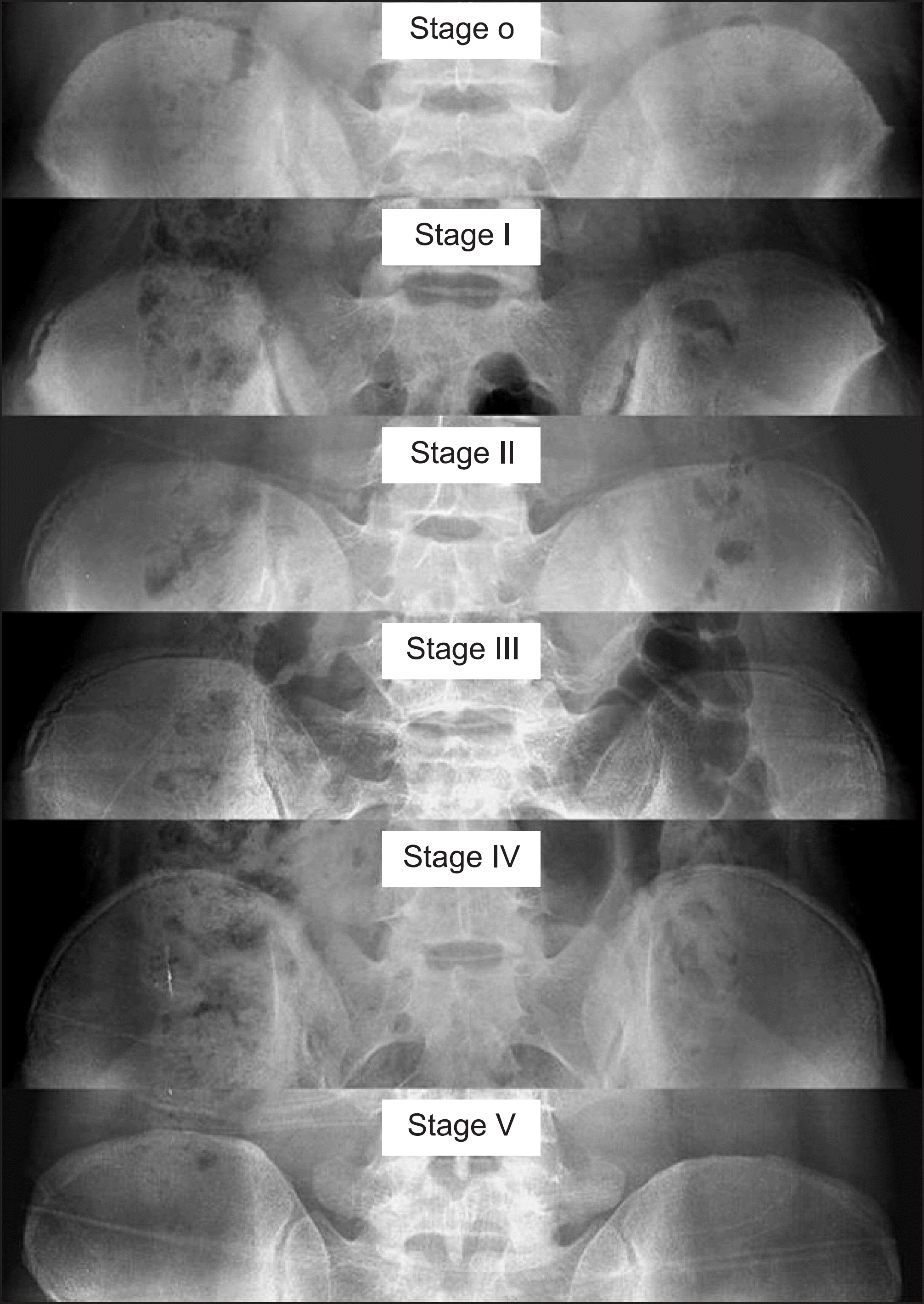
Figure 6.
This showing contributions of the various growth plates to the length of the lower limb and its individual bones.
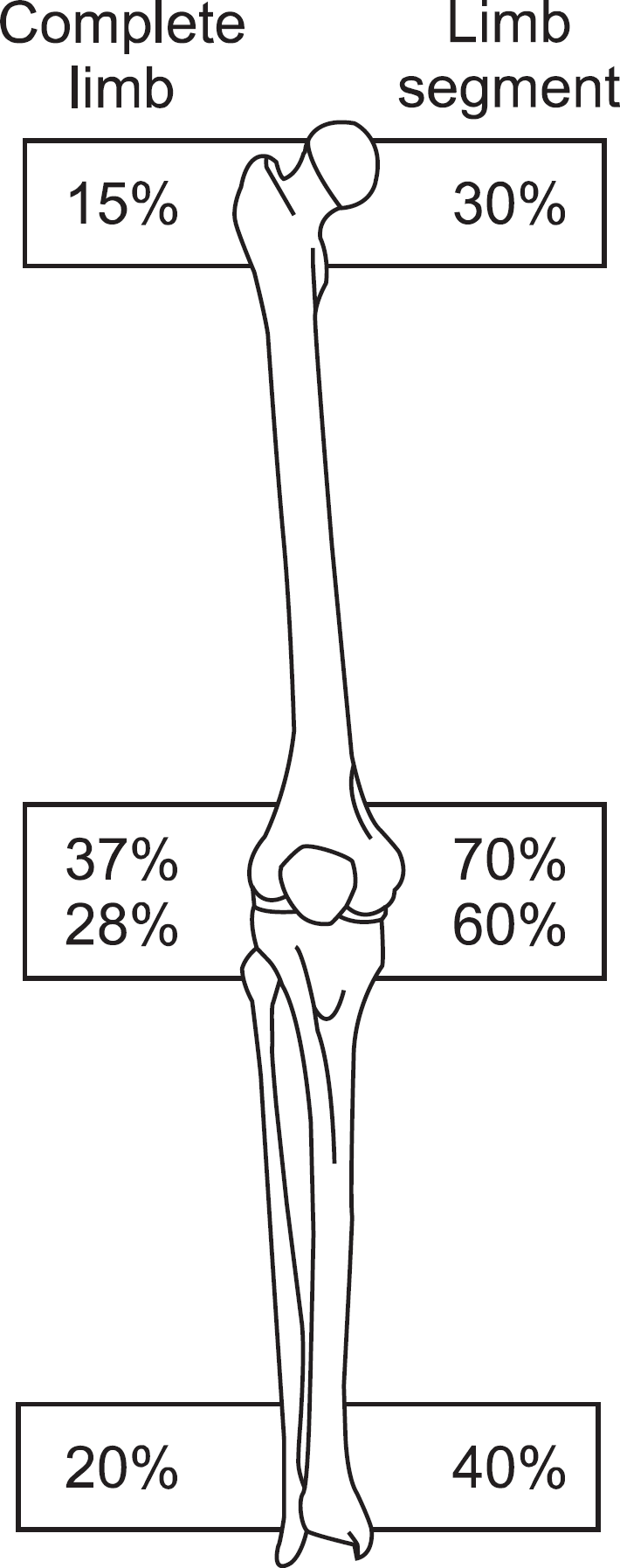
Figure 7.
This showing an average total length of the femur and tibia with a 1 and 2 standard deviation range in girls based on chronologic age provided by Green and Anderson.
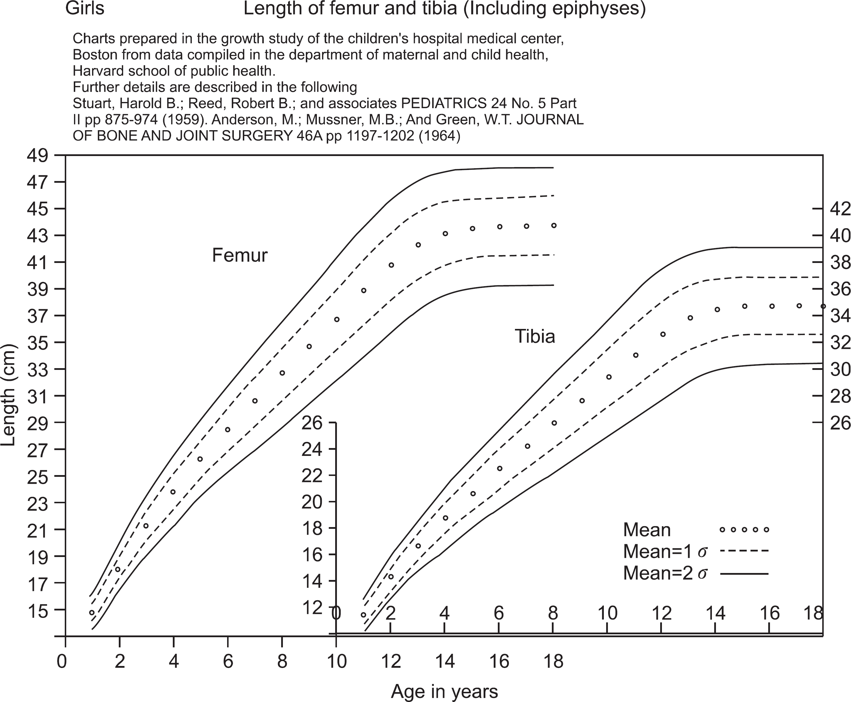
Figure 8.
Plain radiograph showing osteosarcoma on right proximal femur and reconstruction with tumor prosthesis of 7.6 years old patient.
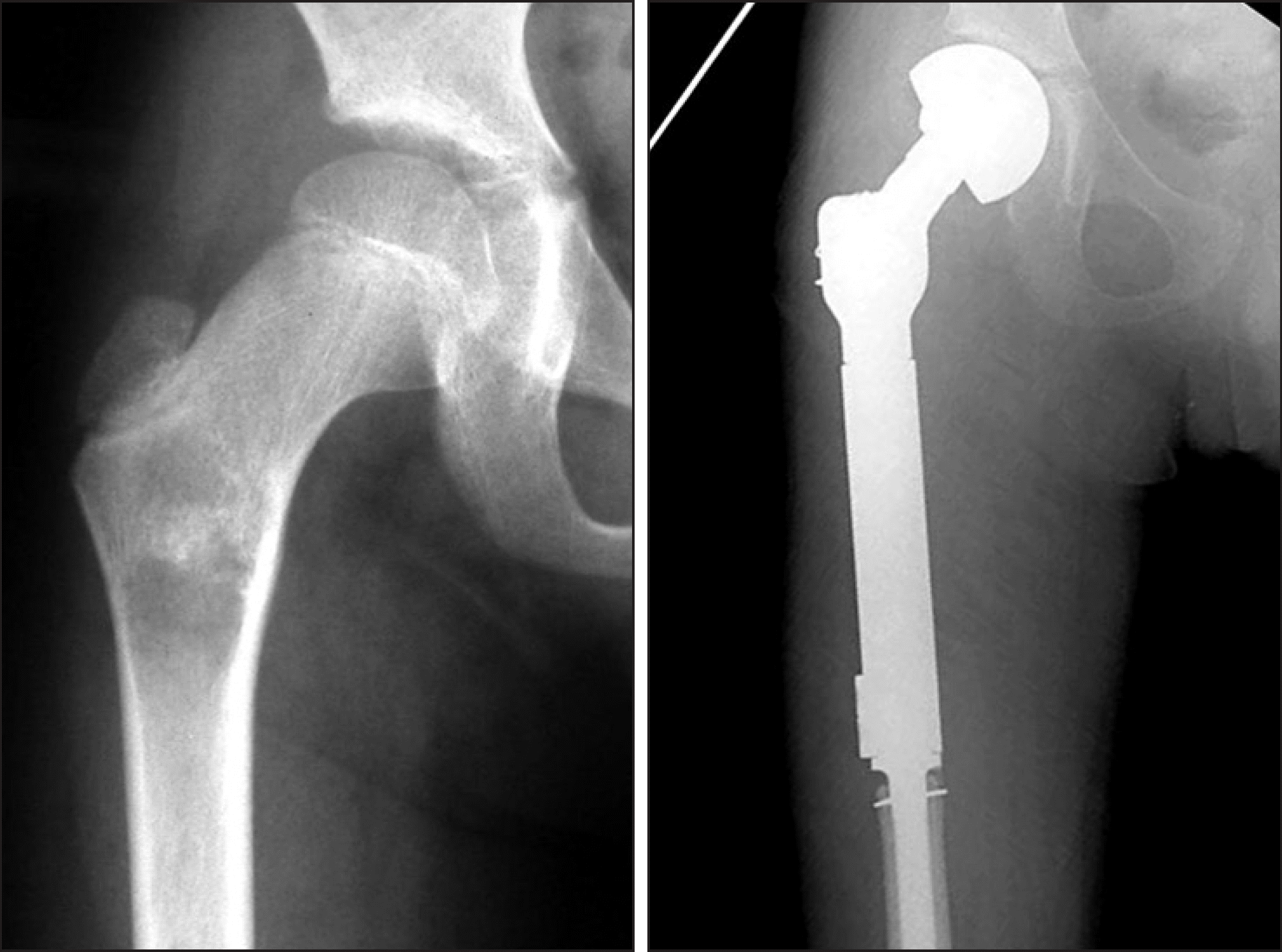
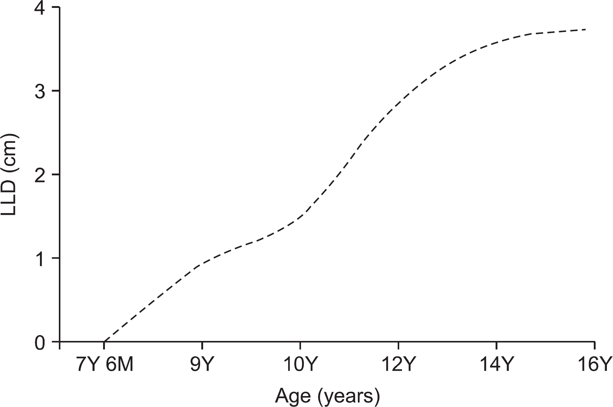
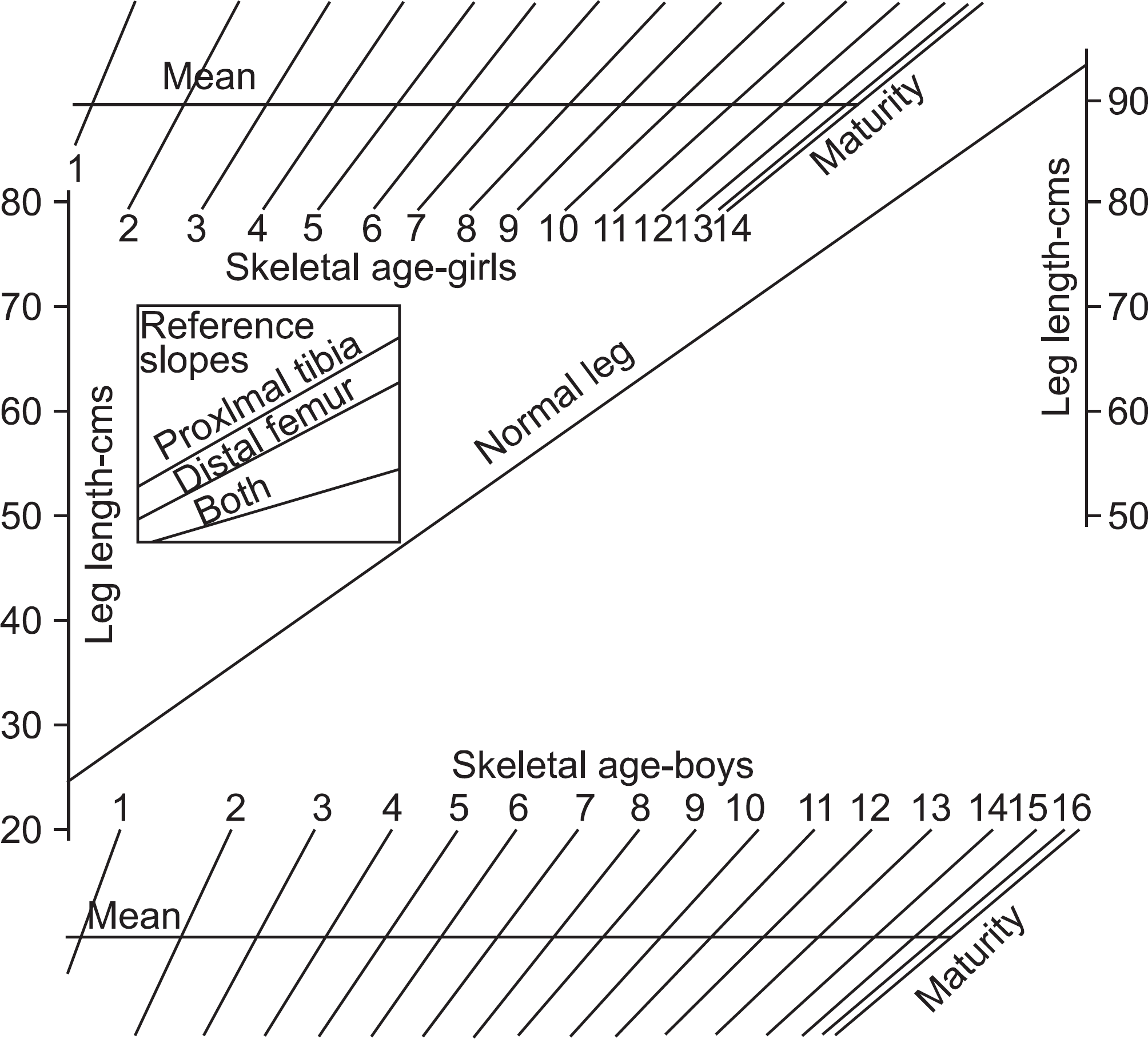
Figure 11.
Plain radiographs showing osteosarcoma on left distal femur, reconstruction with allograft, and about 7 cm LLD after 4 years follow up.
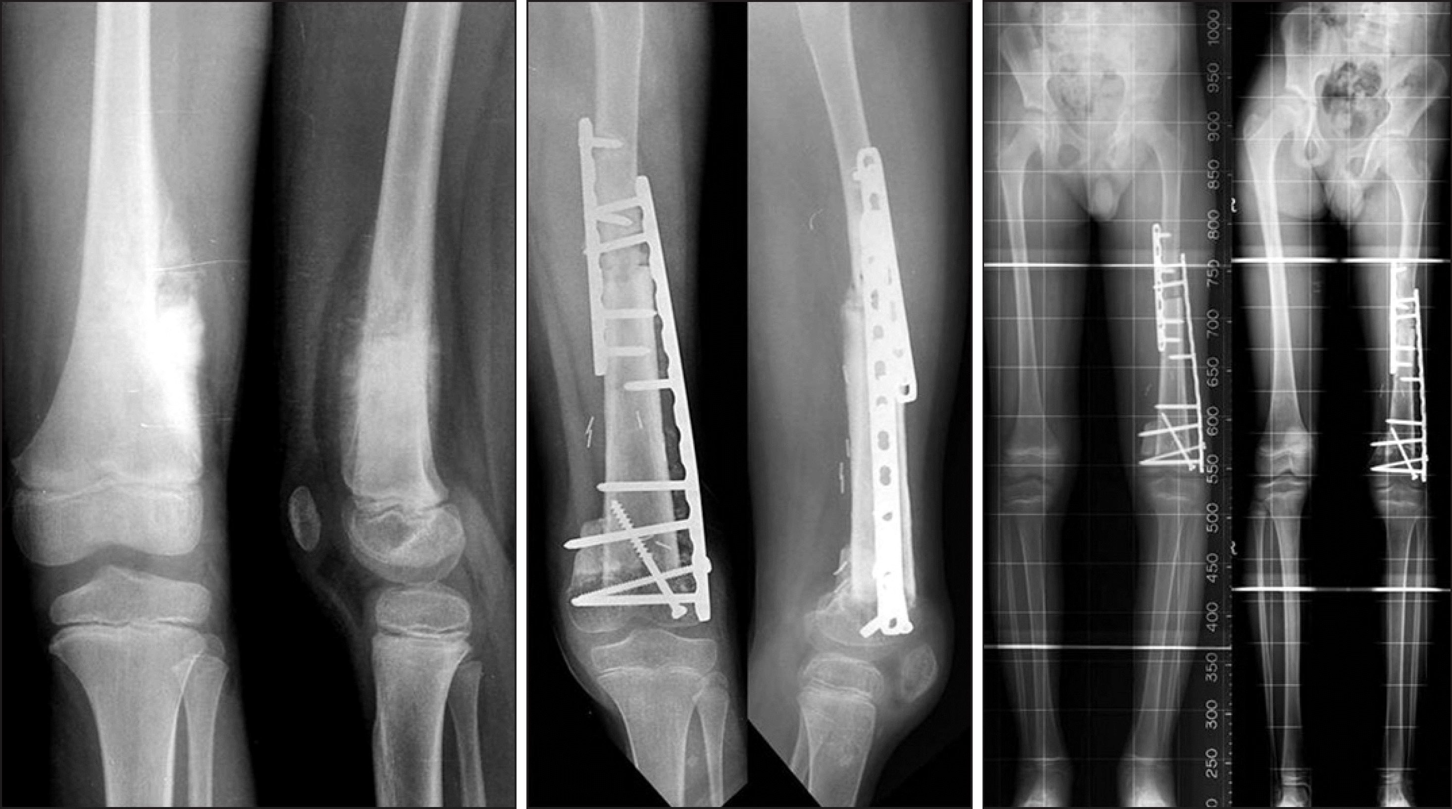
Table 1.
Characteristics of Growth Pattern
Table 2.
Length of Femur and Tibia
| Femur | Tibia | ||
|---|---|---|---|
| At birth | 9 cm | 7 cm | |
| At growth maturation | Girls | 47 cm | 37 cm |
| Boys | 44 cm | 34 cm |
Table 3.
Lower-limb Multipliers
| Age | Multiplier | Age | Multiplier | ||
|---|---|---|---|---|---|
| (yrs∗+mos†) | Boys | Girls | (yrs+mos) | Boys | Girls |
| Birth | 5.080 | 4.630 | 7+6 | 1.520 | 1.370 |
| 0+3 | 4.550 | 4.155 | 8+0 | 1.470 | 1.330 |
| 0+6 | 4.050 | 3.725 | 8+6 | 1.420 | 1.290 |
| 0+9 | 3.600 | 3.300 | 9+0 | 1.380 | 1.260 |
| 1+0 | 3.240 | 2.970 | 9+6 | 1.340 | 1.220 |
| 1+3 | 2.975 | 2.750 | 10+0 | 1.310 | 1.190 |
| 1+6 | 2.825 | 2.600 | 10+6 | 1.280 | 1.160 |
| 1+9 | 2.700 | 2.490 | 11+0 | 1.240 | 1.130 |
| 2+0 | 2.590 | 2.390 | 11+6 | 1.220 | 1.100 |
| 2+3 | 2.480 | 2.295 | 12+0 | 1.180 | 1.070 |
| 2+6 | 2.385 | 2.200 | 12+6 | 1.160 | 1.050 |
| 2+9 | 2.300 | 2.125 | 13+0 | 1.130 | 1.030 |
| 3+0 | 2.230 | 2.050 | 13+6 | 1.100 | 1.010 |
| 3+6 | 2.110 | 1.925 | 14+0 | 1.080 | 1.000 |
| 4+0 | 2.000 | 1.830 | 14+6 | 1.060 | |
| 4+6 | 1.890 | 1.740 | 15+0 | 1.040 | |
| 5+0 | 1.820 | 1.660 | 15+6 | 1.020 | |
| 5+6 | 1.740 | 1.580 | 16+0 | 1.010 | |
| 6+0 | 1.670 | 1.510 | 16+6 | 1.010 | |
| 6+6 | 1.620 | 1.460 | 17+0 | 1.000 | |
| 7+0 | 1.570 | 1.430 | |||
 XML Download