

 PDF
PDF ePub
ePub Citation
 Print
Print


Abstract
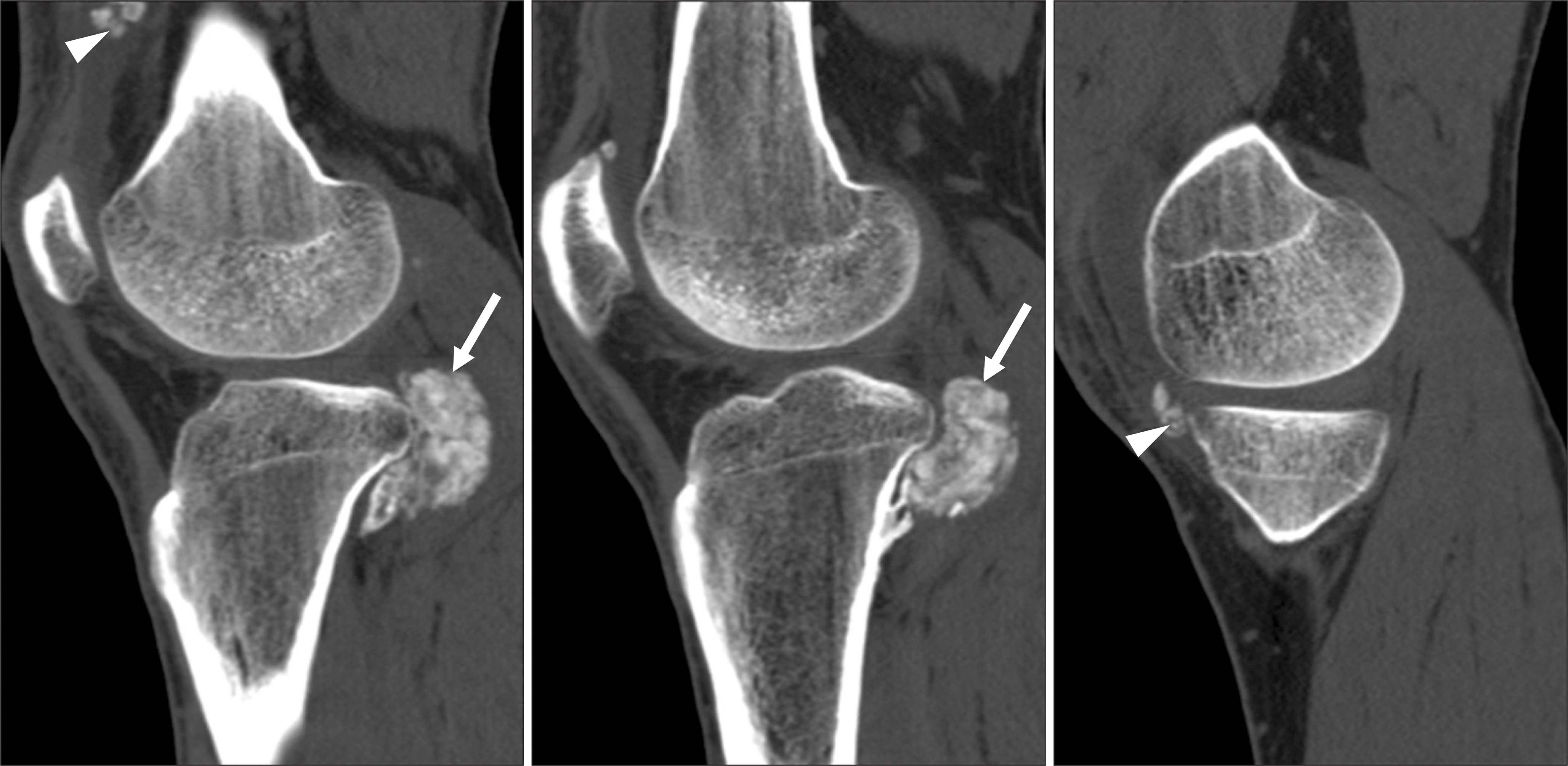
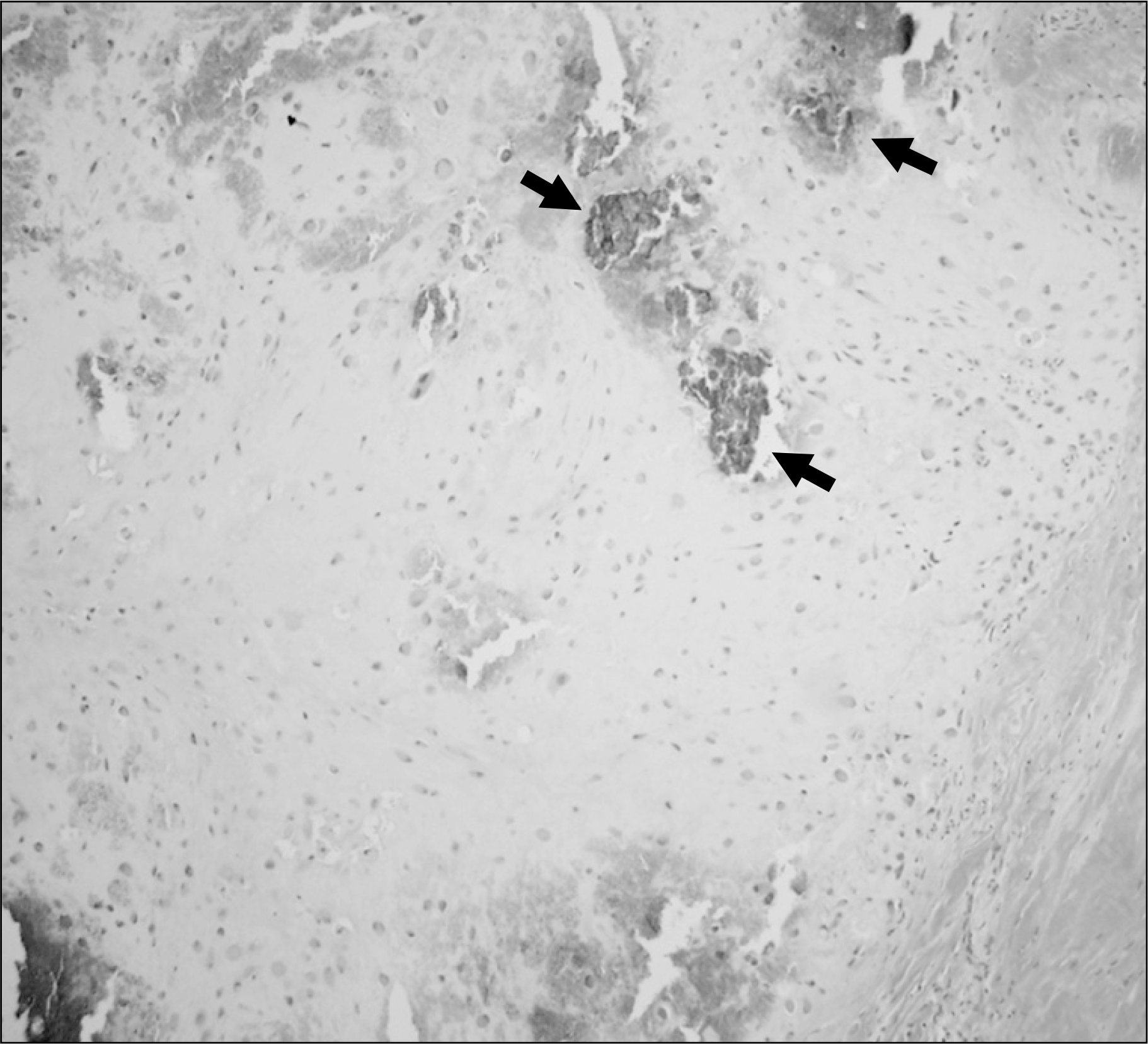
Synovial chondromatosis is a benign nodular cartilaginous proliferation arising in the synovium of joints. The radiolographic features of this condition are variable. Rarely, it would be confused with malignancy such as chondrosarcoma, osteosarcoma or synovial sarcoma. We report a case of primary synovial chondromatosis of the posterior aspect of the proximal tibia mimicking a parosteal osteoarcoma on the radiography, which showed a homogeneously radiopaque juxtacortical mass. However, subsequent computed tomography (CT) showed multiple intra-articular masses containing chondroid mineralization, suggesting synovial chondromatosis.
Go to : 
REFERENCES
1. Davis RI, Hamilton A, Biggart JD. Primary synovial chondromatosis: a clinicopathologic review and assessment of malignant potential. Hum Pathol. 1998; 29:683–8.

2. Milgram JW, Hadesman WM. Synovial osteochondromatosis in the subacromial bursa. Clin Orthop Relat Res. 1988; 236:154–9.
3. Norman A, Steiner GC. Bone erosion in synovial chondromatosis. Radiology. 1986; 161:749–52.
4. Villacin AB, Brigham LN, Bullough PG. Primary and secondary synovial chondrometaplasia: histopathologic and clinicoradiologic differences. Hum Pathol. 1979; 10:439–51.
5. Murphey MD, Vidal JA, Fanburg-Smith JC, Gajewski DA. Imaging of synovial chondromatosis with radiologic-pathologic correlation. Radiographics. 2007; 27:1465–88.
6. Edeiken J, Edeiken BS, Ayala AG, Raymond AK, Murray JA, Guo SQ. Giant solitary synovial chondromatosis. Skeletal Radiol. 1994; 23:23–9.
7. Levine E, De Smet AA, Huntrakoon M. Juxtacortical osteosarcoma: a radiologic and histologic spectrum. Skeletal Radiol. 1985; 14:38–46.
8. Lorentzon R, Larsson SE, Boquist L. Parosteal (juxtacortical) osteosarcoma. A clinical and histopathological study of 11 cases and a review of the literature. J Bone Joint Surg Br. 1980; 62-B:86–92.
9. Okada K, Frassica FJ, Sim FH, Beabout JW, Bond JR, Unni KK. Parosteal osteosarcoma. A clinicopathological study. J Bone Joint Surg Am. 1994; 76:366–78.
Go to :
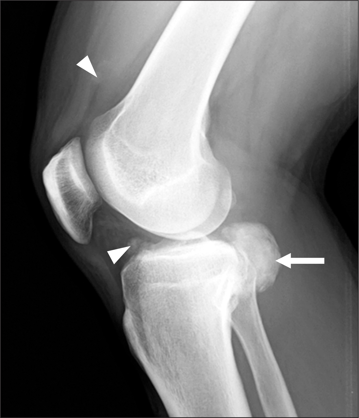 | Figure 1.Lateral radiograph shows a soft tissue mass (arrow) containing dense mineralization extending into the popliteal fossa. Partial attachment to the cortex as well as radiolucent cleavage plane was evident. Two small radiodense soft tissue masses (arrowheads) were initially missed on the radiograph. |
 XML Download