

 PDF
PDF ePub
ePub Citation
 Print
Print


Abstract
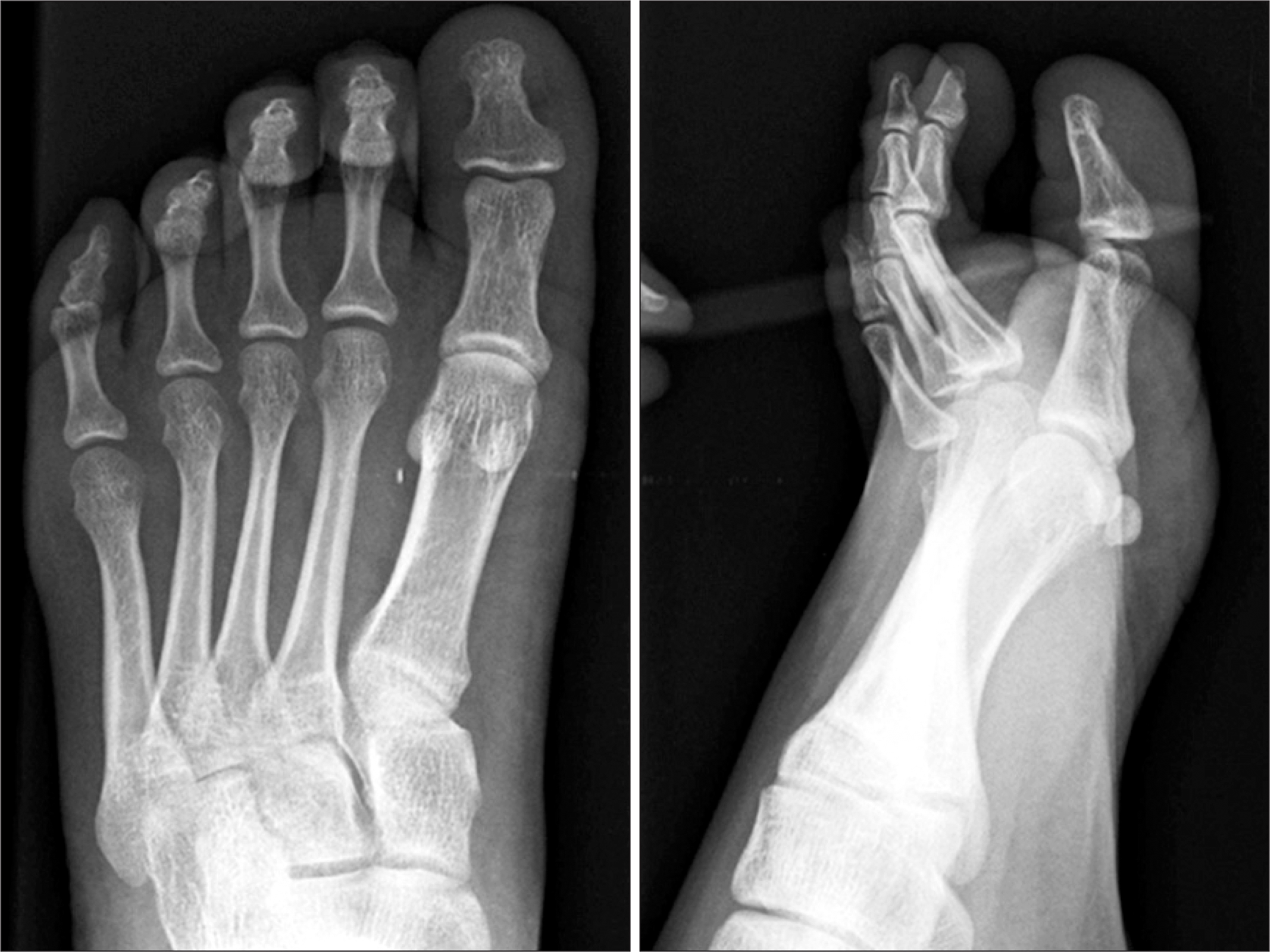
Fibroosseous pseudotumor is an extremely rare benign lesion which is fast-growing and painful. It is often misdiagnosed as a malignancy, but it is a noninvasive entity and can be cured by simple resection. We report a case of fibroosseous pseudotumor of the distal phalanx of great toe in 20-year-old female patient who present with painful mass.
Go to : 
REFERENCES
1. Dupree WB, Enzinger FM. Fibro-ossoeous pseudotumor of the digitis. Cancer. 1986; 58:2103–9.
2. Moosavi CA, Al-Nahar LA, Murphey MD, Fanburg-Smith JC. Fibrosseous pseudotumor of the digit: a clinicopathologic study of 43 new cases. Annals of Diagnostic Pathology. 2008; 12:21–8.

3. Nishio J, Iwasaki H, Soejima O, Naito M, Kikuchi M. Rapidly growing fibroosseous pseudotumor of the digits mimicking extraskeletal osteosarcoma. J Orthop Sci. 2002; 7:410–3.
4. Schutte HE, van der Heul RO. Pseudomalignant non-neoplastic osseous soft tissue tumors of the hand and foot. Radiology. 1990; 176:149–53.
5. Porter AR, Tristan TA, Rudy FR, Eshbach TB. Florid reactive periostitis of the phalanges. AJR Am J Roentgenol. 1985; 144:617–8.
6. De Silva MVC, Reid R. Myositis ossificans and fibroosseous pseudotumor of digits. Int J Surg Pathol. 2003; 11:187–95.
7. Kempson RL, Fletcher CD, Evans HL, Hendrickson MR, Sibley RK. Tumors of the soft tissue. Washington, DC: Armed forces Institute of Pathology;2001. p. 409–13.
8. Kransdorf MJ. Benign soft-tissue tumors in a large referral population: distribution of diagnosis by age, sex and location. AJR Am J Roentgenol. 1995; 164:395–402.
9. Park IS, Han JY, Han HS, Kim YB, Chu YC. Fibroosseous pseudotumor of the digits: a case report. Korean J Pathol. 1999; 33:540–3.
10. Chaudhry IH, Kazakov DV, Michal T, Luzar B, Calonje E. Fibroosseous pseudotumor of the digit: a clinicopathologic study of 17 cases. J Cutan Pathol. 2010; 37:323–9.
Go to :
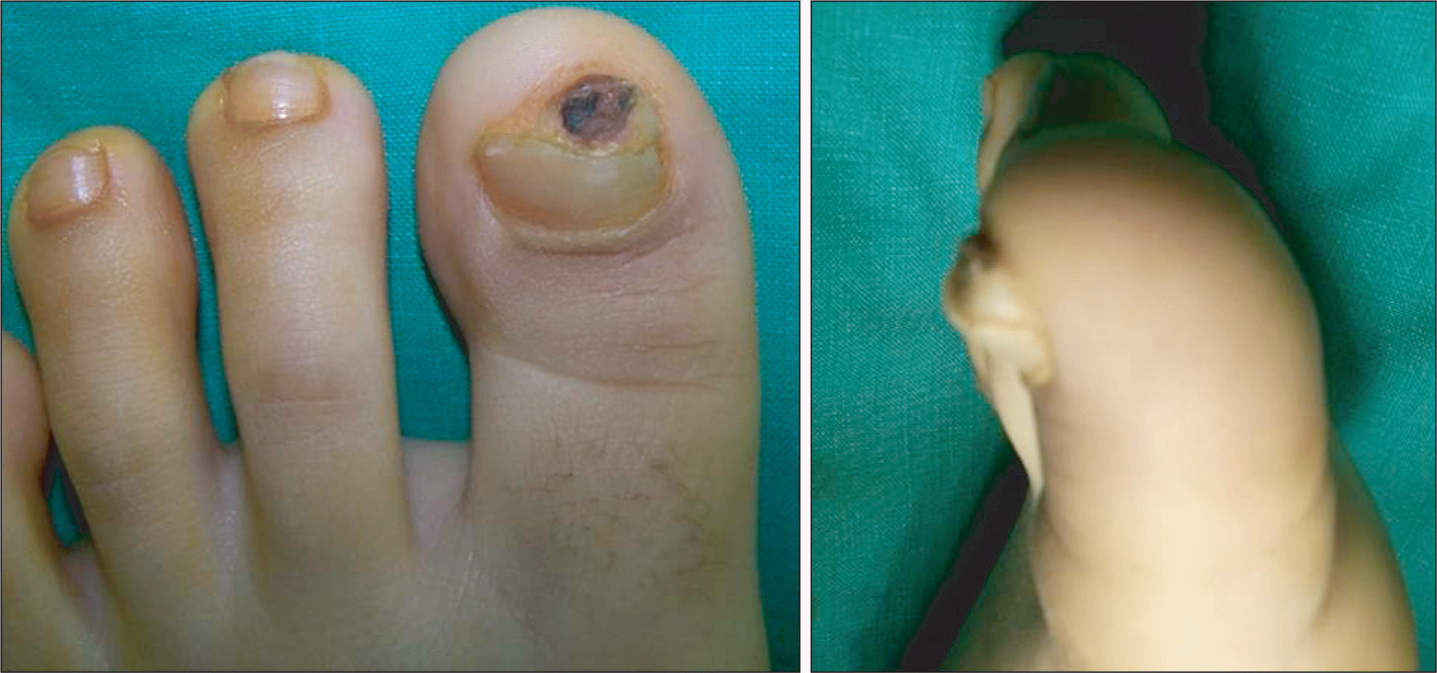 | Figure 1.Initial anteroposterior and lateral views of left great toe show a brown-colored solid mass with granulation tissue. |
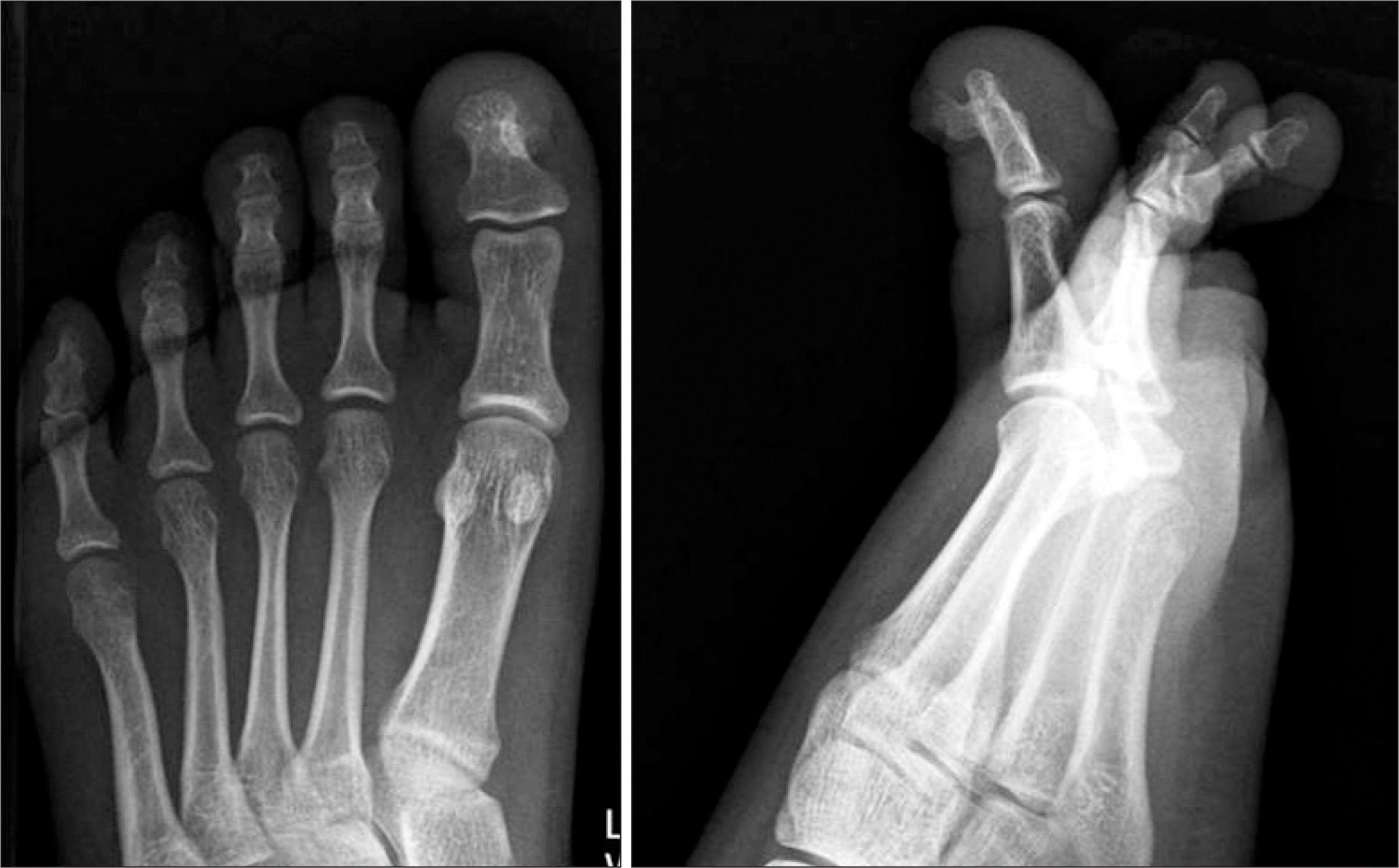 | Figure 2.The anteroposterior and lateral radiographs of the left foot show that bony projection arose from the subungual portion of the distal phalanx of the great toe without periosteal reaction. |
 XML Download