Abstract
Enchondroma is a benign tumor mainly developed in the hand and uncommon in the foot. Even if it is in the foot, most are in the phalanges and distal metatarsals of the foot. Enchondroma in the calcaneus is very rare. A 44-year-old male suffered from left heel pain for several months, authors treated it with curettage and bone graft, it was histologically confirmed as an enchondroma in the calcaneus. The authors presented a rare case presentation of an enchondroma in the calcaneus with pain.
REFERENCES
1. Burns AE. Enchondroma: two case reports. J Foot Surg. 1990; 29:551–6.
2. Dahlin DC, Unni KK. Bone tumors: general aspects and data on 11,087 cases. 5th ed.Philadelphia: Lippincott-Raven;1996. p. 22–45.
3. Mirra JM. Bone Tumors. Clinical, radiologic and pathologic correlations. Philadelphia: Lea and Febiger;1989. 480-1. 487:p. 515–6.
4. Perlman MD, Gold ML, Schor AD. Enchondroma: a case report and literature review. J Foot Surg. 1988; 27:556–60.
5. Mirra JM, Gold R, Downs J, Eckardt JJ. A new histologic approach to the differentiation of enchondroma and chondrosarcoma of the bones. A clinicopathologic analysis of 51 cases. Clin Orthop Relat Res. 1985; 201:214–37.
6. Gamble FO, Yale I. Clinical foot roentgenology. An illustrated handbook. Baltimore: The Williams and Wilkins;1966. 106.
7. Flemming DJ, Murphey MD. Enchondroma and chondrosarcoma. Semin Musculoskelet Radiol. 2000; 4:59–71.

8. Hasselgren G, Forssblad P, Tornvall A. Bone grafting unnecessary in the treatment of enchondromas in the hand. J Hand Surg Am. 1991; 16:139–42.

9. Kang E, Rho K, Yoo J. Comparative study of the simple curettage and the curettage with bone graft in enchondroma of the hand. J Korean Orthop Assoc. 1997; 32:156–62.
10. Song S, Lee J, Yoon H. Treatment of enchondroma of the hand with curettage and dehydrated alcohol instillation. J Korean Soc Surg Hand. 2004; 9:148–52.
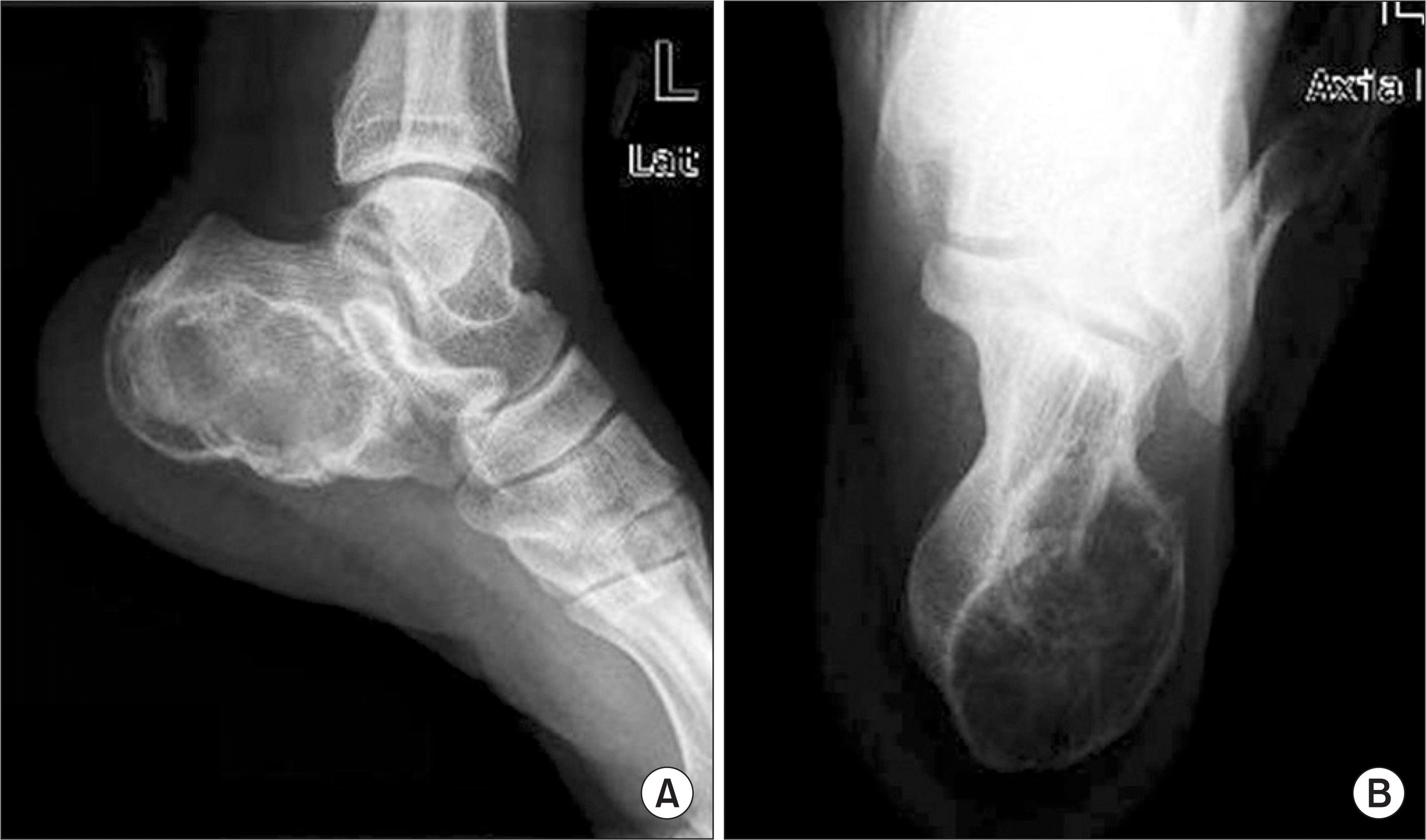
Figure 1.
(A) Lateral preoperative radiograph of the left calcaneus revealed osteolytic lesion with thin sclerotic cortex and internal ground glass opacity at posterior and plantar aspect. (B) Axial preoperative radiograph showed osteolytic lesion mainly at medial aspect of calcaneus.

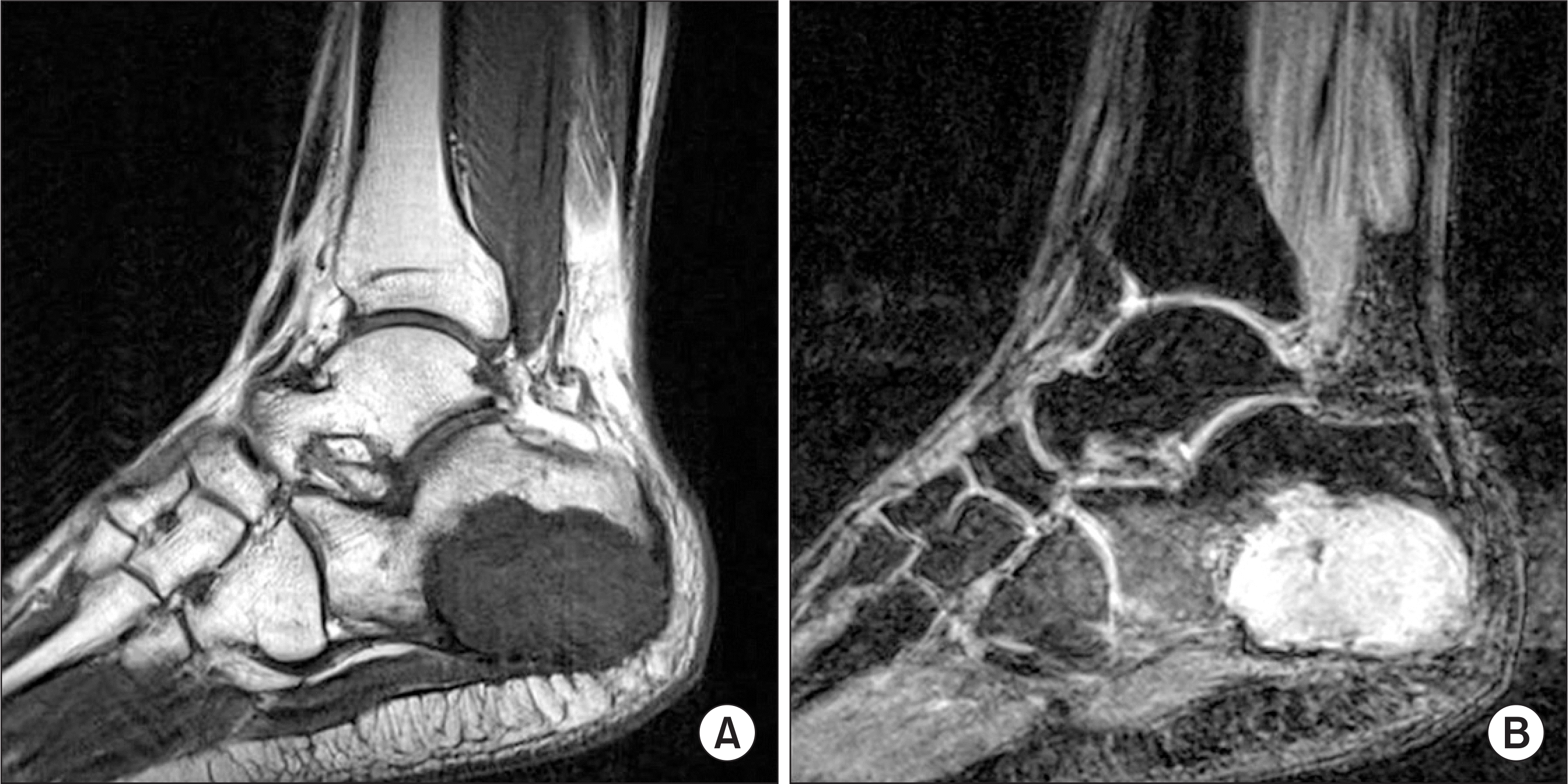
Figure 2.
(A) T1-weighted sagittal plane image showed a well-circumscribed, lobulated lesion displaying intermediate signal intensity. (B) Enhanced T2-weighted sagittal plane image showed a mixed-intensity signal of the high signal areas represent cartilaginous tumor and low signal areas show calcification.


 PDF
PDF ePub
ePub Citation
 Print
Print


 XML Download