

 PDF
PDF ePub
ePub Citation
 Print
Print


Abstract
Purpose
The skeleton is commonly affected by metastatic cancer. The purpose of this study was to evaluate the results of treating metastatic pathologic fractures in lower extremities using locking plates.
Materials and Methods
Between 2004 and 2010, we evaluated 12 patients (13 cases) of metastatic pathologic fractures in lower extremities, treated with the locking plate. Mean patient age was 62.2 years (range, 50-81 years), the locations of the fractures were; proximal femur in 2 cases, femoral mid-shaft in 3, distal femur in 3, proximal tibia in 4, and distal tibia in 1 case. The interval to wheelchair ambulation, pain relief and complications were evaluated. Additionally, we assessed operation time and postoperative blood loss.
Results
Mean time from operation to wheelchair ambulation was 3.2 days (range, 1-6 days). Mean VAS scores improved from a preoperative score of 8.1 points (range, 7-9 points) to a score of 2.7 points (range, 2-4 points) at 1 week postoperatively. No early complications associated with surgery were encountered. Mean operation time was 88.4 minutes (range, 70-105 minutes), and mean postoperative blood loss was 246.5 ml (range, 130-320 ml).
Conclusion
Internal fixation of metastatic pathologic fractures using a locking plate in the lower extremity can be an effective treatment option in the meta- or diaphyseal area of long bones with massive bony destruction or poor bone stock by offering early ambulation, pain relief and low postoperative complications.
REFERENCES
1. Coleman RE. Skeletal complications of malignancy. Cancer. 1997; 80:1588–94.

2. Dahlin DC, Unni KK. Bone tumours. 4th ed.Springfield, Illinois: Thomas;1986.
3. Varadhachary GR, Abbruzzese JL, Lenzi R. Diagnostic strategies for unknown primary cancer. Cancer. 2004; 100:1776–85.
4. Biermann JS, Holt GE, Lewis VO, Schwartz HS, Yaszemski MJ. Metastatic bone disease: diagnosis, evaluation, and treatment. Instr Course Lect. 2010; 59:593–606.
5. Siegel HJ, Lopez-Ben R, Mann JP, Ponce BA. Pathological fractures of the proximal humerus treated with a proximal humeral locking plate and bone cement. J Bone Joint Surg Br. 2010; 92:707–12.
6. Bashore CJ, Temple HT. Management of metastatic lesions of the humerus. Orthop Clin North Am. 2000; 31:597–609.
7. Bauer HC. Controversies in the surgical management of skeletal metastases. J Bone Joint Surg Br. 2005; 87:608–17.
8. Frassica FJ, Frassica DA. Evaluation and treatment of metastases to the humerus. Clin Orthop Relat Res. 2003; 415:S212–8.
9. Ward WG, Holsenbeck S, Dorey FJ, et al. Metastatic disease of the femur: surgical treatment. Clin Orthop. 2003; 415:S230–44.
10. Bickels J, Kollender Y, Wittig JC, et al. Function after resection of humeral metastases: analysis of 59 consecutive patients. Clin Orthop. 2005; 437:201–8.
11. Harrington KD. Impending pathologic fractures from metastatic malignancy: evaluation and management. Instr Course Lect. 1986; 35:357–81.
12. Jung ST, Ghert MA, Harrelson JM, et al. Treatment of osseous metastases in patients with renal cell carcinoma. Clin Orthop. 2003; 409:223–31.
13. Kay PR. Cement augmentation of pathological fracture fixation. J Bone Joint Surg Br. 1989; 71:702.
14. Mercadante S. Malignant bone pain: pathophysiology and treatment. Pain. 1997; 69:1–18.
15. Mundy GR. Mechanisms of bone metastasis. Cancer. 1997; 80:1546–56.
16. Friedl W. Indication, management and results of surgical therapy for pathological fractures in patients with bone metastases. Eur J Surg Oncol. 1990; 16:380–96.
17. Friedl W, Mieck U, Fritz T. Surgical therapy of bone metastases of the upper and lower extremity. Chirurg. 1992; 63:897–911.
18. Graupe F, Heitmann C, Becker M, et al. Palliative surgical treatment of bone metastases. Improved quality of life by early intervention? Dtsch Med Wochenschr. 1996; 121:393–7.
19. Hardy DC, Descamps PY, Krallis P, et al. Use of an intramedullary hip-screw compared with a compression hip-screw with a plate for intertrochanteric femoral fractures. A prospective, randomized study of one hundred patients. J Bone Joint Surg Am. 1998; 80:618–30.
20. Brumback RJ, Toal TR Jr, Murphy-Zane MS, et al. Immediate weight-bearing after treatment of a comminuted fracture of the femoral shaft with a statically locked intramedullary nail. J Bone Joint Surg Am. 1999; 81:1538–44.
21. Anglen J, Kyle RF, Marsh JL, et al. Locking plates for extremity fractures. J Am Acad Orthop Surg. 2009; 17:465–72.
22. Hage WD, Aboulafia AJ, Aboulafia DM. Incidence, location, and diagnostic evaluation of metastatic bone disease. Orthop Clin North Am. 2000; 31:515–28.
23. Heinz T, Stoik W, Vécsei V. Treatment and results of pathologic fractures. A collaborative study from 1965 to 1985 of 16 Austrian hospitals. Unfallchirurg. 1989; 92:477–85.
24. Barwood SA, Wilson JL, Molnar RR, et al. The incidence of acute cardiorespiratory and vascular dysfunction following intramedullary nail fixation of femoral metastasis. Acta Orthop Scand. 2000; 71:147–52.
25. Cole AS, Hill GA, Theologis TN, et al. Femoral nailing for metastatic disease of the femur: a comparison of reamed and unreamed femoral nailing. Injury. 2000; 31:25–31.
26. Sharma H, Bhagat S, McCaul J, Macdonald D, Rana B, Naik M. Intramedullary nailing for pathological femoral fractures. J Orthop Surg (Hong Kong). 2007; 15:291–4.
27. Moholkar K, Mohan R, Grigoris P. The Long Gamma Nail for stabilization of existing and impending pathological fractures of the femur: an analysis of 48 cases. Acta Orthop Belg. 2004; 70:429–34.
Figure 1.
A 70-year-old man with a fracture of the distal femur secondary to metastasis of lung cancer. (A) Anteroposterior (AP) radiograph of the distal femur shows a displaced pathologic fracture. (B) The patient underwent an open reduction and internal fixation using a locking plate with cement augmentation.
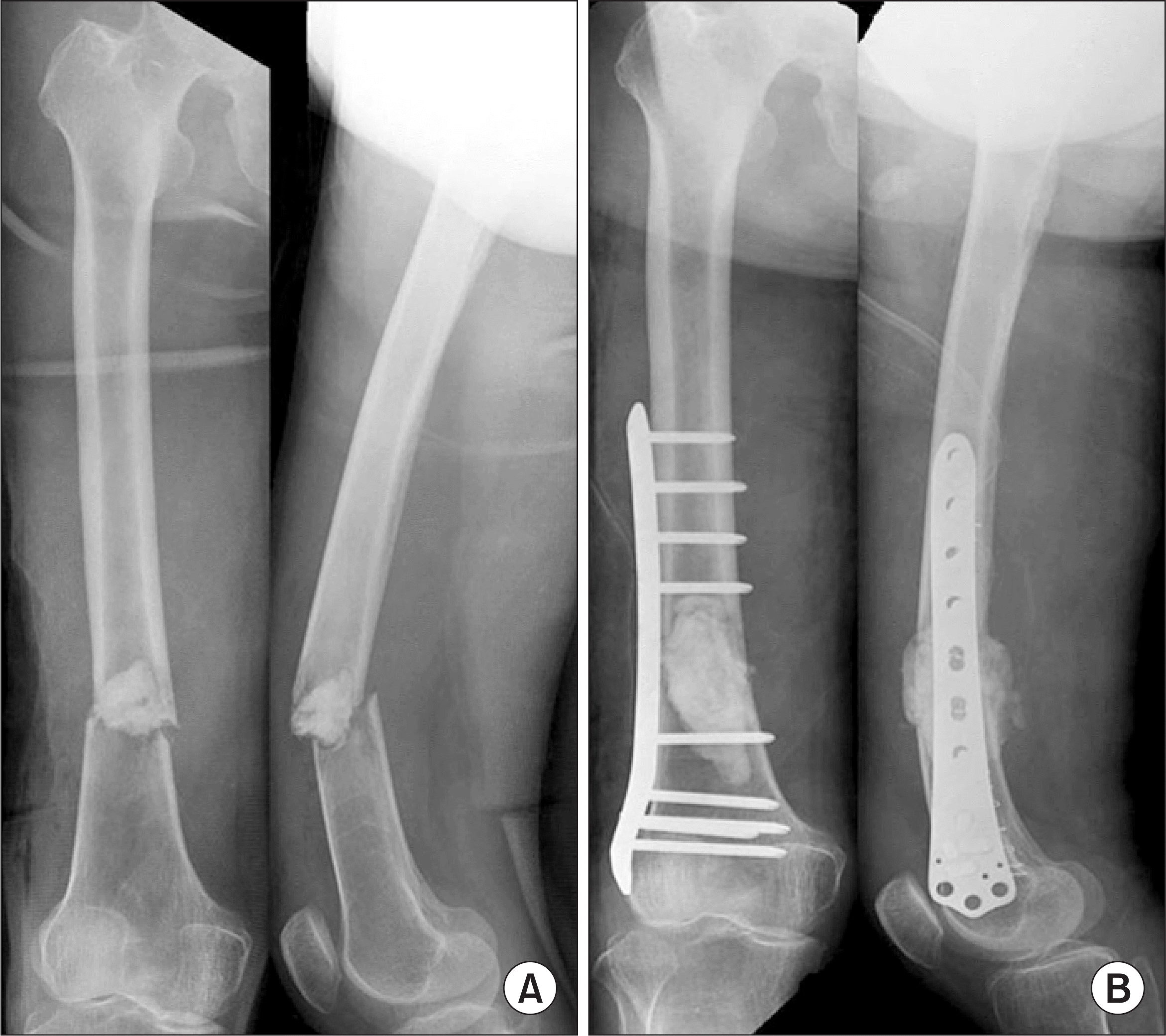
Figure 2.
An 81-year-old man with thyroid cancer showing a pathologic fracture of the femoral mid-shaft. (A) Preoperative radiograph shows an osteolytic lesion of the femoral shaft. (B) Coronal plane of preoperative CT scan demonstrates a cortical discontinuity around the lesion of the femoral shaft. (C) A minimally invasive plate osteosynthesis was performed with a locking plate.

Figure 3.
A 58-year-old man with a pathologic fracture of the distal femur due to lung cancer. (A) AP radiograph shows an osteolytic lesion of the distal femur with a pathologic fracture. (B) The distal femur with a pathologic fracture was stabilized by using locking plate with cement augmentation. (C) AP and lateral radiograph obtained 3 months after internal fixation shows loss of reduction and failure of the internal fixation.
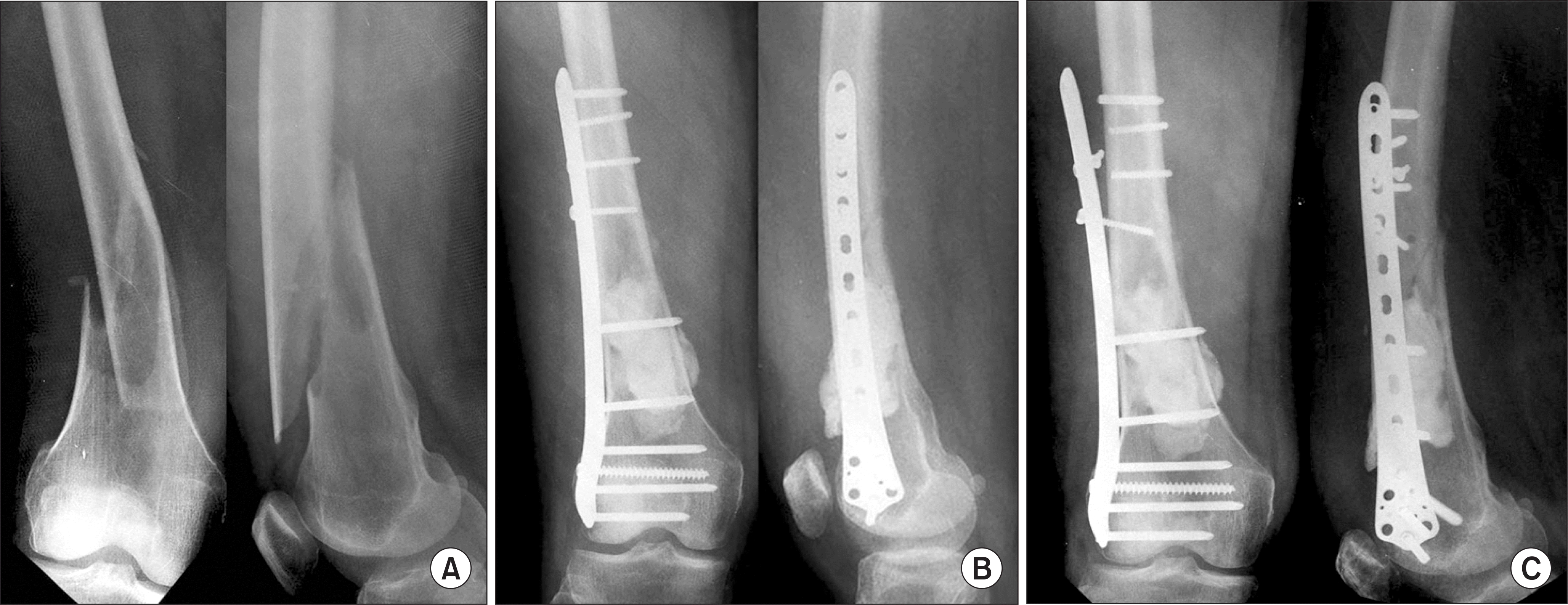
Table 1.
Demographic Data of Patients with an Impeding or Pathologic Fracture of the Lower Extremity Caused by Metastatic Cancer
| Cases | Gender | Age (year) | Primary tumor | Metastases | Pathologic fracture site | Operation name | Survival after fracture (day) | Interval from operation to ambulation (day) | Preoperative VAS (points) | Postoperative VAS (points) | Postoperative blood loss (ml) | Operation time (minute) |
|---|---|---|---|---|---|---|---|---|---|---|---|---|
| 1 | M | 81 | Thyroid | Multiple (skeleton, Lymph nodes) | Femur mid-shaft | ORIF with LCP | 84 | 1 | 8 | 3 | 260 | 70 |
| 2 | M | 61 | Lung | Multiple (skeleton) | Proximal femur | ORIF with LCP | 14 | No∗ | 8 | 3 | 200 | 65 |
| 3 | M | 61 | Lung | Multiple (skeleton) | Proximal tibia | ORIF with LCP & cementing | 14 | No∗ | 8 | 2 | 315 | 80 |
| 4 | M | 70 | Stomach | Multiple (Skeleton, Brain) | Distal tibia | ORIF with LCP & cementing | 32 | 3 | 7 | 4 | 130 | 65 |
| 5 | M | 70 | Lung | Multiple (Skeleton, Lymph nodes) | Distal femur | ORIF with LCP & cementing | 44 | 2 | 9 | 3 | 320 | 85 |
| 6 | M | 58 | Lung | Multiple (Skeleton, Brain) | Distal femur | ORIF with LCP & cementing | 220 | 6 | 9 | 2 | 200 | 90 |
| 7 | M | 50 | Lung | Multiple (Skeleton, Brain) | Distal femur | ORIF with LCP & cementing | 378 | 4 | 8 | 2 | 300 | 95 |
| 8 | F | 58 | Thyroid | Multiple (Skeleton) | Proximal femur | ORIF with LCP & cementing | 38 | 4 | 7 | 4 | 225 | 105 |
| 9 | M | 60 | Lung | Multiple (Skeleton) | Mid-shaft femur | ORIF with LCP & cementing | 65 | 2 | 8 | 2 | 255 | 105 |
| 10 | F | 66 | Stomach | Multiple (Skeleton) | Proximal tibia | ORIF with LCP & cementing | 149 | 2 | 8 | 3 | 230 | 100 |
| 11 | F | 56 | Kidney | Multiple (Skeleton) | Proximal tibia | ORIF with LCP | 241 | 5 | 9 | 3 | 295 | 95 |
| 12 | M | 57 | Lung | Multiple (Skeleton, Brain) | Mid-shaft femur | ORIF with LCP & cementing | 74 | 3 | 7 | 2 | 265 | 95 |
| 13 | M | 62 | Lung | Multiple (Skeleton) | Proximal tibia | ORIF with LCP | 107 | 3 | 9 | 2 | 210 | 100 |
| ORIF, open reduction and internal fixation; LCP, locking compression plate. | ||||||||||||
Table 2.
Results Compared with Intramedullary Nailing for the Treatment of Pathologic Fractures
| Authors | Cases | Operation time (minute) | Blood loss (ml) | Ambulation (day) | Complications (numbers) |
|---|---|---|---|---|---|
| Sharma et al.26) | 21 | 140 | 900 | 3∗ | Superficial wound infection (2) |
| Pneumonia (2) | |||||
| Loss of reduction (1) | |||||
| Moholkar et al.27) | 48 | 98 | 400 | 7† | Chest infection (4) |
| Urinary track infection (2) | |||||
| Superficial wound infection (2) | |||||
| Deep wound infection (1) | |||||
| Renal failure (1) | |||||
| Death (2) | |||||
| Present study | 13 | 88.4 | 246.5 | 3.2 | Screw breakage (1) |
 XML Download