

 PDF
PDF ePub
ePub Citation
 Print
Print


Abstract
Purpose
We analyzed disease free survival and the prognostic factors of liposarcoma in the extremity.
Materials and Methods
Between 1994 and 2005, of 44 patients who were diagnosed and treated for liposarcoma of the extremity, 40 patients were restrospectively analysed. 13 out of 40 patients got postoperative radiotherapy. We examined local recurrence, distant metastasis and disease free 5-year survival rate. We also analyzed clinical prognostic factors, such as age, gender, size of tumor, prior unplanned excision, histologic type, surgical excision margin and postoperative radiotherapy respectively.
Results
There were 3 cases of local recurrence and 4 cases of distant metastasis. The disease free 5-year survival rate was 85.0%. 26 patients presented with myxoid, 8 well differentiated, 4 round cell, 1 pleomorphic and 1 dedifferentiated histology. The disease free 5-year survival rate of mixoid, well differentiated and round cell liposarcoma were 100.0%, 84.6% and 75.0% (p=0.419). The 5-year disease free survival rate was 90.6% in negative surgical margin (n=25) and 62.5% in positive surgical margin (n=15) (p=0.003).
Go to : 
REFERENCES
1. Fletcher CDM, Unni KK, Mertens F. World Health Organization classification of tumors: pathology and genetics of tumours of soft tissue and bone. 4th ed.Lyon: IARC Press;2002. p. 35–46.
2. Kim HS, Lee J, Yi SY, et al. Liposarcoma: exploration of clinical prognostic factors for risk based stratification of therapy. BMC Cancer. 2009; 9:205.

3. Lee SY, Jeon DG, Kim SS. Clinical analysis of liposarcoma. J Korean Orthop Assoc. 1993; 28:454–8.
4. Singer S, Antonescu CR, Riedel E, Brennan MF. Histologic subtype and margin of resection predict pattern of recurrence and survival for retroperitoneal liposarcoma. Ann Surg. 2003; 238:358–70.
5. Linehan DC, Lewis JJ, Leung D, Brennan MF. Influence of biologic factors and anatomic site in completely resected liposarcoma. J Clin Oncol. 2000; 18:1637–43.
6. Evans HL. Liposarcoma: a study of 55 cases with a reassessment of its classification. Am J Surg Pathol. 1979; 3:507–23.
7. Chung PW, Deheshi BM, Ferguson PC, et al. Radiosensitivity translates into excellent local control in extremity myxoid liposarcoma: a comparison with other soft tissue sarcomas. Cancer. 2009; 115:3254–61.
8. Fiore M, Grosso F, Lo Vullo S, et al. Myxoid/round cell and pleomorphic liposarcomas: prognostic factors and survival in a series of patients treated at a single institution. Cancer. 2007; 109:2522–31.
9. Jemal A, Siegel R, Ward E, Hao Y, Xu J, Thun MJ. Cancer statistics, 2009. CA Cancer J Clin. 2009; 59:225–49.
10. Dei Tos AP. Liposarcoma: new entities and evolving concepts. Ann Diagn Pathol. 2000; 4:252–66.
11. Loubignac F, Bourtoul C, Chapel F. Myxoid liposarcoma: a rare soft-tissue tumor with a misleading benign appearance. World J Surg Oncol. 2009; 7:42.
12. Weiss SW, Goldblum JR. Eizinger and Weiss's soft tissue tumours. 4th ed.St. Louis: Mosby;2001.
13. Ng YCS, Tan MH. Liposarcoma of the extremities: a review of the cases seen and managed in a major tertiary hospital in Singapore. Singapore Med J. 2009; 50:857–61.
14. Smith TA, Easley KA, Goldblum JR. Myxoid/round cell liposarcoma of the extremities. A clinicopathologic study of 29 cases with particular attention to extent of round cell liposarcoma. Am J Surg Pathol. 1996; 20:171–80.
15. Antonescu CR, Tschernyavsky SJ, Decuseara R, et al. Prognostic impact of P53 status, TLS-CHOP fusion transcript structure, and histological grade in myxoid liposarcoma: a molecular and clinicopathologic study of 82 cases. Clin Cancer Res. 2001; 7:3977–87.
16. Issakov J, Soyfer V, Kollender Y, et al. Liposarcoma in adult limbs treated by limb-sparing surgery and adjuvant radiotherapy. J Bone Joint Surg Br. 2006; 88:1647–51.
17. Chang HR, Gaynor J, Tan C, Hajdu SI, Brennan MF. Multifactorial analysis of survival in primary extremity liposarcoma. World J Surg. 1990; 14:610–8.
18. Yang JC, Chang AE, Baker AR, et al. Randomized prospective study of the benefit of adjuvant radiation therapy in the treatment of soft tissue sarcomas of the extremity. J Clin Oncol. 1998; 16:197–203.
19. Pisters PW, Harrison LB, Woodruff JM, Gaynor JJ, Brennan MF. A prospective randomized trial of adjuvant brachytherapy in the management of low-grade soft tissue sarcomas of the extremity and superficial trunk. J Clin Oncol. 1994; 12:1150–5.
Go to :
 | Figure 1.The round cell liposarcoma was diagnosed in the right thigh of 24 year old male patient. (A) T1-weighted axial MR image. (B) T2-weighted fat suppressed axial MR image. (C, D) Gross photos of liposarcoma. (E) Cross section of liposarcoma. |
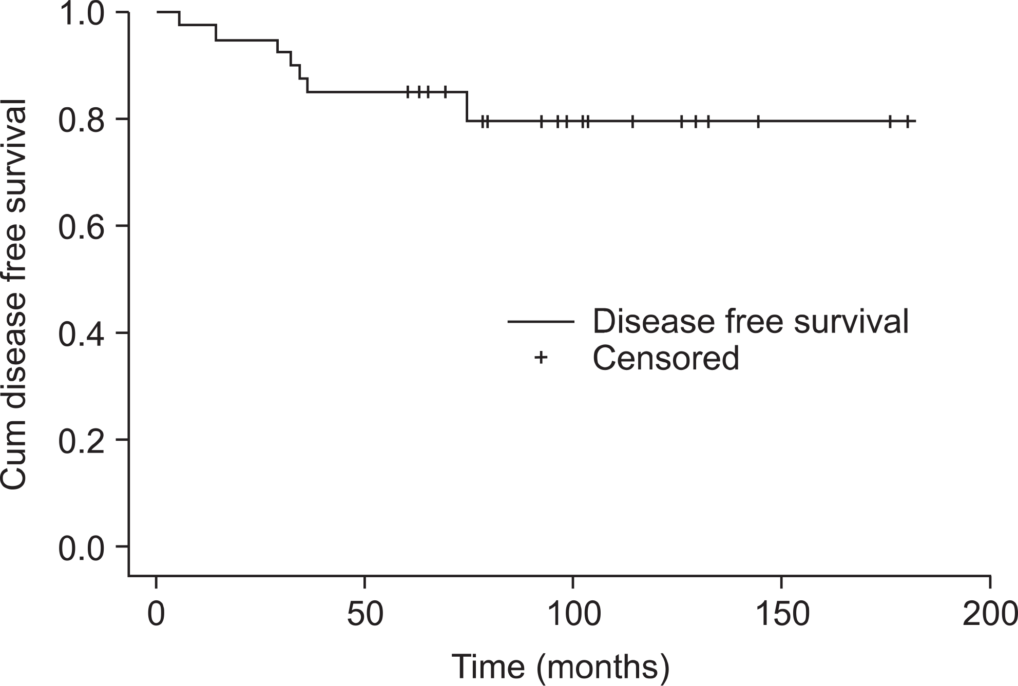 | Figure 2.The graph shows the cumulative disease free survival of patients of liposarcoma in extremities. The 5-year disease free survival rate was 85.0%. |
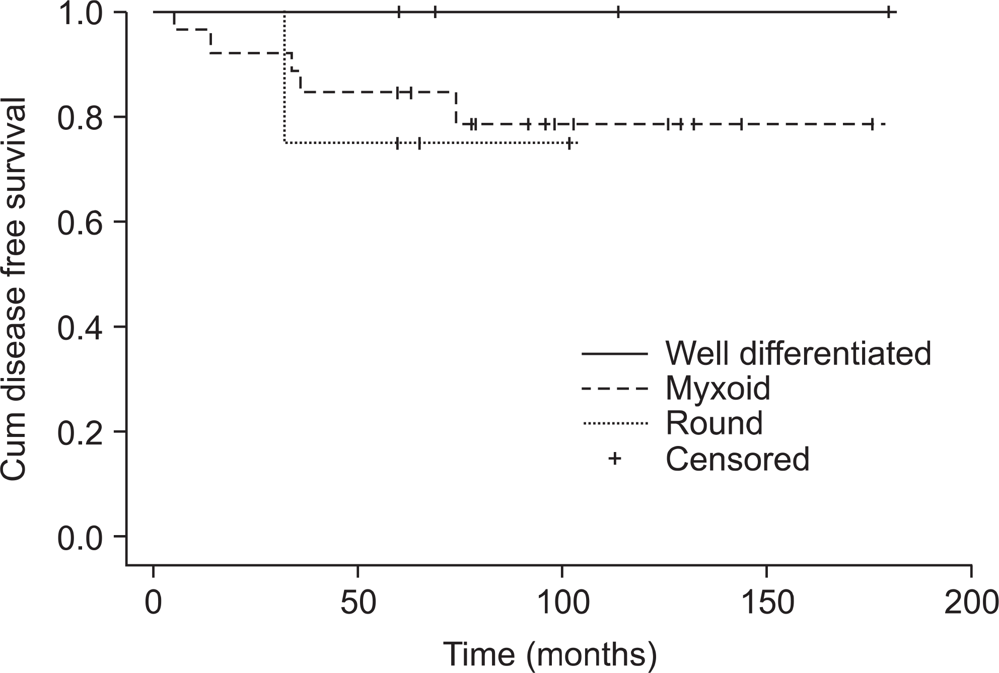 | Figure 3.The graph shows disease free survival curve according to histological type of liposarcoma. The 5-year disease free survival rate of well-differentiated liposarcoma, mixoid liposarcoma and round cell liposarcoma were 100%, 84.6% and 75.0% respectively. The p-value was 0.419 in the log rank test. |
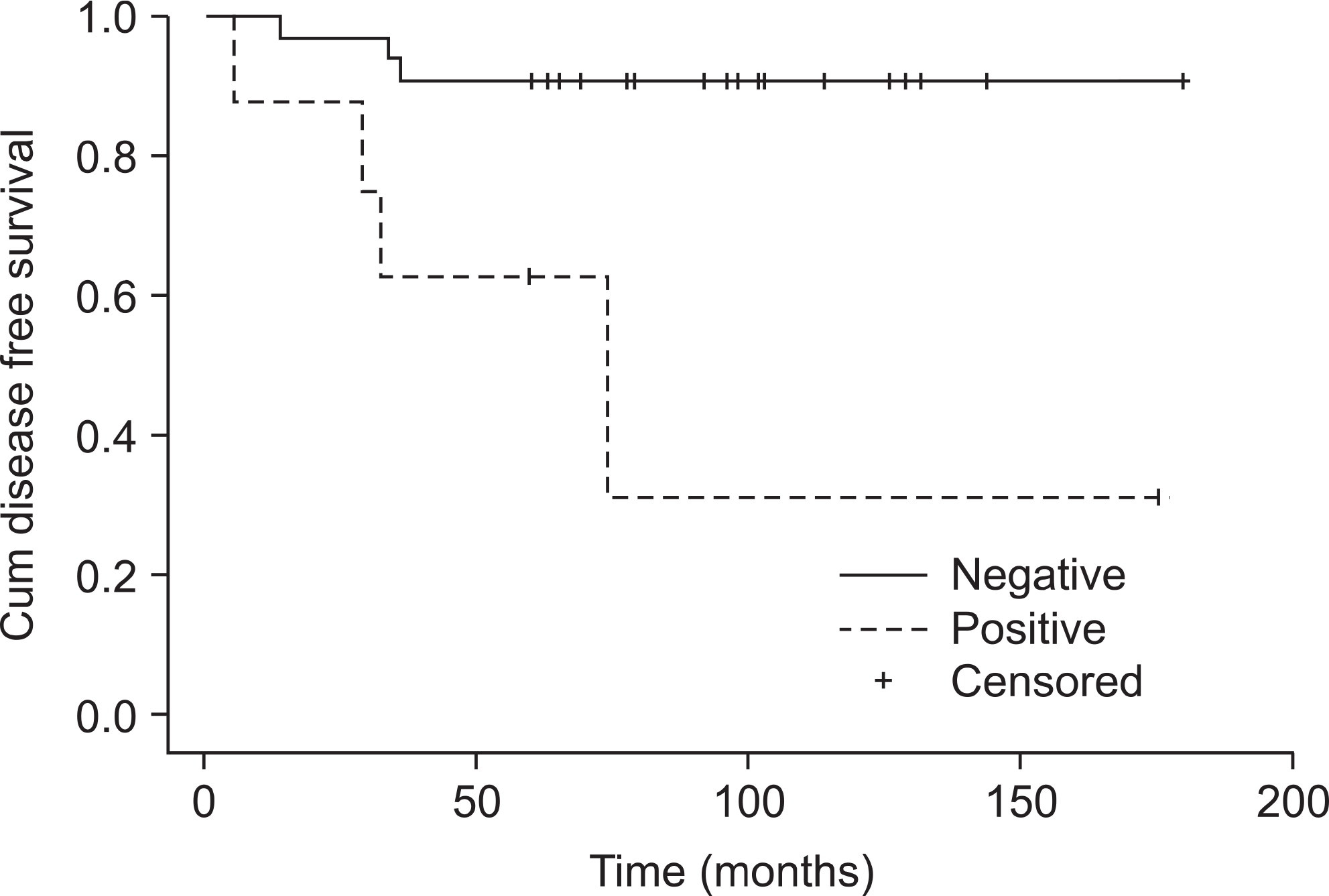 | Figure 4.The graph shows disease free survival curve according to surgical margin. The 5-year disease free survival rate of the negative margin was 90.6% and that of the positive margin was 62.5%. The p-value was 0.003 in the log rank test. |
Table 1.
Patient Characteristics
 XML Download