Abstract
Purpose
Pigmented villonodular synovitis (PVNS) is a rare soft tissue tumor, which usually arises in larger joints, such as the knee. It has a high recurrence rate after surgical treatment. The purpose of this study is to evaluate and analyze the clinical results of diffuse-type pigmented villonodular synovitis cases that were treated with open total synovectomy.
Materials and Methods
Between 1994 and 2006, 21 patients who had diffuse-type pigmented villonodular synovitis were selectively reviewed. Among the 21 cases studied, 14 patients presented at the knee, 5 at the ankle, and 2 at the shoulder and elbow. The mean follow up period was 5.5 years (range, 36-157 months). The average age of the patients was 34 years consist of 7 men and 14 women. Clinical outcomes were analyzed retrospectively, including range of motion and complications.
Results
Open total synovectomy and adjuvant electrocautrization were done in all cases except one. During the regular followup period after the surgery, two patients showed symptoms of recurrence. After re-operation, only one case was pathologically confirmed as a recurrence. The patient who had partial synovectomy and the other patient who had second operation due to recurrence received additional radiation therapy. Clinical outcome scores were improved in every aspect (p<0.0001). 2 out of 14 Patients who had pigmented villonodular synovitis at the knee developed stiff knee after the surgery.
Go to : 
REFERENCES
1. Jaffe HL, Lichtenstein L, Sutro CJ. Pigmented villonodular synovitis, bursitis and tenosynovitis. A discussion of the synovial and bursal equivalents of the tenosynovial lesion commonly denoted as xanthoma, xanthogranuloma, giant-cell tumor of myeloplasioma of the tendon sheath, with some consideration of this tendon sheath lesion itself. Arch Pathol. 1941; 31:731–65.
2. Byers PD, Cotton RE, Deacon OW, et al. The diagnosis and treatment of pigmented villonodular synovitis. J Bone Joint Surg Br. 1968; 50:290–305.

3. Granowitz SP, D'Antonio J, Mankin HL. The pathogenesis and longterm end results of pigmented villonodular synovitis. Clin Orthop Relat Res. 1976; 114:335–51.
4. Myers BW, Masi AT. Pigmented villonodular synovitis and tenosynovitis: a clinical epidemiologic study of 166 cases and literature review. Medicine (Baltimore). 1980; 59:223–38.
6. McMaster PE. Pigmented Villonodular Synovitis with Invasion of Bone: Report of Six Cases. J Bone Joint Surg Am. 1960; 42:1170–83.
7. Scott PM. Bone lesions in pigmented villonodular synovitis. J Bone Joint Surg Br. 1968; 50:306–11.

8. Jergesen HE, Mankin HJ, Schiller AL. Diffuse pigmented villonodular synovitis of the knee mimicking primary bone neoplasms. A report of two cases. J Bone Joint Surg Am. 1978; 60:825–9.

9. Johansson JE, Ajjoub S, Coughlin LP, Wener JA, Cruess RL. Pigmented villonodular synovitis of joints. Clin Orthop Relat Res. 1982. 159–66.

10. Rao AS, Vigorita VJ. Pigmented villonodular synovitis (giant-cell tumor of the tendon sheath and synovial membrane). A review of eighty-one cases. J Bone Joint Surg Am. 1984; 66:76–94.

11. Schwartz HS, Unni KK, Pritchard DJ. Pigmented villonodular synovitis. a retrospective review of affected large joints. Clin Orthop Relat Res. 1989; 247:243–55.
12. Wiss DA. Recurrent villonodular synovitis of the knee. Successful treatment with yttrium-90. Clin Orthop Relat Res. 1982; 169:139–44.
13. Atmore WG, Dahlin DC, Ghormley RK. Pigmented villonodular synovitis; a clinical and pathologic study. Minn Med. 1956; 39:196–202.
14. Jones FE, Soule EH, Coventry MB. Fibrous xanthoma of synovium (giant-cell tumor of tendon sheath, pigmented nodular synovitis). A study of one hundred and eighteen cases. J Bone Joint Surg Am. 1969; 51:76–86.
15. Flandry FC, Hughston JC, Jacobson KE, Barrack RL, McCann SB, Kurtz DM. Surgical treatment of diffuse pigmented villonodular synovitis of the knee. Clin Orthop Relat Res. 1994; 300:183–92.

16. Kotwal PP, Gupta V, Malhotra R. Giantcell tumour of the tendon sheath. Is radiotherapy indicated to prevent recurrence after surgery? J Bone Joint Surg Br. 2000; 82:571–3.

17. Chin KR, Barr SJ, Winalski C, Zurakowski D, Brick GW. Treatment of advanced primary and recurrent diffuse pigmented villonodular synovitis of the knee. J Bone Joint Surg Am. 2002; 84A:2192–202.

18. Bickels J, Isaakov J, Kollender Y, Meller I. Unacceptable complications following intra-articular injection of yttrium 90 in the ankle joint for diffuse pigmented villonodular synovitis. J Bone Joint Surg Am. 2008; 90:326–8.

19. Wu CC, Pritsch T, Bickels J, Wienberg T, Malawer MM. Two incision synovectomy and radiation treatment for diffuse pigmented villonodular synovitis of the knee with extra-articular component. Knee. 2007; 14:99–106.

20. Ogilvie-Harris DJ, McLean J, Zarnett ME. Pigmented villonodular synovitis of the knee. The results of total arthroscopic synovectomy, partial, arthroscopic synovectomy, and arthroscopic local excision. J Bone Joint Surg Am. 1992; 74:119–23.

21. Sharma H, Jane MJ, Reid R. Pigmented villonodular synovitis of the foot and ankle: forty years of experience from the Scottish bone tumor registry. J Foot Ankle Surg. 2006; 45:329–36.

22. Sharma H, Rana B, Mahendra A, Jane MJ, Reid R. Outcome of 17 pigmented villonodular synovitis (PVNS) of the knee at 6 years mean followup. Knee. 2007; 14:390–4.
23. Bertoni F, Unni KK, Beabout JW, Sim FH. Malignant giant cell tumor of the tendon sheaths and joints (malignant pigmented villonodular synovitis). Am J Surg Pathol. 1997; 21:153–63.

24. Oda Y, Takahira T, Yokoyama R, Tsuneyoshi M. Diffuse-type giant cell tumor/pigmented villonodular synovitis arising in the sacrum: malignant form. Pathol Int. 2007; 57:627–31.

25. Finis K, Sultmann H, Ruschhaupt M, et al. Analysis of pigmented villonodular synovitis with genomewide complementary DNA microarray and tissue array technology reveals insight into potential novel therapeutic approaches. Arthritis Rheum. 2006; 54:1009–19.

Go to : 
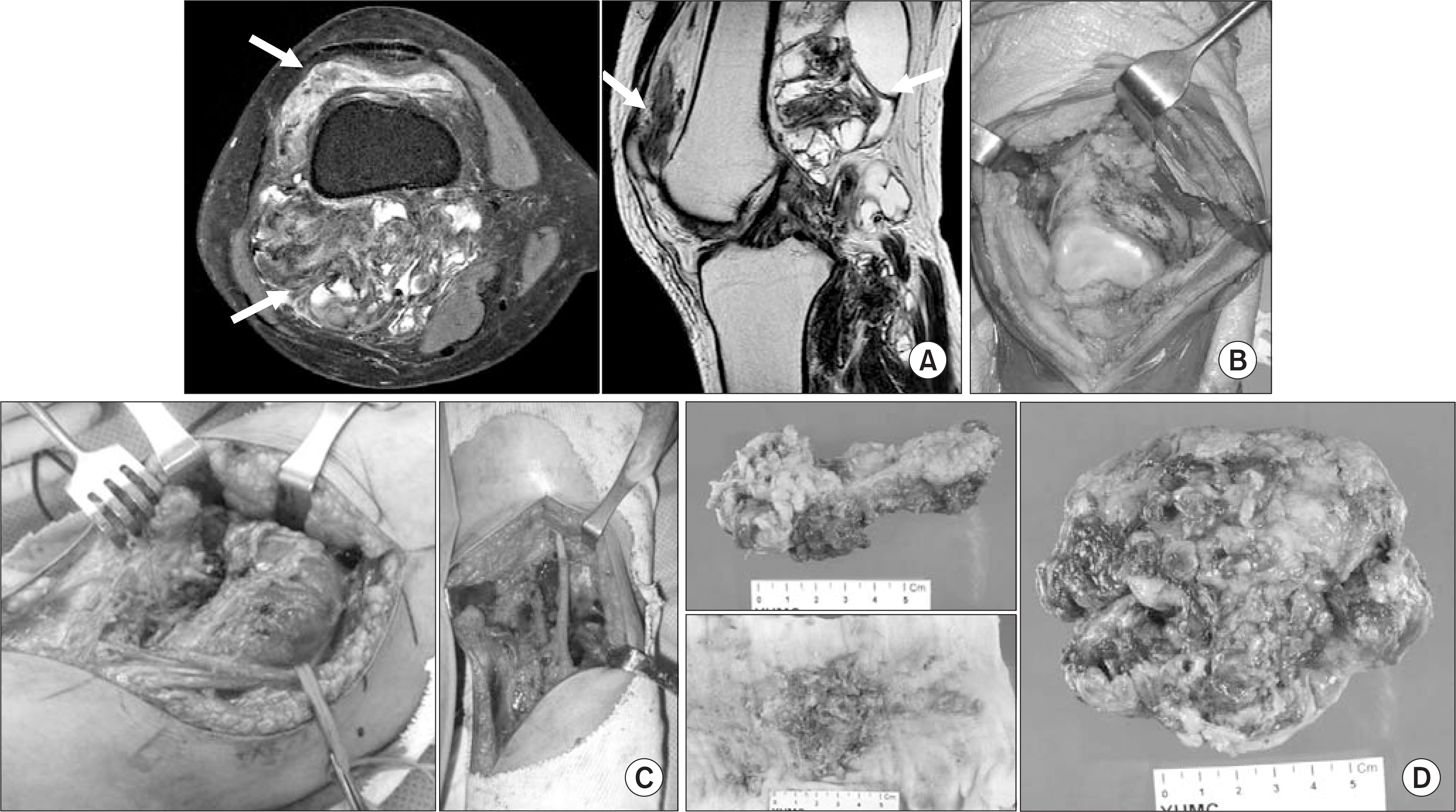
 | Figure 1.MRI and gross photos representing the diffuse type PVNS located in anterior and posterior aspect of the knee. (A) Pre-operative MRI revealing diffuse mass on anterior and posterior aspect of the knee. (B) Intraoperative findings after an anterior medial para patella approach. (C) Intraoperative findings after a posterior popliteal approach. (D) Gross photo of pathology. |
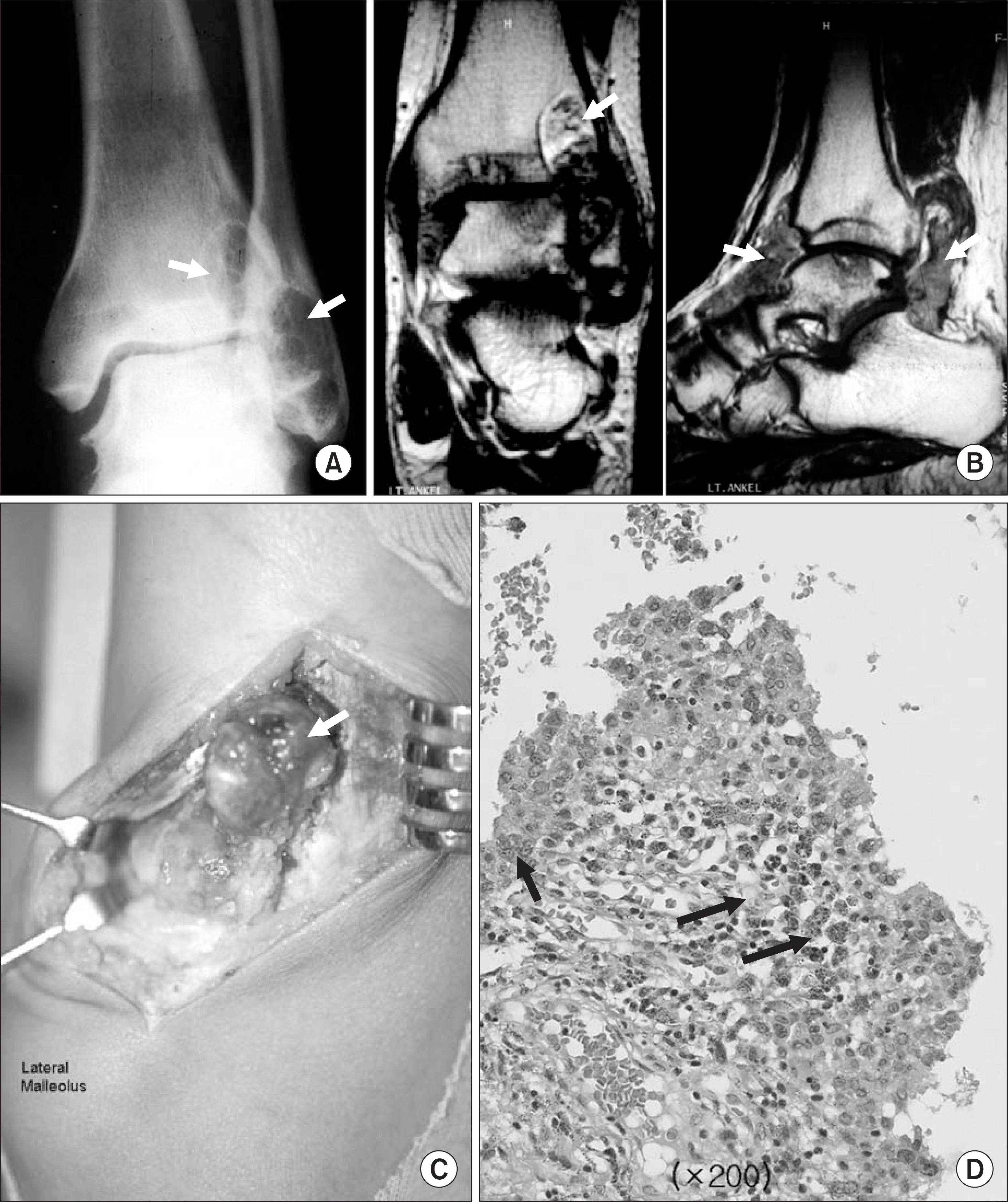
 | Figure 2.A 36 year old male who had diffuse-type PVNS in his left ankle. (A) Preoperative x-ray presenting radiolucent lesions on distal fibular and tibia. (B) Preoperative MRI images showing diffuse mass on anterior and posterior aspect of the ankle. (C) Intraoperative gross findings after a posterolateral approach. (D) Microscopic findings present synovial-like mononuclear cells, hemosiderin laden macrophages, foam cells and giant cells which are consistent findings of PVNS (H&E stain). |
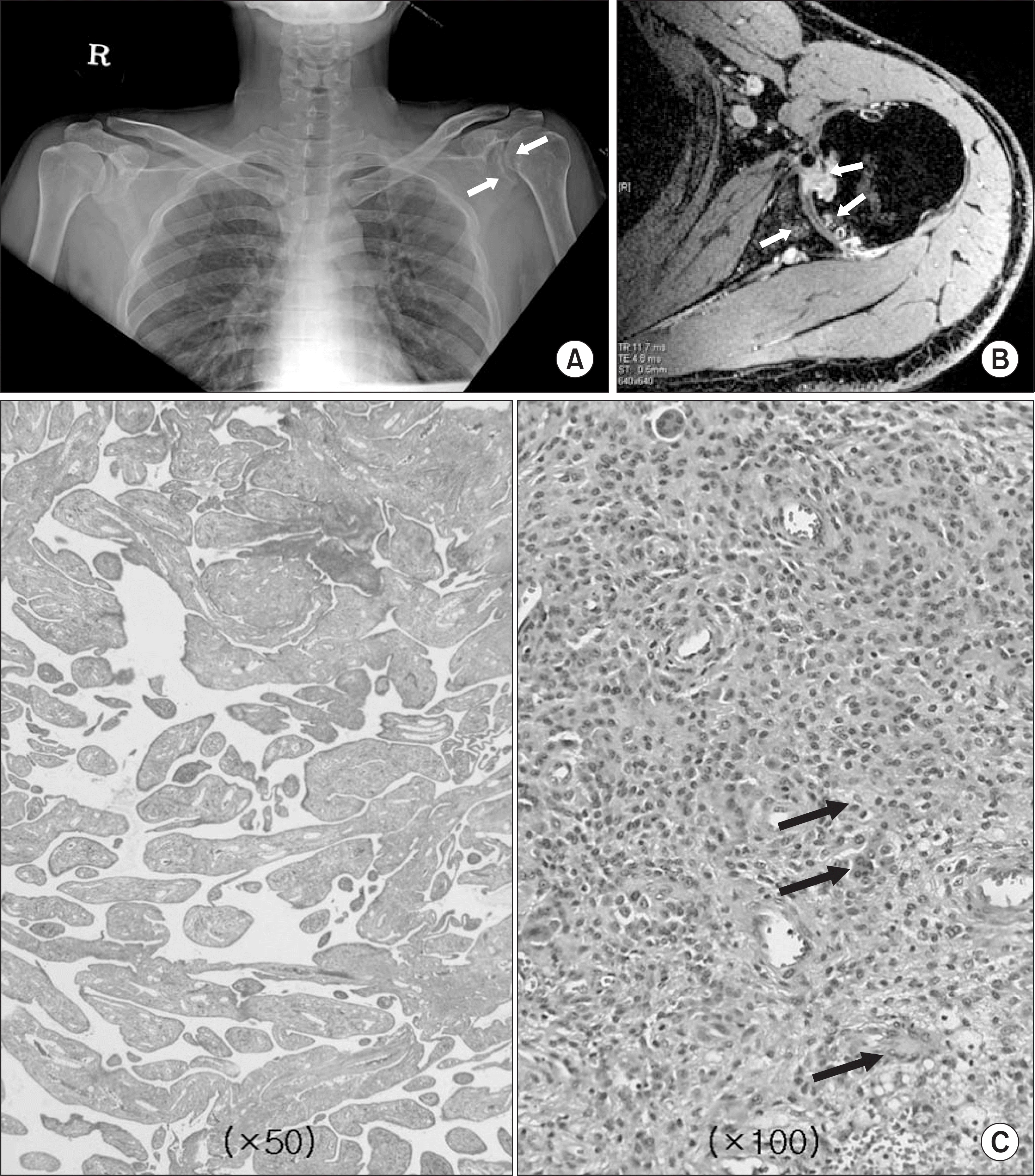
 | Figure 3.Representive plain radiography, MRI and Microscopic findings of diffuse-type PVNS in shoulder joint. (A) Preoperative plain radiography presenting radiolucent lesion and bony erosions on glenohumeral joint. (B) Pre-operative MRI images (axial) presenting infiltrative diffuse mass on glenohumeral joint with bony erosion and cysts. (C) Microscopic findings present hypertrophied synovium, foam cells, giant cells which are consistent findings of PVNS (H&E stain). |
Table 1.
Summary of Cases
| No | Age/ Sex | Site | Trauma | Previous surgery | Surgical method (approach) | RTx. | Complication | F/U (month) | Recurrence |
|---|---|---|---|---|---|---|---|---|---|
| 1 | 40/F | Left shoulder | r No | Once: arthroscopy | Open total synovectomy, | (-) | None | 43 | No |
| 92-11(YUMC) | Electrocautrization (Anterior) | ||||||||
| 2 | 38/F | Right elbow | No | Once: arthroscopy | Open total synovectomy, | (-) | None | 62 | No |
| 92-07 (YUMC) | Electrocautrization | ||||||||
| (Anterior+posterior) | |||||||||
| 3 | 7/F | Left knee | No | (-) | Open total synovectomy, | (-) | None | 41 | No |
| Electrocautrization | |||||||||
| (Popliteal fossa) | |||||||||
| 4 | 43/F | Right knee | No | ∗Twice: arthroscopy | Open total synovectomy, | (-) | Pain | 41 | No |
| 92-10 1st op | Electrocautrization | swelling | |||||||
| 97-04 2nd op | (Anterior+popliteal fossa) | ||||||||
| 5 | 20/M | Left knee | No | (-) | Open total synovectomy, | (-) | Pain | 44 | No |
| Electrocautrization (Anterior) | |||||||||
| 6 | 37/F | Left knee | No | (-) | Open total synovectomy, | (-) | None | 54 | No |
| Electrocautrization | |||||||||
| (Popliteal fossa) | |||||||||
| 7 | 38/F | Left knee | No | (-) | Open total synovectomy, | (-) | None | 78 | No |
| Electrocautrization (Anterior) | |||||||||
| 8 | 25/F | Left knee | No | (-) | Open total synovectomy, | (-) | None | 87 | Re-operation |
| Electrocautrization | pathology | ||||||||
| (Anterior+popliteal fossa) | (-) | ||||||||
| 9 | 47/F | Left knee | No | ∗Once: open | Open total synovectomy, | (-) | None | 75 | No |
| excision 01-12 | Electrocautrization | ||||||||
| (Anterior+popliteal fossa) | |||||||||
| 10 | 31/F | Right knee | No | (-) | Open total synovectomy, | (+) after | LOM | 36 | Yes |
| Electrocautrization | 2nd op. | ||||||||
| (Anterior+popliteal fossa) | |||||||||
| 11 | 44/F | Left knee | No | Once: arthroscopy | Open total synovectomy, | (-) | None | 44 | No |
| 04-11 (YUMC) | Electrocautrization | ||||||||
| (Popliteal fossa) | |||||||||
| 12 | 15/M | Left knee | No | ∗Three times: | Open total synovectomy, | (-) | Stiff knee | 38 | No |
| arthroscopy | Electrocautrization | (0-45) | |||||||
| 03-11 1st op | (Anterior+popliteal fossa) | ||||||||
| 04-02 2nd op | |||||||||
| 04-10 3rd op | |||||||||
| 13 | 42/M | Left knee | No | Twice: open excision | Open total synovectomy, | (-) | Stiff knee | 36 | No |
| (unknown) | Electrocautrization (Anterior) | (15-70) | |||||||
| 14 | 13/M | Right knee | No | (-) | Open total synovectomy, | (-) | Swelling | 41 | No |
| Electrocautrization (Anterior) | |||||||||
| 15 | 39/M | Left knee | No | (-) | Open total synovectomy, | (-) | None | 36 | No |
| Electrocautrization (Popliteal fossa | a) | ||||||||
| 16 | 54/F | Right knee | No | Once: open excision | Open partial synovectomy, | (+) | LOM | 37 | No |
| (unknown) | Electrocautrization (Popliteal fossa | a) | |||||||
| 17 | 42/F | Right ankle | No | ∗Once: open | Open total synovectomy, | (-) | None | 157 | No |
| excision 95-6 | Electrocautrization | ||||||||
| (Anterolateral+posteromedial) | |||||||||
| 18 | 42/F | Left ankle | No | Once: open excision 92-09 (at local hospital) | Open total synovectomy, Electrocautrization (Anterolateral+posterolateral) | (-) | None | 133 | No |
| 19 | 36M | Left ankle | Yes | (-) | Open total synovectomy, | (-) | LOM | 135 | No |
| Electrocautrization | |||||||||
| (Anterolateral+posterolateral) | |||||||||
| 20 | 43/F | Right ankle | No | (-) | Open total synovectomy, | (-) | None | 38 | No |
| Electrocautrization | |||||||||
| (Anterolateral+posterolateral) | |||||||||
| 21 | 19/M | Right ankle | No | (-) | Open total synovectomy, | (-) | Pain | 58 | No |
| Electrocautrization | |||||||||
| (Anterolateral+posteromedial) |
Table 2.
Criteria for Assessment Proposed by Ogilvie-Harris
Table 3.
Preoperative Symptoms
| Symptom | No. of patients |
|---|---|
| Palpable mass | 14 (66%) |
| Pain | 11 (52%) |
| Swelling | 7 (33%) |
| LOM | 2 (9%) |
Table 4.
Result of Clinical Assessment


 PDF
PDF ePub
ePub Citation
 Print
Print


 XML Download