

 PDF
PDF ePub
ePub Citation
 Print
Print


INTRODUCTION
Diabetes mellitus is a metabolic disease that manifests due to insulin insufficiency and/or insulin resistance and has become a serious health problem worldwide. In 2010, approximately 285 million adults between 20 and 79 years of age in the world were affected by diabetes and it is expected that 439 million adults will have diabetes by 2030 [1]. The primary goals for the management of diabetes include the tight regulation of glucose levels in the blood and the prevention of diabetic complications [2]. Hyperglycemic control is crucial for the prevention and delay of the progression of diabetic complications [3].
One approach used to control hyperglycemia in patients with diabetes is the use of agents such as acarbose, which inhibits α-glucosidase activity and subsequently impairs the digestion of carbohydrates and delays glucose absorption after a meal [4]. α-Glucosidase inhibitors such as acarbose are prescribed as an oral hypoglycemic agent for patients with type 2 diabetes [5]. α-Glucosidase inhibitors are also used to control hyperglycemia in conjunction with insulin therapy in type 1 diabetic patients [6]. However, acarbose can induce undesirable side effects such as flatulence, vomiting, and diarrhea [5]. Therefore, studies have focused on the identification of plants that may contain substances that inhibit α-glucosidase activity to develop possible hypoglycemic agents and establish functional foods for patients with diabetes [7]. Among them, guava leaf showed α-glucosidase inhibitory activity in vitro [8,9]. Consumption of guava leaf extract at 250 mg/kg effectively flattened postprandial glucose response in normal mice, demonstrating its inhibitory effect against α-glucosidase in vivo [10]. Chronic consumption of guava leaf extract at 250 mg/kg alleviated fasting hyperglycemia in db/db mice, an animal model of type 2 diabetes [10]. Guava leaf polyphenol, polymerized polyphenols composed of ellagic acid, cyanidin, and other low-molecular-weight polyphenols, has been identified as the active component responsible for α-glucosidase inhibition [11]. Guava leaf extract has been accepted as health functional food in Korea [12].
In diabetic patients, hyperglycemia induces oxidative stress, which, in turn, plays a pathological role in the development and progression of diabetic complications, including macro- and microvascular symptoms [13]. Thus, the identification of natural sources of agents, such as edible plants, that can inhibit α-glucosidase activity and act as antioxidants will aid in the development of beneficial dietary guidelines for diabetic patients as well as potential anti-diabetic agents with fewer side effects.
Actinidia arguta is a perennial vine that belongs to the family Actinidiaceae and bears grape-sized edible fruits (hardy kiwi-fruits) [14,15]. The shoot of A. arguta (Daraesoon) is a traditional Korean food; raw Daraesoon is collected only in the spring but it is difficult to store for a long period of time due to its high moisture content. Therefore, the majority of raw Daraesoon is blanched and then air-dried to make mucknamul, which can be stored for extended periods and is commercially available year round [16]. Mucknamul is soaked in water, boiled, and then re-soaked in cold water before being prepared as namul, a cooked Korean vegetable dish with seasonings.
The polyphenol fraction of A. arguta inhibits the activity of α-glucosidase both in vitro and in vivo through the action of isoquercitrin, one of its active components [17]. Chronic consumption of isoquercitrin at 0.15% of the diet improved fasting hyperglycemia via α-glucosidase inhibition in KK-Ay mice, an animal model of type 2 diabetes [17]. The stem of A. arguta, which is not used as a food ingredient, inhibits α-glucosidase and exerts antioxidant effect in vitro [18]. Other study has found that Daraesoon inhibits α-glucosidase activity in vitro [19]. In addition, Daraesoon acts as a 2,2-diphenyl-1-picrylhydrazyl (DPPH) radical scavenger in vitro and is a good source of phenolic compounds [20]. Thus, Daraesoon has the potential to benefit patients with diabetes via its inhibition of α-glucosidase and antioxidant effects, but these capabilities have yet to be fully characterized using an in vivo model. Therefore, the purpose of the present study was to investigate the hypoglycemic and antioxidant effects of Daraesoon in rodent models of diabetes mellitus. The effect of Daraesoon on postprandial hyperglycemia was determined in rats with streptozotocin (STZ)-induced diabetes. Previously, the extracts of guava leaf [9] and Saururus chinensis Baill with α-glucosidase inhibitory activity in vitro administered at a dose of 500 mg/kg improved postprandial hyperglycemia in STZ-induced diabetic rats [21]. Oral administration of the extract of seed of Holarrhena antidysenterica (400 mg/kg) was reported to suppress the blood glucose response after carbohydrate loading in normal rats [22]. We examined the acute effect of Daraesoon extract (400 mg/kg) on blood glucose response after carbohydrate loading in STZ-induced diabetic rats and compared its effect with that of acarbose (40 mg/kg) to investigate the inhibitory effect of Daraesoon on α-glucosidase in vivo. The effect of chronic consumption of Daraesoon on fasting hyperglycemia and oxidative status was determined in C57BL/6J mice fed the high-fat/high-sucrose (HFHS) diet, an animal model of type 2 diabetes that shows insulin resistance and hyperglycemia [23,24].
MATERIALS AND METHODS
Preparation of the Daraesoon extract
Daraesoon in the form of mucknamul was purchased from National Forestry Cooperative Federation in Yeoju, Korea in 2013. Daraesoon was prepared using a standard procedure prior to cooking, as described previously by Ahn et al. [16]. Briefly, the Daraesoon was soaked in cold water for 16 h, boiled in water for 30 min, re-soaked in cold water for 1 h, and then the excess water was removed by squeezing. The pretreated Daraesoon was freeze-dried, powdered, and then twice extracted using 20 volumes of 70% ethanol at room temperature over a 24-h period. The solution was filtered (Whatman No. 1 paper; GE Healthcare, UK) and the filtrate was removed with a rotary evaporator (Eyela; Tokyo, Japan).
Determination of total polyphenol content
The total polyphenol content of the Daraesoon extract was measured by the method developed by Folin and Denis [25] using tannic acid as standard. Briefly, 1 mg of the extract was dissolved in 1 mL of methanol, and centrifuged at 1,500 g for 10 min. The supernatant (400 µL) was mixed with 50% Folin-Ciocalteu's phenol reagent (200 µL), and then incubated for 3 min. After the solution was mixed with 2% Na2CO3 (400 µL) and allowed to incubate for 60 min, the absorbance at 760 nm was measured.
Oral carbohydrate loading in diabetic rats
Male Sprague-Dawley rats weighing 220-250 g (Bio Genomics, Inc.; Seoul, Korea) were fed with commercial chow (Samyang Co.; Korea) during an initial 2-week adaptation period. The animals were individually housed in a room maintained at 21 ± 2℃ and 55 ± 5% relative humidity under a 12 : 12 h light : dark cycle. To induce diabetes, the rats were intraperitoneally injected with STZ (65 mg/kg) in a citrate buffer (pH 4.5). One week later, fasting blood samples were obtained from the tip of the tail and glucose levels were measured using a glucometer (Glucotrend, Roche Diagnostics; Lewes, UK). Rats with blood glucose levels between 200 and 400 mg/dL (n = 21) were included in the present study.
For the carbohydrate-loading test, the rats were administered soluble starch (1 g/kg body weight) with or without the extract of Daraesoon (400 mg/kg) or acarbose (40 mg/kg; Glucobay; Bayer Korea Ltd.; Seoul, Korea) by gastric intubation after an overnight fast. Blood samples were collected from the tip of the tail at 0, 30, 60, 120, 180, and 240 min after the administration of the starch. Glucose levels in the blood were measured using a glucometer (Glucotrend) and the incremental blood glucose level at each time point and area under the response curve (AUC) were calculated.
Assessments of fasting blood glucose levels and antioxidant status in mice with insulin resistance
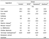
Five-week-old male C57BL/6J mice (n = 28) were obtained (Bio Genomics, Inc.; Seoul, Korea) and allowed a 1-week adaptation period. The mice were then randomly divided into four groups: the control group was fed a basal diet, the high-fat/high-sucrose (HFHS) group was fed a HFHS diet, the Daraesoon group was fed a HFHS diet containing 0.4% Daraesoon extract, and the acarbose group was fed a HFHS diet containing 0.04% acarbose ad libitum for 12 weeks (Table 1). The basal diet contained 5.0% corn oil and 65.0% cornstarch. The HFHS diet was formulated based on previous studies showing that the long-term consumption of this diet resulted in the development of type 2 diabetes in C57BL/6J mice [23,24]. Consumption of HFHS diet containing 3.0 corn oil, 33.0% lard, 10.1% cornstarch, 27.0% sucrose, 5.0% AIN-76 mineral mixture, and 1.4% AIN-76 vitamin mixture for 12 weeks induced insulin resistance and hyperglycemia in these animals [24]. Following the 12th week from the initiation of their respective diets, the mice were sacrificed by a heart puncture procedure after an overnight fast and blood, small intestine, and liver samples were collected.
The blood was centrifuged at 1,500 g for 15 min and serum glucose levels were measured by enzymatic methods [26] using an assay kit (Yeongdong Co.; Seoul, Korea) while insulin levels were measured using an enzyme-linked immunosorbent assay (ELISA) kit (Mercodia; Uppsala, Sweden). The homeostasis model assessment for insulin resistance (HOMA-IR) index, a surrogate indicator of insulin resistance, was calculated as follows: fasting glucose (mmol/L) × fasting insulin (µU/mL)/22.5 [27].
After the duodenum was removed, the proximal one-third of the jejuno-ileum was cut longitudinally and the lumen was flushed out with ice-cold saline. The mucosa was scraped off with a glass slide, homogenized in four volumes of cold saline, and then centrifuged at 12,000 g for 30 min. The maltase activity of the supernatant was determined according to the method described by Dahlqvist [28] and the protein content was measured according to the method described by Lowry et al. [29]. One unit of maltase activity was defined as µmoles of malate substrate hydrolyzed per minute.
Lipid peroxidation in the liver was determined by measuring the production of thiobarbituric acid reactive substance (TBARS) according to the method described by Ohkawa et al. [30]. Briefly, a portion of the liver tissue was homogenized in five volumes of 10 mM sodium phosphate buffer (pH 7.4), and the homogenate was mixed with a solution of 15% trichloroacetic acid (TCA), 0.4% thiobarbituric acid (TBA), and 2.5% HCl. After being heated at 100℃ for 45 min, the reaction mixture was cooled on ice and centrifuged at 1,500 g for 15 min. The absorbance of the supernatant was measured at 532 nm and the results were expressed as nmol of malondialdehyde (MDA)/mg protein. Glutathione (GSH) levels in the liver were measured according to the method described by Ellman [31] and expressed as nmol/mg protein. Briefly, the liver homogenates were prepared with nine volumes of 0.1 mM phosphate buffer (pH 7.4) and centrifuged at 10,000 g for 30 min at 4℃. The supernatant was incubated with 10 mM 5,5-dithiobis-2-nitrobenzonic acid (DTNB) in 0.1 M phosphate buffer (pH 8.0) for 15 min and the absorbance was measured at 534 nm. The protein content of the liver was measured according to the method described by Lowry et al. [29]. All assays were performed in triplicate. All animal experimental procedures were carried out in accordance with the guidelines of animal experimentation as approved by the Animal Resource Center at Inje University, Korea (Approval no. 2013-43 and 2014-18).
RESULTS
Total polyphenol content of the Daraesoon extract
The extraction yield of the Daraesoon extract was 8.1%. The total polyphenol content of the Daraesoon extract was 99.7 ± 1.0 mg tannic acid equivalent/g extract.
Effects of Daraesoon on postprandial hyperglycemia in carbohydrate-loaded diabetic rats
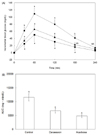
The changes in blood glucose levels at each timepoint after a single oral administration of only starch (control group), starch with Daraesoon extract (400 mg/kg), or starch with acarbose (40 mg/kg) in STZ-induced diabetic rats are provided in Fig. 1. The consumption of Daraesoon resulted in a significant reduction of blood glucose levels at 30, 60, and 120 min (P < 0.05) compared to the control group. The blood glucose levels of the acarbose group exhibited significant decreases at 30, 60, 120, and 180 min (P < 0.01) compared to the control group, but these values did not differ significantly from those of the Daraesoon group. Additionally, the AUCs for the glucose responses were significantly suppressed in the Daraesoon and acarbose groups compared to the control group (P < 0.01; Fig. 1).
Effects of Daraesoon on fasting hyperglycemia and oxidative stress in diet-induced diabetic mice
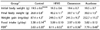
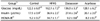
The C57BL/6J mice that were fed a HFHS diet for 12 weeks exhibited significantly increased final body weight, enhanced weight gain, and heightened feed efficiency ratios (FERs) compared to the control group (P < 0.01; Table 2). However, there were no significant differences in final body weight, weight gain, food intakes, or the FERs among the HFHS, Daraesoon, and acarbose groups.
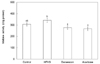
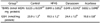
The serum glucose and insulin levels and HOMA-IR values were significantly elevated in the HFHS group compared with the control group (P < 0.05; Table 3). However, the mice that consumed either Daraesoon or acarbose along with the HFHS diet showed significant reductions in insulin levels and HOMA-IR values compared with the mice that were fed a HFHS diet (P < 0.05). Additionally, compared to the HFHS group, there were significant decreases in the serum glucose levels of the Daraesoon and acarbose groups to levels that approached those of the control group. The maltase activities of the Daraesoon and acarbose groups exhibited significant reductions compared to the HFHS group (P < 0.05) but these values did not differ significantly from those of the control group (Fig. 2).
The mice that were fed a HFHS diet exhibited significantly increased hepatic TBARS levels and decreased GSH concentrations compared with the mice that were fed a basal diet (P < 0.05; Table 4). The Daraesoon group had significantly lower TBARS levels and significantly higher GSH levels than the HFHS and acarbose groups (P < 0.05), but these levels were not significantly different from the control group.
DISCUSSION
The ingestion of a 70% ethanol extract of Daraesoon (400 mg/kg) effectively flattened the increase in postprandial blood glucose after starch-loading and reduced the AUC in STZ-induced diabetic rats; the hypoglycemic effects of Daraesoon were as effective as those of acarbose (40 mg/kg). Because α-glucosidase in the small intestinal brush-border catalyzes the final step of digestion of carbohydrates, α-glucosidase inhibitors can attenuate postprandial hyperglycemia [4]. Control of both fasting and postprandial hyperglycemia is crucial in achieving overall glycemic control [32]. Daraesoon strongly inhibits α-glucosidase activity in vitro via its active compound, pinoresinol diglucoside [19]. Thus, Daraesoon may be beneficial in management of diabetes by exerting α-glucosidase inhibitory activity.
The chronic consumption of a HFHS diet induces obesity and causes the downregulation of insulin signaling which results in insulin resistance and type 2 diabetes in C57BL/6J mice [33]. Mice that were fed a HFHS diet in the present study exhibited obesity, hyperinsulinemia, and hyperglycemia, which agree with the findings of previous studies [23,24,33]. However, the consumption of Daraesoon extract (0.4%) or acarbose (0.04%) reduced the serum insulin levels and HOMA-IR values of mice fed a HFHS diet; this is indicative of improved insulin resistance. In the present study, the average daily intakes of Daraesoon extract and acarbose in the Daraesoon and acarbose groups were 404.3 and 41.8 mg/kg, respectively. The Daraesoon extract also reduced fasting serum glucose levels in mice with HFHS diet-induced diabetes to levels similar to those of the control group. Importantly, the ameliorating effect of Daraesoon (0.4%) on fasting hyperglycemia was as effective as those of acarbose (0.04%). The consumption of Daraesoon or acarbose also decreased maltase activity in the small intestine. Liu et al. reported that STZ-induced diabetic rats exhibited reductions in small intestinal α-glucosidase activity following the chronic consumption of acarbose [34]. They suggested decreased activities of disaccharidases could contribute to a decrease of fasting blood glucose in these animals.
Acarbose decreases the demand for insulin by delaying the rise in blood glucose levels after a meal and the attenuated postprandial glucose and insulin responses are thought to improve insulin sensitivity [35]. The chronic consumption of an α-glucosidase inhibitor improves insulin sensitivity in an animal model of diabetes [36] and patients with diabetes [37]. Therefore, in the present study, Daraesoon could have improved in vivo insulin resistance and fasting hyperglycemia via the inhibition of α-glucosidase activity.
The mice that were fed a HFHS diet exhibited elevations of lipid peroxide levels in the liver compared with the control group but Daraesoon reduced hepatic TBARS to levels comparable with those of the control group. Daraesoon has been shown to effectively scavenge DPPH free radicals in vitro [20] and the Daraesoon extract exerted antioxidant effects in the present in vivo investigation. Additionally, although there was a reduction in hepatic GSH levels due to the ingestion of a HFHS diet, they were restored to values comparable to non-diabetic mice by the consumption of Daraesoon extract. GSH is thiol-containing tripeptide that can protect the body against reactive oxygen species (ROS) [38]. Daraesoon extract contained toal polyphenol at 99.7 mg tannic acid equivalent/g. Polyphenols are strong antioxidant agents which can directly scavenge free radicals and induce antioxidant enzyme systems [39]. Antoxidant effect of Daraesoon could have been mediated by polyphenols in this study. Moskaug et al. reported that dietary plant polyphenols, particularly flavonoids, can induce the expression of γ-glutamylcysteine synthetase (γGCSh), which is the rate-limiting enzyme in the synthesis of GSH [40]. Daraesoon is a good source of total polyphenols and flavonoids [20] and, thus, Daraesoon may increase GSH levels via the induction of γGCSh. Future studies investigating the effects of Daraesoon on the gene expression of γGCSh could be necessary.
In the present study, Daraesoon improved postprandial hyperglycemia via the inhibition of α-glucosidase in STZ-induced diabetic rats. In addition, chronic consumption of Daraesoon alleviated fasting hyperglycemia and oxidative stress in mice fed a HFHS diet. Thus, Daraesoon could be considered a potential anti-diabetic agent.
 XML Download