

 PDF
PDF ePub
ePub Citation
 Print
Print


INTRODUCTION
According to a report of the World Health Organization (W.H.O.) in 2008, 80% of the populations in the Asia region depend on traditional medicine for primary health care. In some communities, medicinal plants have been used as the primary sources of health care for thousands of years. Medicinal plants contain powerful compounds in any of the parts, including the bark, stem, resin, leaf, root, flower, fruit, and seed. Such healing properties of plants have been found to cure various health problems. In the middle of the last century, synthetic medicines were preferred in treatment of various diseases for their composability, rapidity, and efficaciousness.
However, synthetic medicines can cause serious side effects [1,2]. Unlike synthetic medicines, natural medicinal plants promote the natural functions of the body. In recent decades, study of natural medicines has been highly suggested by an increasing number of scientific observers in biological activity research [3,4,5,6,7,8,9].
Biological activity describes the beneficial or adverse effects of a drug on living organisms in pharmacology. Biological properties relate to the antioxidant, anti-aging, anticancer, anti-anxiety, hypocholesterolemic, anticoagulant, antithrombotic, anti-diabetes, antifungal, antihistaminic, anti-inflammatory, antihistaminic, immunosuppressive, anti-leishmanial, insecticidal, antibacterial, and cytoprotective activities [10,11,12,13,14]. Medicinal plants possess various biological activities that may contribute to the therapeutic effects for various diseases.
The aim of this study was to investigate the different medicinal plants of Asia for their associated antioxidant, anti-α-glucosidase, α-tyrosinase inhibitory, anti-inflammatory, and anticancer activities using the in vitro models.
MATERIALS AND METHODS
Sample preparation
One hundred methanol extracts (Table 1) were purchased from the Plant Extract Bank (Korea). Different parts of various plants were dried in the dark for five days and then ground to a powder. Each sample was dipped in HPLC methanol at 50℃, and the solvent was evaporated under reduced pressure using a vacuum rotary evaporator at 45℃. The dried extracts used in the experiments were dissolved in 0.05% DMSO.
2,2-diphenyl-2-picrylhydrazyl (DPPH) free radical scavenging activity assay
DPPH free radical scavenging activity was evaluated according to the method described by Zhang et al. [15], with modifications. Each sample of 100 µg/ml was well-mixed with 0.1 mM of DPPH solution and incubated at room temperature for 30 min. Absorbance was measured at 515 nm using an enzyme-linked immunosorbent assay (ELISA) plate reader (Bio-Tek, Winooski, VT, USA). The scavenging ratio for each sample was calculated using the following equation:
DPPH scavenging activity (%) = [Acontrol - (Asample - Ablank)]/Acontrol × 100.
Where, Acontrol indicates the absorbance of the mixture without sample (use 0.05% DMSO instead of sample); Asample indicates the absorbance of the reacted mixture; Ablank indicates the absorbance of the mixture without DPPH (use methanol instead of DPPH).
α-glucosidase inhibitory activity assay
The α-glucosidase inhibitory activity was determined as described by Nishioka et al. [16], with a slight modification. Each sample of 100 µg/ml was mixed with 0.075 units of α-glucosidase. The reaction of the mixture was started with substrate 3 mM p-nitrophenyl glucopyranoside (pNPG) in a phosphate buffer. The reaction was stopped by addition of 2 ml of 0.1 M Na2CO3, after incubation at 37℃ for 30 min. The α-glucosidase activity was determined by measuring the p-nitrophenol release from pNPG at 400 nm. The percent α-glucosidase inhibitory activity was calculated as:
Percent inhibition (%) = [Acontrol - (Asample - Ablank)]/Acontrol × 100.
Where, Acontrol indicates the absorbance of the mixture without sample (use 0.05% DMSO instead of sample); Asample indicates the absorbance of the reacted mixture; Ablank indicates the absorbance of the mixture without pNPG (use PBS instead of pNPG).
α-tyrosinase inhibitory activity assay
Results of the tyrosinase inhibition assay were determined spectrophotometrically using an ELISA plate reader according to the method described by Alam et al. [17], with modifications. Briefly, a 40 µl sample, 40 µl of 125 U/l tyrosinase enzyme, and 80 µl of 67 nM phosphate-buffered saline (PBS, pH 6.8) were mixed and pre-incubated at 37℃ for 5 min. To the substrate 40 µl of 25 nM L-3,4-dihydroxyphenylalanine (L-DOPA) was added. Then, tyrosinase activation was tested by measuring the absorbance at 492 nm. The inhibition of the enzyme activity was calculated as follows:
Percent inhibition (%) = [Acontrol - (Asample - Ablank)]/Acontrol × 100.
Where, Acontrol indicates the absorbance of the mixture without sample (use 0.05% DMSO instead of sample); Asample indicates the absorbance of the reacted mixture; Ablank indicates the absorbance of the mixture without L-DOPA (use PBS instead of L-DOPA).
Cell lines and Cell culture
Murine macrophage RAW 264.7 cell and human colon cancer cell (HT-29) lines were purchased from the Korean Cell Line Bank (Seoul, Korea). RAW 264.7 and HT-29 cells were grown in Roswell Park Memorial Institute medium 1640 (RPMI 1640) and supplemented with 10% fetal bovine serum, 100 U/ml penicillin, and 100 µg/ml streptomycin. Cells were cultured in a humidified atmosphere with 5% CO2 at 37℃.
3-(4,5-dimethylthiazol-2-yl)-2,5-diphenyltetrazolium bromide (MTT) assay
RAW 264.7 and HT-29 cells were seeded in 96-well plates (4 × 10⁴ cells/well) with 200 µl RPMI for 24 h. Both cell lines were pretreated with 50 µg/ml of different samples. In addition, RAW 264.7 cells were treated with lipopolysaccharide (LPS, 2 µg/ml), following exposure to samples for 30 min. After incubation for 24 h, cell viability was measured using the MTT (2 µg/ml in PBS) assay. After incubation for 4 h, the supernatant was removed, and the purple crystals were dissolved in dimethyl sulfoxide (DMSO). Absorbance was measured at 550 nm using an ELISA plate reader.
Nitric oxide assay
The level of NO production in the pre-incubation of RAW 264.7 cell supernatants was determined according to the quantity of the nitrite indicator, using a colorimetric assay based on the Griess reaction; 100 µl of cell culture medium was mixed with 100 µl Griess reagent (1% sulfanilamide in 5% phosphoric acid and 0.1% naphthyl-ethylenediamine dihydrochloride). Subsequently, the absorbance of the mixture was measured at 550 nm using an ELISA plate reader, after incubation at room temperature for 10 min.
RESULTS
DPPH radical-scavenging activity
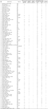
The scavenging properties of the methanol extracts are shown in Table 1. Except for the samples of No. 1, 3, 11, 15, 39, 53, 59, 69, 74, and 96, all other plants showed fairish DPPH scavenging activities. The samples of No. 2, 12, 16, 18, 21, 34, 40, 49, 77, 89, 92, 95, 97, 99, and 100 worked as good scavengers, eliminating more than 90% of free radicals.
α-glucosidase inhibitory activity
Among all of the extracts, the extracts of No. 18 and 21 showed a strong inhibitory effect on α-glucosidase. In addition, No. 12, 16, 34-37, 40, 58-60, 62, 72, 77, 81, 82, 84, and 100 exhibited considerable α-glucosidase inhibition (Table 1).
α-tyrosinase inhibitory activity
In this study, tyrosinase inhibition activity was determined using L-DOPA as the substrate. The extracts of No. 21, 63, 88, 92, and 94 (Table 1) showed a tyrosinase inhibitory effect; and, therein, 100 µg/ml of No. 63 and 94 inhibited 51.46% and 67.74% of tyrosinase activity, respectively.
Cytotoxicity effect
Cytotoxicity of the extract was measured using the MTT assay on RAW264.7 cells. As shown in Table 1, cells incubated with the extracts of No. 1, 2, 8, 9, 13, 14, 16-18, 21, 23, 24, 27, 29, 31-39, 42, 45, 51-53, 56, 58, 61-69, 73, 75-77, 79, 80, 83-87, 89, and 97 showed high viability. Thus, these extracts were considered safe and valuable for further research.
Anti-inflammatory activity
The NO content in RAW264.7 cells was considered to be a marker in anti-inflammatory testing. The extracts of No. 12, 14, 30, 43, 44, 48, 49, 63, 71, 72, 78, 79, 91, 93, 94, and 95 induced significant inhibition of the NO product. The extracts of No, 1, 3, 7, 8, 10, 20, 22, 25, 28, 29, 39-41, 56-58, 61, 66-70, 73, 74, 76, 77, 81, 82, 85, 86, and 92 exhibited a considerable NO inhibitory effect (Table 1).
Anticancer activity
The anticancer activity of 100 extracts was determined using the MTT assay. Most of the extracts showed weak anticancer activity on HT-29 cells (Table 1), except No 1-3, 22, 25, 40, 57, 58, 60, 69, and 95. In particular, No. 57 killed more than 90% of HT-29 cells after incubation for 24 h.
DISCUSSION
Since the beginning of mankind, plants, described as grains, fruits, and vegetables have been used as basic foodstuffs In addition, plants have formed the basis of traditional medicine systems that have been in existence for thousands of years. For traditional medicine, humans usually drank water or spirit dripped from herbs. In recent years, attention has been paid to natural plant use in analytical chemistry and pharmacology. Investigation of potential biological activities and analysis of effective compounds are common methods. Research studies have found that methanol is the best reagent for extraction because it can easily penetrate the cell wall. In addition, many useful compounds have been found in methanol extracts [18]. In this study, we conducted a major investigation of the preliminary biological activity of the methaol extracts from the 100 samples.
Evaluation of the antioxidant activity of the 100 methanol extracts was performed using a rapid, simple, and inexpensive method, the DPPH assay. Due to its changeable color, DPPH acts as an unstable free radical compound in this assay. In this study, most of the plants showed fairish DPPH scavenging activities. The ever-present free radical scavenging activity may contribute to the various antioxidant compounds in the plants. Plants, the primary sources of natural antioxidants that trap free radicals, have sourced antioxidants, such as vitamin C, vitamin E, carotenes, phenolic acids, phytate, and phytoestrogens [19]. Samples of No. 12, 18, and 21 used in this study have been reported to have antioxidant contents, including phenolic, flavonoid, and tannic [20,21]. Thus, the samples of No. 2, 12, 16, 18, 21, 34, 40, 49, 77, 89, 92, 95, 97, 99, and 100, which exhibited very good antioxidant activity, may contribute to the contents of natural antioxidant compounds. Reactive oxygen species (ROS), highly-reactive free radicals in the human body, such as superoxide radical anion (O2·), contribute to redox imbalance in cells, with harmful physiological consequences. ROS-induced damage is closely associated with oxidative damage, which is likely related to the etiology of various chronic diseases, such as cardiovascular disease, diabetes mellitus, aging, gastric ulcers, cancer, arthritis, Alzheimer's disease, Parkinson's disease, and inflammation [22]. Antioxidants have been the subject of intensive scientific research and have been suggested to reduce the risks of various chronic diseases. However, in our study, no obvious relationship was found between antioxidant activity and other biological activities.
Diabetes mellitus (DM) is a serious health problem with an increased incidence worldwide, particularly in Asia. This disease is the result of a metabolic disorder characterized by absolute (type I) or relative (type II) deficiencies in insulin [23]. Without insulin, the cells are unable to take in glucose, which leads to a high blood sugar level. Several oral antidiabetic agents, such as biguanides and sulphonylurea, are available for treatment of DM [24]. However, due to their side effects, increasing interest has been invested in various herbal remedies. More than 1,200 species of plants have been used in empirical treatment of diabetes for their alleged hypoglycemic activities [25]. As recorded in the traditional Korean medical book, No. 12 has been used in the anti-diabetes prescription [26]. The extracts from No. 34 and 77 were also investigated as anti-diabetes objects in a study conducted in Korea in 2010 [27]. In our study, the anti-diabetes property was analyzed by the inhibition of the α-glucosidase enzyme, which breaks down starch and disaccharides to glucose. Because α-glucosidase inhibitor contributes to impairment of the carbohydrates of blood sugar for patients with type II diabetes, plants No. 16, 18, 21, 35-37, 40, 58-60, 62, 72, 81, 82, 84, and 100, with an α-glucosidase inhibitory effect, may protect against the symptoms of diabetes.
Tyrosinase is a rate-limiting, essential enzyme in the biosynthesis of the pigment melanin in plants, microorganisms, and mammalian cells [28]. In human melanogenesis, tyrosinase plays a key role in catalyzing the hydroxylation of monophenols (tyrosine) to ο-diphenols and their subsequent oxidation to ο-quinones [29]. In addition, tyrosinase was reported to be related to cancer and some neurodegenerative diseases [30]. Researchers have studied the tyrosinase inhibitors for cosmetic and pharmaceutical applications. Vaibhav and Lakshaman [31] reported that No. 63 has significant tyrosinase enzyme inhibitory activity in selected Indian herbs; 100 µg/ml of No. 63 inhibited 23.54% of the enzyme. However, in our study, the extracts of No. 21, 63, 88, 92, and 94 showed a tyrosinase inhibitory effect; and, therein, 100 µg/ml of No. 63 and 94 inhibited 51.46% and 67.74% of tyrosinase activity, respectively. Thus, No. 63 and 94 may contain potential skin-whitening agents that inhibit melanogenesis. In addition, there may be possible effects of No. 63 and 94 on the anticancer and anti-neurodegenerative activities.
Symptoms of inflammation usually occur when the vascularized living tissues attempt to counteract pathogenic, biochemical, or pharmacological damage [32]. Macrophage, working as a key player, plays an important role in inflammation, providing an immediate defense against foreign elements prior to leukocyte migration. In this study, a murine macrophage cell line (RAW 264.7) was used for measurement of the anti-inflammatory activity of methanol extracts from traditional medicinal plants. Lipopolysaccharide (LPS), in the inflammatory stimuli, activates immune cells to up-regulate inflammatory products, such as nitric oxide (NO) [33]. The extracts of No. 12, 14, 30, 43, 44, 48, 49, 63, 71, 72, 78, 79, 91, 93, 94, and 95 induced significant inhibition of the NO product. The extracts of No, 1, 3, 7, 8, 10, 20, 22, 25, 28, 29, 39-41, 56-58, 61, 66-70, 73, 74, 76, 77, 81, 82, 85, 86, and 92 exhibited a considerable NO inhibitory effect. Because NO is an important mediator of inflammatory responses and an oxidative damage-inducer in related diseases, NO inhibitors may have potential properties of preventing/treating inflammation or NO-related diseases. Most of the extracts showed very weak or no cytotoxicity to RAW 264.7 cells. However, No. 3-7, 10-12, 15, 19, 20, 22, 25, 26, 28, 30, 40, 41, 43, 44, 46-50, 54, 55, 57, 59, 60, 70-72, 74, 78, 81, 82, 88, 90-96, and 98-100 showed a substantive cytotoxicity to the cells (Table 1). Thus, No. 1, 8, 14, 29, 39, 56, 58, 61, 63, 66-69, 73, 76, 77, 79, 85, and 86 were considered safe and valuable for further anti-inflammatory research.
Cancer is a class of diseases characterized by unregulated cell growth. The World Health Organization has reported that approximately 13% of all deaths in the world are caused by cancer each year. Death from colon cancer has risen to be the fourth highest among all cancer-related deaths. In our study, the human colon cancer cell (HT-29) line was used as the target. Sample No. 57 showed significant anticancer activity. More than 90% of HT-29 cells met cell-death by incubation with 50 µg/ml of No. 57 for 24 h. In addition, No. 1-3, 22, 25, 40, 58, 60, 69, and 95 also exhibited a considerable anticancer activity. Patil and Magdum [34] and Sidambaram et al. [35] reported that Euphorbia hirta L. (No. 57) showed anticancer activity on a mouse lymphoma cell line (EL-4) and a human larynx epithelioma cell line (Hep-2), respectively. However, No. 57 also showed cytotoxicity on RAW 264.7 cells (Table 1). Thus, the side-effects of No. 57 cannot be ignored. Further analyses of the effective compounds are necessary.
In summary, among the 100 selected traditional medicinal extracts, No. 1, 2, 8, 9, 13, 14, 16-18, 21, 23, 24, 27, 29, 31-39, 42, 45, 51-53, 56, 58, 61-68, 73, 75-77, 79, 80, 83-87, 89, and 97 showed low toxicity on normal cells. The low toxicity activity of the extracts seems to be safe for organisms. Thus, No. 2, 16, 18, 21, 34, 77, and 89, which exhibited significant antioxidant activity, may be used for further antioxidant compound analysis. In addition, No. 18, 21, 34-37, 58, 62, 77, 84; No. 63; No. 1, 8, 14, 29, 39, 56, 58, 61, 63, 66-69, 73, 76, 77, 79, 85, 86; and No. 1-3, 69, 95 (in Table 1) may have potential value for research on diabetes, cosmetic skin-whitening, inflammation, and cancer, respectively. These findings may contribute to nutrition and pharmacological studies. In further study, we will conduct a detailed investigation on the significant biological activity of the outstanding samples. By analytical chemistry, some effective compounds, like β-carotene and vitamins, might be found and used in foodstuff, nutrition, and pharmacy.
 XML Download