

 PDF
PDF ePub
ePub Citation
 Print
Print


INTRODUCTION
Cholecystokinin (CCK), a neuropeptide, is secreted in response to intraluminal nutrients by enteroendocrine I-cells of the intestine and has important physiological actions related to appetite regulation and satiety [1]. Postprandial release of CCK is key in activation of intestinal feedback control of gastrointestinal function, comprising short-term inhibition of gastric emptying and acid secretion, stimulation of the exocrine pancreas and gallbladder, and inhibition of food intake [2]. Consequently, the stimulation on CCK secretion from the intestine is of potential relevance for body weight management.
Increase in intracellular Ca2+ concentrations ([Ca2+]i) is coupled to CCK release from enteroendocrine cells [3]. One mechanism involved in CCK-coupled increases in [Ca2+]i is the entry from the extracellular milieu through activation of the transient receptor potential (TRP) ankyrin 1 (A1) channel in enteroendocrine cells [4].
The flavanone naringenin (4',5,7-trihydroxyflavanone) is present in citrus fruits and tomatoes as its glycoside form, naringin (naringenin 7-rhamnoglucoside) [5]. Orally administered naringin is metabolized into naringenin in the gut [6]. Naringenin has been reported to have antioxidant [7], anticancer [8], and antiatherogenic [9] activity, and to have effects on lipid metabolism [10,11], plasma glucose levels [11,12,13], and metabolic syndrome [14]. Naringin has also been reported to have anticancer, anti-inflammatory, and cardioprotective activity [15], and to have effects on plasma glucose levels [16], lipid metabolism [17], and metabolic syndrome [18]. Based on this information, we hypothesized that naringenin and naringin have a wide spectrum of biological activities and might stimulate CCK secretion and contribute to inhibition of food intake.
Therefore, the aim of the current study was to investigate the question of whether naringenin and naringin exerted stimulatory effects on CCK release in the murine CCK-producing enteroendocrine cell line STC-1. Then involvement of changes in [Ca2+]i in naringenin- or naringin-induced CCK release, and association of TRPA1 with naringenin- or naringin-induced cellular responses (CCK release and changes in [Ca2+]i) in cells were examined.
MATERIALS AND METHODS
Materials
Naringenin, naringin, allyl isothiocyanate (AITC), ruthenium red (RR), and HC-030031 were purchased from Sigma-Aldrich (St. Louis, MO, USA). Dulbecco's modified Eagle medium (DMEM), fetal bovine serum (FBS), penicillin/streptomycin, and trypsin-ethylenediaminetetraacetic acid (EDTA) were purchased from Gibco Co. (Grand Island, NY, USA). CCK enzyme immunoassay (EIA) kit was purchased from Phoenix Pharmaceuticals Inc. (Belmont, CA, USA). Calcium 5 Assay Kit was purchased from Molecular Devices (Sunnyvale, CA, USA).
Cell culture
Cultures of STC-1 cells, and measurements of CCK release and changes in [Ca2+]i were performed as reported previously [19]. The cells were received by Professor S.-D. Moon (The Catholic University of Korea College of Medicine) under the permission of Dr. D. Hanahan (University of Califonia, San Francisco, CA, USA.), and were maintained in DMEM with 10% FBS, 50 U/mL penicillin, and 50 µg/mL streptomycin at 37℃ in 5% CO2/air humidity.
Determination of CCK release
The cells were seeded in 24-well plates (2 × 10⁵ cells/well) until reaching 80-90% confluency. On the day of the experiments, cells were washed with HEPES buffer (Ca2+ concentration, 1.25 mM) and exposed to test agents dissolved in the same buffer for 60 min at 37℃. HEPES buffer without Ca2+ was used for the secretion study under extracellular Ca2+-free condition. After incubation for 60 min, supernatants from the plates were collected and stored at -70℃ until measurement of CCK concentration using a commercial EIA kit. The concentrations of CCK in the samples were calculated by supplied CCK standard (ng/mL), and the result was expressed as fold of the untreated control. AITC was used as a positive control. A TRP antagonist (RR, 100 µΜ) and a TRPA1 antagonist (HC-030031, 50 µΜ) were preincubated for 30 min before addition of the test agents [19].
Measurement of changes in [Ca2+]i in STC-1 cells
The cells were seeded in 96-well plates (80,000 cells/well). Cells were washed with the assay buffer Hanks' balanced salts solution (HBSS) containing 1.25 mM CaCl2, and 20 mM HEPES, pH 7.4. For the assay in Ca2+-free condition, CaCl2 was omitted. Cells were loaded with a calcium-indicator dye from the FLIPR Calcium 5 Assay Kit by dilution with the assay buffer. The cells were incubated for 60 min at 37℃, after which measurements were performed using a Flex Station 3 (Molecular Devices) set at 27℃. Fluorescence changes (i.e., excitation at 485 nm and emission at 525 nm with a cutoff at 515 nm) were monitored at 2-s intervals. A 100-µl aliquot of the assay buffer supplemented with 2 × test agents was added at 20 s, and scanning was continued for an additional 100 s. The response of each well was determined as ΔRFU (delta relative fluorescent units), calculated as [(maximum fluorescent value) - (minimum fluorescent value)]. The data are reported as the mean ± SEM of the ΔRFU. A TRP antagonist (RR, 100 µΜ) and a TRPA1 antagonist (HC-030031, 50 µΜ) were co-incubated with the calcium-indicator dye for 60 min [19].
Statistical Analysis
Experiments were performed at least three separate experiments, and each experiment was performed in triplicate. Data are expressed as the mean ± standard error of mean (SEM). The groups were analyzed using Student's t-test, one-way ANOVA followed by Duncan's multiple range test, and correlation analysis using SAS software (SAS Institute, Inc., USA). P value of less than 0.05 was considered statistically significant.
RESULTS
Effects of naringenin and naringin on CCK secretion
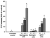
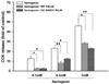
First, we examined the question of whether naringenin and naringin stimulated CCK secretion. Treatment of STC-1 cells with naringenin resulted in a significant (P < 0.05) and dose-dependent increase in secretion of CCK 12.5-fold at 0.1 mM, 23.0-fold at 0.5 mM, and 45.9-fold at 1.0 mM, compared to the untreated control (Fig. 1). The activity obtained with 0.5 mM naringenin was comparable to the activity of 10 mM AITC (a positive control). However, treatment with naringin did not stimulate CCK release in the cells.
Effects of naringenin and naringin on increases
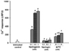
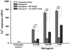
To investigate the involvement of increases in [Ca2+]i in naringenin-induced CCK secretion, we examined the effects of naringenin and naringin on [Ca2+]i under normal (1.25 mM) extracellular Ca2+ conditions. As shown in Fig. 2, naringenin significantly (P < 0.05) stimulated [Ca2+]i 8.99-fold at 0.1 mM, 20.0-fold at 0.5 mM, and 21.3-fold at 1.0 mM, compared to the untreated control. Naringenin at 1 mM stimulated [Ca2+]i by 3.64 times to that produced by 10 mM AITC, an agonist of TRPA1. Meanwhile, exposure to naringin in STC-1 cells did not induce any significant increase in [Ca2+]i (Fig. 2). The results of correlation analysis between CCK release and [Ca2+]i induced by naringenin and naringin showed a Pearson coefficient of 0.930 (P < 0.0001).
Effects of extracellular Ca2+-free conditions on naringenin-induced responses
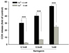
To determine the contribution of Ca2+ influx from the extracellular medium to the CCK release and the increase in [Ca2+]i induced by naringenin in the cells, the extracellular Ca2+-free conditions were applied to the naringenin-induced responses. First, the extracellular Ca2+-free conditions caused significant inhibition of naringenin-induced CCK release by 92.2% at 0.1 mM, 82.5% at 0.5 mM, and 54.6% at 1.0 mM naringenin compared to CCK release under normal Ca2+ conditions (Fig. 3). Second, the removal of extracellular Ca2+ resulted in significant attenuation of naringenin-induced increases in [Ca2+]i by 81.8% at 0.1 mM, 91.5% at 0.5 mM, and 88.9% at 1.0 mM naringenin compared to the increases in [Ca2+]i under normal Ca2+ conditions (Fig. 4).
Effects of TRP antagonist and TRPA1 antagonist on naringenin-induced responses
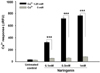
A TRP antagonist, RR, and a TRPA1 antagonist, HC-030031, were applied for examination of the involvement of TRP and TRPA1 channels in naringenin-induced CCK release and increases in [Ca2+]i. In the presence of RR, CCK release in response to naringenin was significantly inhibited by 81.7% at 0.1 mM, 65.9% at 0.5 mM, and 39.2% at 1.0 mM naringenin compared to CCK release without RR (Fig. 5). In the presence of HC-030031, naringenin-induced CCK release was significantly suppressed by 80.2% at 0.1 mM, 46.4% at 0.5 mM, and 50.3% at 1.0 mM naringenin compared to CCK release without HC-030031 (Fig. 5).
RR significantly inhibited naringenin-induced increases in [Ca2+]i by 93.1% at 0.1 mM, 88.6% at 0.5 mM, and 86.6% at 1.0 mM naringenin compared to increases in [Ca2+]i without RR (Fig. 6). HC-030031 significantly inhibited naringenin-induced increases in [Ca2+]i by 77.6% at 0.1 mM, 66.7% at 0.5 mM, and 46.7% at 1.0 mM naringenin compared to the increases in [Ca2+]i without HC-030031 (Fig. 6).
DISCUSSION
As the role of CCK in appetite regulation and satiety has been established, the stimulatory activity on CCK secretion is a potential means for management of body weight. Among major nutrients, protein and fat are strong stimuli for CCK release [20]. However, a limited study has reported that non-nutritive compounds can directly stimulate CCK secretion. A few carbohydrates, such as a sulphated polysaccharide from abalone, have been found to increase CCK release in STC-1 cells [20]. Some bitter compounds stimulate CCK secretion in STC-1 cells [3]. The steroid glycoside H.g.-12 from Hoodia gordonii (an appetite-suppressant plant) was reported to induce CCK release [21]. Flavonoids have not been reported to induce CCK secretion.
In the current study, we report for the first time that naringenin induces CCK release from STC-1 cells. In contrast to the results obtained with naringenin, naringin did not induce any detectable stimulation of CCK release. Flavonoids are mainly present in plants as glycosides. Aglycones (forms lacking sugar moieties) occur less frequently in plants. One important site of flavonoid metabolism is the gastrointestinal lumen, cells of the intestinal wall. Orally administered naringin can be metabolized into naringenin [6]. For native naringin in foods that are subjected to biotransformation following oral ingestion in the gastrointestinal tract, the bioavailability of naringenin is dependent on gastrointestinal biotransformation from naringin into naringenin. Our in vitro results showing that naringenin, not naringin, can stimulate CCK release could suggest that naringenin stimulates CCK secretion in vivo, as orally ingested naringin interacts with enteroendocrine cells as a form of naringenin. Further research is needed in order to explore the anti-obesity effect of naringenin through CCK release and appetite regulation in vivo. The research should include the biotransformation of ingested naringin in the gut and the bioavailability of naringenin.
We showed that naringenin induced a dose-dependent increase in [Ca2+]i, which seems to be coupled with CCK release. However, naringin showed no effect on [Ca2+]i increase. These findings demonstrate a close relationship between CCK release and [Ca2+]i increase in cells upon stimulation by naringenin and naringin.
To determine the origin of Ca2+ responsible for CCK release and increase in [Ca2+]i induced by naringenin, Ca2+ in the extracellular media was removed. The deletion of extracellular Ca2+ resulted in inhibition of naringenin-induced CCK release and increases in [Ca2+]i, suggesting that Ca2+ influx is responsible for CCK release. This also suggests that the naringenin-induced increase in [Ca2+]i occurs through extracellular medium, and is not mobilized from intracellular Ca2.
TRP channels, specifically TRPA1, have been reported to mediate influx of extracellular Ca2+ into STC-1 cells [4,19]. CCK release mediated by naringenin was significantly decreased by the presence of RR (a TRP antagonist) and HC-030031 (a TRPA1 antagonist). The increases in [Ca2+]i induced by naringenin were significantly blocked by the presence of RR and HC-030031, suggesting that activation of TRP channels, including TRPA1, on the cell surface is involved in Ca2+ influx into STC-1 cells, increases in [Ca2+]i, and then CCK release upon stimulation by naringenin. While HC-030031 exerted a weaker inhibitory effect on [Ca2+]i increase compared to RR, HC-030031 exerted an inhibitory effect on CCK release with similar potency of RR. This result suggests that the influx of extracellular Ca2+ mediated by TRPA1 is more specific in mediating CCK release.
In conclusion, we showed that naringenin causes significant stimulation of CCK release in enteroendocrine STC-1 cells. CCK release involves extracellular Ca2+ influx, at least in part, and activation of TRP channels, including TRPA1. These findings provide a novel biological activity of naringenin in regard to CCK release, and demonstrate the potential role of naringenin in appetite regulation and satiety.
 XML Download