

 PDF
PDF ePub
ePub Citation
 Print
Print


Introduction
Al-induced damage to the body organs was mostly elucidated by a dialysis encephalopathy, which was associated to chronic renal failure [1]. Long-term exposure to dialysis fluids and parenteral solutions containing aluminum, and especially orally administered aluminum containing phosphate binders for the control of secondary hyperparathyroidism, were among the first suspected causes. Therefore, despite its efficiency, the use of aluminum based-phosphate binders has been limited and replaced by newer and more costly agents, including calcium based binders [2]. However, since aluminum is cheap, effective and well tolerated, it continues to be used as a binder in many countries, despite its risk of aluminum-related neurological and bone disease [2]. We tried to search for a diet supplement that could reduce the toxicity associated to renal failure during chronic exposure to aluminum. Several plants have been reported to be effective in the treatment of spontaneous or xenobiotic-induced kidney diseases; however, studies regarding the healing effects of plants against Al-induced nephrotoxicity are lacking. Trigonella foenum-graecum is one of the well-known Mediterranean plants, originating from the Middle-East and India, whose seeds are widely used in folk medicine to treat lithiasis patients, especially in Morocco and Arabie Saoudite [3]. Trigonella is also known for its multiple pharmacological effects, including its antidiabetic, antioxidative, antineoplastic, anti-inflammatory, antiulcerogenic, antipyretic, antitumor and immunomodulatory effects [4]. The active components of fenugreek seeds behind their most common properties have been described as polyphenolic flavonoids, steroid saponins, polysaccharides mainly galactomannans and 4-hydroxyisoleucine [4].
Thus, the goal of this study is to assay, via a biochemical and histological analysis, the curative effect of fenugreek (Trigonella foenum-graecum) seeds supplementation on AlCl3-injured kidney and the consequent effects on the brain and bone in rats.
For this purpose, aluminum itself was used to induce nephrotoxicity. Indeed, experimental animals exposed to chronic aluminum intoxication, either with or without normal renal function, showed aluminum accumulation not only in tissues traditionally associated with Al toxicity, such as brain, liver and bone, but also in the kidney [5]. This accumulation led to degeneration in renal tubular cells, inducing nephrotoxicity and consequently renal failure [6].
Materials and Methods
Reagents
Aluminum chloride hexahydrated (AlCl3, 6H2O) was purchased from Sigma-Aldrich Chemical Co. (St. Louis, USA). All other chemicals were of analytical grade.
Animals
Fourty female Wistar rats (weighting 208-220 g) were obtained from the Central Pharmacy (SIPHAT, Tunis, Tunisia). They were fed with a pellet diet purchased from the Industrial Society of Rodents' Diet (SICO, Sfax, Tunisia) and tap water ad libitum. Animals were kept in an air-conditioned room (temperature 22 ± 3℃ and relative humidity of 40%) with a 12 h light/dark cycle. The experimental procedures were carried out according to the guidelines of the Tunisian Society for the Care and Use of Laboratory Animals and were approved by the University of Tunisia Ethical Committee (The Tunisian Association of Laboratory Animals Sciences (ATSAL, Visa 2007T02602APSF1 J.O.R.T. 27 April 2007 n°34. p 2115)).
Plant material
Preparation of fenugreek seeds powder (FSP)
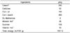
Trigonella seeds, purchased from the local market, were finely powdered and mixed at 5% with ground standard rat feed (i.e. 5 g of dry ground Trigonella seeds in 95 g of ground rat food). The composition of standard rat feed is detailed in Table 1.
Chemical composition of fenugreek seeds
The main constituents of fenugreek seeds have yet been identified through the literature [7-9]. They contain 20-45% carbohydrates, mainly mucilaginous fiber (galactomannans); 27% proteins rich in lysine and tryptophan and free amino acids, including a rare but dominant amino acid: 4-hydroxyisoleucine (30-50%, which corresponds to 0.1 to 0.3% of the dry weight of the seed); fatty oils (6-10%) rich in unsaturated fatty acids and phytosterols; steroidal saponins releasing especially diosgenin and yamogenin (0.1 to 2.2%) after hydrolysis, as well as a dozen of other aglycones; flavonoids (> 100mg/g), including: naringenin quercetin, vitexin, tricin and tricin-7-O-β-D-glucopyranoside; and alkaloids, mainly choline (0.5%) and trigonelline (from 0.2 to 0.36%). There are also other phosphorus compounds, such as lecithin (1-2%) and phytin.
Study design
Rats were treated according to the modified protocol established by Gong et al. [10]. In brief, rats were randomly distributed into four groups of ten animals each: control; AlCl3 daily during 5 months at the level of 500 mg/kg bw i.g for one month then 1600 ppm via drinking water; AlCl3 plus fenugreek seed powder at 5% in the diet (FSP) during the last 2 months and FSP alone. The dose of powdered fenugreek seeds was equated to the therapeutic dose suggested for humans and has been subjected to nutritional and safety evaluation [11]. At the end of the experimental period, animals were sacrificed by decapitation. Blood and tissue collection.
Rats of each group were weighed and samples of blood were collected under anesthesia by cardiac puncture in the heparinized tubes. Plasma was separated from the blood cells by centrifuging the blood at 3,000 × g for 15 min at 4℃ and stored in aliquots at -20℃ until analysis. The two kidneys were removed quickly from animals, washed with ice-cold physiological saline and weighed. Then, one kidney from each rat was cut out, fixed in Bouin's fluid and embedded in paraffin.
Brain tissue was minced and homogenized (10% wt/v) in ice-cold 0.1 M phosphate buffer (pH 7.4) in a Potter-Elvehjem type homogenizer, then centrifuged at 10,000 × g for 30 min at 4℃; the resultant supernatant was used for different enzyme assays.
Multiple lobes of the liver from each rat were cut out, minced and homogenized (10% w/v) separately in ice-cold 1.15% KCl-0.01mol/L sodium, a potassium phosphate buffer (pH 7.4) in a Potter-Elvehjem type homogenizer. The homogenate was centrifuged at 10,000 × g for 20 min at 4℃, and the resultant supernatant was stored at -80℃ to be used for different enzyme assays.
Biochemical assays
Determination of plasma markers
Urea, creatinine, ALP and glucose levels were assayed using commercial reagent kits from Roche Diagnostics.
Lactate dehydrogenase activity
The activity of LDH in the plasma, liver and brain was measured using commercial reagent kits (Randox-Antrim, UK).
Total Antioxidant Status evaluation
TAS evaluation in the blood and brain was performed using commercial tests manufactured by Randox Laboratories (UK, Antrium) in a Randox RX Daytona Chemistry Analyzer.
Lipid Peroxidation estimation
The extent of lipid peroxidation was assessed by measuring the content of thiobarbituric acid reactive substances (TBARS), following the method of Yoshioka et al. [12] in the plasma and the method of Buege and Aust [13] in the liver and brain. TBARS were expressed as an malondialdehyde (MDA) amount, using a freshly diluted malondialdehyde bisdimethylacetal as the standard.
Histopathologic study
Longitudinal sections of the Paraffin-embedded kidneys were cut (5 µm thicknesses) and stained with hematoxylin and eosin (H&E) for a light microscopy examination to determine aluminum toxicity and the restoration possibility by fenugreek seeds [14].
Histochemical study
Paraffin sections were stained by Periodic acid Schiff's reagent (PAS) to visualize the brush-border and membranes in the renal proximal tubules [15]. The silver nitrate stain was also used to accentuate the collagenous structures.
Statistical Analysis
Data were expressed as the mean ± standard deviation (SD) and analyzed using either an analysis of variance (ANOVA) followed by post Hoc Tukey test or a Student's t-test. Values were considered statistically significant when P < 0.05. Statistics were done using IBM SPSS Statistics 19.
Results
Kidney and body weights
No significant difference of the body weight was observed as compared to the control group. On the contrary, absolute and relative kidney weights were significantly decreased in the AlCl3-intoxicated group. Treatment with FSP alone had no effect on the weight of the kidneys' but succeeded to reestablish the weight loss in the AlCl3-treated groups (Table 2).
Blood markers of renal toxicity
Data presented in Table 3 illustrates that the treatment with AlCl3 caused a significant increase in the plasma glucose and creatinine levels, while the urea level significantly decreased as compared with the control. However, the treatment with FSP alone did not cause any significant change in these parameters as compared to the control. The use of FSP with AlCl3 maintained the levels of all the above parameters closer to its normal values.
ALP activity as a bone marker
AlCl3 reduced remarkably ALP activity in plasma (P < 0.001) but fenugreek administration maintained the level of this parameter closer to the normal value (Table 3).
Lactate dehydrogenase activity
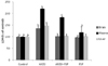
As shown in Fig. 1, AlCl3 treatment induced an important increase in plasmatic LDH (P < 0.001) and relatively similar increase in the liver and brain (P < 0.001 and P < 0.05, respectively). FSP administration significantly decreased the LDH level in the plasma and re-established the values close to its normal values in the liver and brain.
Total Antioxidant Status evaluation
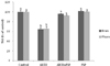
The total antioxidant status was decreased remarkably in the plasma and brain following Al intoxication (P < 0.001 for both); however, FSP succeeded to re-increase it significantly (P < 0.001) (Fig. 2).
Lipid Peroxidation estimation
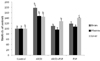
After exposure to AlCl3, a significant increase in the TBARS levels was recorded in the plasma, liver and brain (P < 0.05, P < 0.05 and P < 0.001, respectively); whereas no significant changes were noted in (AlCl3 + FSP) and (FSP) treated rats (Fig. 3).
Histopathological results
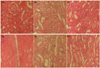
Histopathological changes were firstly assessed by H&E staining at two levels: the cortex and the medulla (Fig. 4). The control kidney cortex sections (Fig. 4a) revealed a normal appearance of the renal corpuscles, glomeruli, surrounded by narrow Bowman's spaces and cortical tubules with the distal and proximal convoluted tubules. Alterations observed in the AlCl3-treated rat kidney (Fig. 4b) included swelling of both the distal and proximal tubules, degeneration of cells in the lining epithelium of the convoluted tubules with a partial replacement of their simple cuboidal epithelium by a simple squamous one and the disappearance of Bowman's spaces in almost all glomeruli. On the other hand, Fig. 4d shows a normal aspect of the medulla in control animals. It consists of longitudinal sections of collecting ducts and loops of the Henle. Cuboidal epithelial cells lining collecting ducts have more distinct cell boundaries and clearer cytoplasm than the cuboidal epithelial cells lining thick segments of loops of the Henle. As a result of AlCl3 toxicity, we noticed a retraction of the cuboidal epithelial cells, which became squamous leading to an enlargement of both the collecting ducts and ascendant thick segments of loops of the Henle. In addition, pyknotic nuclei (intensely basophilic) were observed in the collecting duct epithelium along with the loss of cytoplasm definition (Fig. 4e). Treatment with FSP showed a partial improvement of the kidney histological aspect at two levels (Fig. 4c, 4f) by restoration of the cuboidal form of the different tubules epithelium, retrieval of Bowman's spaces and decrease of the pyknotic cells.
Histochemical results
PAS staining
Light microscopical analyses of PAS stained sections of the kidneys allowed us to grasp an idea regarding the state of the basement membranes, especially the aspect about the brush border which is intensively PAS-positive and this property distinguishes the proximal tubule from the distal tubules in the cortex. The proximal tubules of all control mice had a normal structure. Fig. 5a shows a cross-section through the convoluted tubules of the cortex. All tubules are surrounded by a continuous purple line that is the basement membrane. The brush border can clearly be seen as a continuous darker purple area in the middle of the proximal tubule. As a consequence of AlCl3 treatment, we noticed a decrease in the brush border density, which disappeared in some points leading to a swelling aspect of the proximal tubules. A discontinuity in the tubules basement membrane was also noted (Fig. 5b). Following a fenugreek extract administration (Fig. 5c), a clear restitution of the brush border density, as well as the continuity of the tubules basement membrane, was observed.
Silver nitrate staining
Fig. 6 illustrates a silver nitrate stained section of the control rat kidney. In the control sample (Fig. 6a), we can easily discern the collagenous structures, especially the filtration membrane of the glomerulus and the basement membranes of both the tubules and Bowman's capsule. In AlCl3-intoxicated rats, we observed a complete disappearance of Bowman's spaces, which was already proven by H&E staining and a remarkable discontinuity of Bowman's capsule basement membrane, which thickens in some points and thins until it disappears in other points (Fig. 6b). A notable thinning in the tubules basement membrane, which disappears sometimes, was also recorded. Regarding the glomeruli, we noticed the fading of silver staining intensity, probably due to the thinning or may be the disruption of the filtration membrane in some points. The treatment with FSP succeeded to alleviate all observed changes (Fig. 6c).
Discussion
Because excretion in the urine is the primary route by which the aluminum is eliminated from the body [16], chronic Al exposure could increase the risk of developing aluminum retention, and consequently, aluminum toxicity in the kidneys. Our data indicated that 5 months of AlCl3 intoxication significantly decreased the absolute and relative kidney weights; although tendency towards insignificant lower body weight was observed. The lack of data concerning kidney weight evolution after Al intoxication, leads us to think about the ultrastructural and morphological similarities between rat kidneys exposed to Al and senescent rat kidneys [17]. Based on this correlation between Al exposure and aging, kidney's weight loss could be explained by an aging-like effect of Al. Indeed, it has been demonstrated that age significantly decreases the mass of many organs, particularly the brain, kidneys, liver and spleen [18]. On the other hand however, the metabolic profile of AlCl3-treated rat plasma showed an elevated level of both LDH and creatinine, and a significantly decreased level of urea, although increased urea level in Al toxicity was expected. Indeed, even if LDH increase is a general marker of cell injury, it could indicate kidney damage. In parallel, the elevation of both urea and creatinine levels in the plasma, which is considered as a significant marker of renal dysfunction, was observed in AlCl3-treated animals [19]. However, urea, which is the chief nitrogenous endproduct of amino acids and thus protein catabolism, is elaborated in the liver, filtered by the glomerulus, reabsorbed in part by the tubules and excreted in urine. Therefore, plasma urea concentration depends not only on the glomerular filtration, but also on dietary protein intake, organism protein catabolism and liver production aptitude [20]. For these reasons, creatinine, a nitrogenous compound formed as the irreversible endproduct of muscle creatine metabolism, is a more specific indicator of glomerular function. Thus, a urea decrease could be attributed to a reduction of protein synthesis in the liver as a consequence of AlCl3 hepatotoxicity already proven by increased hepatic LDH and TBARS levels. These physiological and biochemical results are concordant with the histopathologiacal data of AlCl3-treated rats. Indeed, kidney tissue alterations have been evidenced by different staining (H&E, PAS and silver nitrate). In this study, we found that AlCl3 affects the Bowman's capsule by thinning its basement membrane, which is the first step in the filtration of blood to form urine. Al also changed the aspect of the glomerulus filtration membrane that acts as a selective barrier and reduced and reduced the Bowman's space into which the glomerular plasma filtrate collects as it leaves the capillaries through the filtration membrane, until it disappears These changes are features of many kidney diseases, like Alport syndrome, which is a glomerular basement membrane (GBM) disease caused by mutations in type IV collagen genes characterized by irregular thickening and thinning of GBM [21], or Focal and segmental glomerulosclerosis (FSGS) known by the formation of adhesions between the glomerular tuft and Bowman's capsule as a consequence of Bowman's space reduction [22]. In addition to renal corpuscle, AlCl3 also affects the renal tubule histology by changing the aspect of their lining epithelia. The proximal convoluted tubules seemed to be the most affected because they represent the initial segment of the renal tubule that restores much of the filtrate into the blood, reabsorbing most minerals and other nutrients. In fact, we found that AlCl3 altered the apical differentiation of the cortex proximal tubule cells by reducing the brush border density, leading to a reduction of the absorption surface area. This result was in agreement with previous studies using other salts of Al in vivo and in vitro [17,23]. The biochemical and histological changes induced in the kidneys by Al chronic exposure could be mediated by a multitude of toxicity mechanisms already reported for other organs. Prooxidant action of Al, proven here by the decrease of TAS and the induction of LPO in plasma, was one of the major elucidated mechanisms [17,23], in addition to an impairment in sodium and water balance [24]. Al nephrotoxicity was also attributed to a relative parathyroid hormone inhibition [24]. Besides, Al associated hyperglycemia could play an important role in renal failure as is the case during diabetes where glucose overutilization in kidney causes several metabolic changes together with impairment in antioxidant capacity, leading to tissue degeneration [25].
Plasmatic ALP activity, used as a bone marker, showed a significant unexpected decrease among the AlCl3-treated groups, but before discussing this result, it is worthwhile to mention that unlike in the human, serum ALP isozymes in rats are mainly of the bone type [26]; besides, that is why we simply used ALP as the bone marker. These enzymes seem to be essential for normal bone mineralization by calcium and phosphate, because the disordered bone turnover is linked to hypophosphatasia [27]. Thus, a decrease of plasmatic ALP activity in the AlCl3-intoxicated groups might be a consequence of an induced state of phosphate deficiency. In fact, it has been reported that hypophosphatemic osteomalacia could appear following a long-term treatment with aluminum-containing antacid [28]. Our result could testify a biphasic activity of ALP following chronic exposure to AlCl3 as is the case for patients with diabetes mellitus where the serum level of ALP activity is elevated in the incipient stage of the disease, and is reduced markedly in the intermediate stage [29].
Moreover, Al has long been known as a neurotoxicant [30], and despite its equivocal role in the etiology of several neurodegenerative diseases, aluminum salts, as AlCl3, were associated to induced brain toxicity that fairly resemble that of AD. However, given that the purpose of this study is far from elucidating Al neurotoxicity, we have chosen to show prooxidant action of Al in this rat model of renal failure, as it is suggested to be the potential pathway through which aluminum chloride exerts its effects in the brain [31]. Effectively, AlCl3 was able to decrease the total antioxidant status in rat brains, leading to the onset of lipid peroxidation and LDH leakage in the brain as a marker of cell membrane deterioration.
Regarding the effects of Trigonella foenum-graecum on AlCl3 toxicity, the current results clearly indicated that the treatment with FSP did not induce any harmful effects on animals, as it was shown through kidney weight, biochemical and histological parameters (data not shown). FSP supplementation had shown its ability to counteract Al toxicity at many levels.
With regard to the effect of fenugreek seeds on the bone, FSP succeeded to abolish ALP inhibition in the AlCl3-treated groups. However, fenugreek seeds have unexpectedly decreased this biochemical parameter when administrated alone to normal rats. The causes of this decrease could not be those described during AlCl3-induced inhibition of plasmatic ALP, since fenugreek supplementation has modulated ALP activity in the presence of AlCl3. Indeed, for centuries, fenugreek has been used in folk medicine to heal infantile rickets characterized by increased activity of ALP, hypocalcemia and hypophosphatemia [32]. This is understandable because fenugreek seeds have been qualified as good sources of calcium and phosphorus [33]. Such a slight decrease in ALP activity was also reported after milk consumption [34]. This study showed that calcium supplementation, via milk, suppressed bone resorption to a greater extent than bone formation, which led to a decrease of serum ALP, derived from osteoblasts in healthy adults. In brief, we can say that the slight decrease in plasmatic ALP activity after FSP supplementation could be attributed to an osteostatic effect due to an increase of calcium intake.
On the other hand, FSP was able to protect the brain from prooxidant insult of Al as it lowered the LDH level, boosted TAS, and consequently, decreased LPO; this is probably owing to the antiradical and antioxidant potential of polyphenolic flavonoids of Trigonella seeds emphasized through in vitro and in vivo experiments [35-39].
Interestingly, FSP was also found to act as a kidney protector. Indeed, in the AlCl3-treated group, FSP succeeded to counterbalance the weight loss of kidneys. Moreover, it has significantly improved the biochemical and histological parameters. The significantly lowered plasma level of accumulated creatinine was attributed to the enhanced glomerular filtration and the anti-lipid peroxidative property of Trigonella seeds on the kidney [40]; whereas, the re-increase of plasma urea level is probably due to the re-establishment of a normal protein synthesis level in the liver as a consequence of the hepatoprotective effect of FSP, as it was shown through LPO inhibition and LDH level re-establishment. The protective action of FSP against Al hepatotoxicity was effectively demonstrated in a recent study [41], where a notable increase of the hepatic total protein level was noticed after Al ingestion. This is because Al, like other toxic metals, may affect intracellular processing of secreted proteins, and also retard their discharge, resulting in an inhibition of protein secretion from the liver parenchymal cells [42]. This result may explain the decrease of some plasmatic proteins in rats intoxicated by AlCl3, like urea. In addition, FSP supplementation elicited a significant improvement in the histology of the kidneys among the AlCl3 + FSP group compared to those of the AlCl3 group. This curative effect on the kidneys could be attributed to various properties of FSP, especially its potential to scavenge the free radicals [35]. The antioxidant activity of fenugreek seeds, mentioned in several reports [35], is attributed to polyphenols particularly flavonoids. Taking into account that flavonoids, particularly quercetin isolated from other nephroprotective medicinal plants, have been reported to inhibit xenobiotic-induced nephrotoxicity in experimental animal models [43], we can postulate that the nephroprotective activity of FSP could be explained by a synergic potent antioxidant and free radicals scavenging effect. This idea has been corroborated recently by Xue et al. [44], who confirmed that Trigonella seed aqueous extract was able to restore the kidney function of diabetic rats, via its antioxidant activity. On the other hand, flavonoids in the FSP could be behind its beneficial influence on collagenous structures, like basement membrane and filtration membrane. Indeed, it has been demonstrated that fenugreek seeds extract improved the properties of collagen and restored the collagen content in the ethanol-injured liver, possibly by exerting control over ROS production [45].
It is also likely that the modulatory effect of FSP on AlCl3 toxicity was due to an improvement in glucose homeostasis of the kidney tissue, as it was confirmed by the brought down of the elevated blood glucose level to the control value. This hypoglycemic property of fenugreek seeds has been well exploited to explain the efficacy of Trigonella seeds in preventing and reverting diabetes-induced physiological, biochemical and histological alterations in the liver and kidney of diabetic rats [25]. It was attributed to the amino acid, 4-hydroxyisoleucine, which demonstrated to have insulinotropic and antidiabetic properties in animal models [46]. Indeed, supplementation of fenugreek seeds in the diets of diabetic rats reduced the hepatic and renal output of glucose by decreasing the levels of the two enzymes glucose-6-phosphatase and fructose-1-6 bisphosphatase in these organs [47]. Other studies have demonstrated that oral administration of fenugreek seed powder to alloxan-diabetic rats could improve glucose homeostasis by normalizing the different glycolytic, lipogenic and related enzyme changes in the liver and kidney [48]. Trigonella seed supplementation in the diet was also reported to partly prevent an increase in the renal glycogen content and a decrease in hepatic glycogen content with diabetes [36].
In this study, the AlCl3-induced renal damage was illustrated not only by a significant increase of the plasma nephrotoxicity markers, but also an altered histological feature in the kidney tissue reminiscent of some known diseases. In this rat model, AlCl3 was also able to cause neurotoxicity and bone damage. FSP supplementation normalized plasma markers and improved histological alterations. In conclusion, the overall results have clearly shown the ability of FSP to offer protection against some aspects of AlCl3 ingestion in the plasma, brain, bone and kidney, probably due to a synergic effect of many compounds.
Thus, fenugreek seeds can be used as a regular nutrient to alleviate the side effects of Al ingestion, not only in the brain and bone, but also in the kidneys, especially for chronic renal failure patients who are more susceptible to developing aluminum toxicity.


 XML Download