

 PDF
PDF ePub
ePub Citation
 Print
Print


Introduction
The prevalence of diabetes mellitus has been increasing at an epidemic rate [1]. The number of people with diabetes worldwide increased from 153 million in 1980 to 347 million in 2008. Most of this increase has been due to a rise in the prevalence of type 2 diabetes, which is characterized by hyperglycemia, primarily due to increased insulin resistance and impaired insulin secretion [2].
The primary goals of diabetes treatment are to control hyperglycemia and prevent diabetic complications, which threaten the lives of diabetic patients, reduce the quality of life, and account for much of the social and financial burdens imposed by the disease [3,4]. Strict control of blood glucose levels is also the most important factor in reducing the risk of diabetic complications [3]. Among oral hypoglycemic agents commonly used by patients with type 2 diabetes, α-glucosidase inhibitors, such as acarbose, are used to inhibit the function of α-glucosidase in the small intestine, resulting in delayed digestion and absorption of dietary complex carbohydrates [5]. Although acarbose has the potential advantage of posing a low risk of hypoglycemia, it may induce side effects including flatulence, abdominal cramping, vomiting, and diarrhea [6]. Therefore, many studies have been undertaken to identify α-glucosidase inhibiting activity in plants, which have minimal side effects [7-9]. Among the plants in which this activity has been found, guava leaf extract has been accepted as a health/functional food item in Korea [10].
Hyperglycemia in a diabetic state induces the overproduction of reactive oxygen species (ROS) and free radicals [11]. The resulting oxidative stress has been proposed to be the root cause of the progression of diabetes and an important mediator of diabetic complications [12]. Accumulating evidence has suggested that antioxidant nutrients and phytochemicals are beneficial in alleviating diabetic complications [13,14]. Thus, agents with hypoglycemic and antioxidant activities could be very promising for the management of diabetes.
Chamnamul [Pimpinella brachycarpa (Kom.) Nakai] is an edible green, leafy vegetable commonly consumed in northeastern Asian regions [15] that belongs to the Pimpinella L. genus, Umbelliferae family [16]. It is used to prepare namul, a seasoned vegetable dish consumed raw or parboiled in Korea. Chamnamul has been reported to have antioxidant activity in vitro [17,18]. It displays 2,2-diphenyl-1-picrylhydrazyl (DPPH) radical-scavenging activity [17] and has been shown to inhibit the production of intracellular ROS induced by hydrogen peroxide [18]. Thus, chamnamul could be helpful in reducing the oxidative stress associated with diabetes. However, the antioxidant effects of chamnamul have not been investigated in vivo. Thus, the purpose of the present study was to investigate the antioxidant effects of chamnamul in an animal model of type 2 diabetes mellitus. In addition, the hypoglycemic activity of chamnamul in vivo and α-glucosidase inhibitory activity in vitro were measured. We investigated the hypoglycemic and antioxidant effects of chamnamul in C57BL/6J mice fed a high-fat, high-sucrose (HFHS) diet. These mice have been reported to become obese and develop insulin resistance and type 2 diabetes when fed such a diet [19-21]. The results will provide the basis for determining the potential benefits of chamnamul in diabetes mellitus.
Materials and Methods
Reagents
Casein, D,L-methionine, a mineral mixture, and a vitamin mixture were purchased from ICN Pharmaceuticals Inc. (Costa Mesa, CA, USA) and tert-butyl hydroquinone was purchased from Fluka Co. (Milwaukee, WI, USA). Cornstarch was obtained from Daesang Co. (Seoul, Korea) and sucrose and soybean oil were purchased from Cheiljedang Co. (Seoul, Korea). Lard was obtained from the Lotte Samgang Co. (Seoul, Korea). Acarbose was purchased from Bayer Korea Ltd. (Seoul, Korea). Assay kits for glucose, triglycerides, and cholesterol were acquired from the Asan Co. (Seoul, Korea) and an insulin assay kit was obtained from the Linco Co. (St. Charles, MO, USA). An enzyme-linked immunosorbent assay (ELISA) kit for adiponectin supplied by BioVendor Research and Diagnostic Products (Modrice, Czech Republic) was used. Yeast α-glucosidase, p-nitropheny-α-D-glucopyranoside, Alphacel, choline bitartrate, and all other reagent-grade chemicals were obtained from the Sigma Chemical Co. (St. Louis, MO, USA).
Preparation of chamnamul extract
Chamnamul was obtained from a local market in Busan, Korea, washed several times with water, drained, freeze dried, and powdered. The chamnamul powder was extracted twice with 20 volumes of 70% ethanol for 24 h at room temperature, and the solvent was removed by rotary evaporation under vacuum [17]. The extraction yield was 13.4%.
Enzyme inhibition assay
Yeast α-glucosidase inhibitory activity was measured using a method described by Watanabe et al. [22]. Yeast α-glucosidase (0.7 U) was dissolved in 100 mM phosphate buffer (pH 7.0) containing 2 g/L bovine serum albumin and 0.2 g/L NaN3 to prepare the enzyme solution. p-Nitrophenyl-α-D-glucopyranoside (5 mM) in the same buffer was used as a substrate solution. The inhibitory activities of 70% ethanol extracts of chamnamul and acarbose, a positive control, against α-glucosidase were measured at concentrations of 0.5 mg/mL using a microplate reader (model 550; Bio-Rad, Hercules, CA, USA). All measurements were performed in triplicate.
Animals and experimental protocol
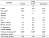
Five-week-old male C57BL/6J mice (n = 21) obtained from Bio Genomics, Inc. (Seoul, Korea) were housed individually in plastic cages and maintained under standard laboratory conditions at a temperature of 24 ± 5℃ and a relative humidity of 55 ± 5% with a regular light cycle (06:00-18:00 light, 18:00-06:00 dark). The mice were divided randomly into three groups after 1 week of adaptation, during which time they had free access to commercial chow. Control, HFHS, and chamnamul groups were fed a basal diet, HFHS diet, and HFHS diet containing a 70% ethanol extract of chamnamul at 0.5% of the diet, respectively, ad libitum for 12 weeks (Table 1). The compositions of the basal and HFHS diets were determined based on previous studies [19-21], in which C57BL/6J mice developed type 2 diabetes when fed a diet high in fat and simple sugar. The basal diet contained 5% corn oil and 65% cornstarch, and the HFHS diet contained 3% corn oil, 33% lard, 11% cornstarch and 27.1% sucrose. The fat-calorie percentages of the basal and HFHS diets were 11.7% and 58.3%, respectively. Body weight and food intake were measured once and three times a week, respectively. All study procedures were approved by the Animal Resource Center at Inje University (2012-51).
Analytical sample preparation
At the end of the feeding trials, the mice were sacrificed by heart puncture following an overnight fast. Blood samples were collected immediately and serum was obtained by centrifugation (1,500 g, 15 min). After collecting the blood, liver samples were taken. Serum and liver samples were stored at -70℃ for further analysis.
Biochemical analyses of serum
Serum glucose [23], triglyceride [24], and cholesterol levels were measured by enzymatic methods [25] using commercial kits. Serum insulin levels were determined by radioimmunoassay using commercial kits. Homeostasis model assessment for insulin resistance (HOMA-IR) values were estimated by dividing the product of fasting insulin (µU/mL) and glucose (mmol/L) levels by 22.5 [26]. Serum adiponectin levels were measured using a commercial ELISA kit.
Measurement of lipid peroxide and antioxidant enzyme activities in the liver
Hepatic lipid peroxidation was evaluated as a thiobarbituric acid reactive substance (TBARS) using the method of Ohkawa et al. [27]. Liver tissue was homogenized in five volumes of 10 mM sodium phosphate buffer (pH 7.4). The homogenate (0.5 mL) was mixed with a solution comprising 15% trichloroacetic acid, 0.4% TBA, and 2.5% HCl (1 mL) and then incubated at 100℃ for 45 min. After cooling, the reaction mixture was centrifuged at 1,500 × g for 10 min and absorbance was then measured at 534 nm. The protein content was measured using the Bradford method [28] with bovine serum albumin as a standard. The level of lipid peroxides was expressed as nmol malondialdehyde (MDA)/mg protein.
To measure the activities of antioxidant enzymes, liver samples were homogenized in 10 volumes of 50 mM phosphate buffer (pH 7.4). The homogenates were centrifuged at 700 g for 10 min, and the supernatant was further centrifuged at 10,000 g for 10 min to collect mitochondrial pellets for the measurement of catalase (CAT) activity. The remaining supernatant was further centrifuged at 100,000 g for 60 min, and the final supernatant was used to measure superoxide dismutase (SOD) and glutathione peroxidase (GSH-Px) activities. CAT activity was determined according to the Aebi method [29]. One unit of CAT activity was defined as µmoles of hydrogen peroxide reduced per minute. Activities of SOD and GSH-Px were measured by the Marklund and Marklund method [30] and a method developed by Paglia and Valentine [31], respectively. One unit of SOD activity was defined as the amount of enzyme that reduced the rate of pyrogallol autoxidation by 50%. One unit of GSH-Px activity was defined as nmoles of NADPH substrate converted to NADP+ per minute. All assays were carried out in triplicate and the enzyme activity was expressed as specific activity (U/mg protein).
Statistical analysis
All data are expressed as means ± standard error of the mean (SEM). One-way analysis of variance with a post hoc Tukey's test was used to evaluate significant differences among groups (P < 0.05). Statistical analysis was performed with SAS version 9.2 (SAS Institute Inc., Cary, NC, USA).
Results
Inhibition of α-glucosidase activity in vitro
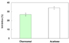
The inhibitory activities of the 70% ethanol extract of chamnamul and acarbose against yeast α-glucosidase in vitro are shown in Fig. 1. The chamnamul extract and acarbose inhibited α-glucosidase by 26.7% and 34.1%, respectively, at 0.5 mg/mL.
Body weight, food intake, and feed efficiency ratio
The body weights, weight gains, food intakes, and feed efficiency ratios (FERs) of the animals are listed in Table 2. The final body weight and weight gain were significantly higher in the HFHS group than in the control group (P < 0.01). The consumption of chamnamul did not significantly influence the final body weight and weight gain in mice fed a HFHS diet. The FER was significantly higher in the HFHS group than in the control group (P < 0.01). The FER tended to be lower in the chamnamul group than in the HFHS group, although this difference was not significant.
Serum glucose, insulin, adiponectin, and lipid concentrations
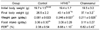
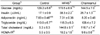
Serum glucose levels were significantly elevated in the HFHS group (170.5 ± 8.9 mg/dL) compared with the control group (129.2 ± 6.6 mg/dL; P < 0.01; Table 3). However, the consumption of chamnamul extract significantly lowered serum glucose levels (144.5 ± 7.0 mg/dL; P < 0.05) compared with those in the HFHS group. No significant difference in serum glucose level was observed between the control and chamnamul groups. Feeding with a HFHS diet increased serum insulin and HOMA-IR values compared with the control group (P < 0.01). Serum insulin and HOMA-IR values in the chamnamul group were significantly lower than those in the HFHS group (P < 0.01) and higher than those in the control group (P < 0.01 and P < 0.05, respectively). Serum adiponectin levels did not differ significantly among the control, HFHS, and chamnamul groups.
Serum cholesterol levels were significantly higher in the HFHS group (144.8 ± 9.1 mg/dL) than in the control group (95.3 ± 6.3 mg/dL; P < 0.01). Chamnamul extract significantly reduced serum cholesterol levels (116.7 ± 7.4 mg/dL; P < 0.05) compared with the HFHS group. Serum cholesterol levels did not differ significantly between the chamnamul and control groups. Serum triglyceride levels did not differ significantly among the three groups.
Hepatic antioxidant parameters
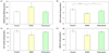
The effects of chamnamul on lipid peroxide levels and the activity of antioxidant enzymes in the liver are shown in Fig. 2. Consumption of a HFHS diet increased hepatic TBARS levels (0.924 ± 0.063 nmol MDA/mg protein) compared with the control group (0.659 ± 0.038 nmol MDA/mg protein; P < 0.01). The consumption of chamnamul extract reduced hepatic TBARS levels in mice fed a HFHS diet (0.644 ± 0.044 nmol MDA/mg protein; P < 0.01) to values comparable with those of the control group.
SOD activity in the liver was significantly lower in the HFHS group than in the control group (P < 0.01). However, the consumption of chamnamul extract elevated SOD activity by 24.3% compared with the HFHS group (P < 0.05), to values that did not differ significantly from those of the control group. The activities of hepatic CAT and GSH-Px were significantly reduced in the HFHS group compared with the control group (P < 0.05). The consumption of chamnamul extract increased the activities of CAT and GSH-Px by 17.5% and 22.8%, respectively, compared with the HFHS group (P < 0.05). The activities of CAT and GSH-Px did not differ significantly between the chamnamul and control groups.
Discussion
We determined the effect of chamnamul on blood glucose status and oxidative stress in vivo and α-glucosidase inhibitory activity in vitro to evaluate its benefit for diabetes mellitus. Consumption of a HFHS diet for 12 weeks produced obesity, insulin resistance, hyperinsulinemia, and hyperglycemia in C57BL/6J mice. This finding is in line with those of previous studies [19-21,32,33], which have demonstrated the inducement of type 2 diabetes following consumption of a HFHS diet in these animals. Yang et al. [32] reported that a HFHS diet induced insulin resistance by the downregulation of genes involved in insulin signaling, resulting in hyperinsulinemia and hyperglycemia.
Consumption of chamnamul extract at 0.5% of the diet did not significantly influence HFHS diet-induced obesity. However, chamnamul effectively alleviated hyperinsulinemia and hyperglycemia (Table 3). Chamnamul extract also decreased the HOMA-IR index, a surrogate index of insulin resistance [34]. In addition, the α-glucosidase inhibitory activity of the chamnamul extract was 78.3% as strong as that of acarbose in vitro (Fig. 1). An α-glucosidase inhibitor, such as acarbose, inhibits the digestion of carbohydrates, resulting in a flattening of postprandial hypoglycemia. Lebovitz [35] reported that acarbose reduced glucose toxicity through the alleviation of postprandial hyperglycemia, resulting in an improvement of overall glycemic control. Chamnamul extract may be able to improve fasting hyperglycemia by reducing glucose toxicity and increasing insulin sensitivity. Determining the effect of chamnamul on the expression of insulin signaling genes would be useful for the investigation of the underlying mechanism of improved insulin resistance. A study aiming to isolate and identify the active compound(s) with α-glucosidase inhibitory activity from chamnamul is currently in progress.
Mice fed a HFHS diet displayed increased serum cholesterol (Table 3), which is consistent with the results of previous studies [32,33]. Chamnamul extract effectively alleviated HFHS diet-induced hypercholesterolemia. This extract has been reported to reduce blood cholesterol levels in rats fed a high-cholesterol diet, and chamnamul has been suggested to exert a hypocholesterolemic effect, partly by inhibiting the reabsorption of bile salts [36]. Lowering blood cholesterol levels reduces the risk of cardiovascular complications in patients with diabetes [37].
We investigated the antioxidant effects of chamnamul by determining lipid peroxide levels and enzyme activity involved in the antioxidative system in the liver. Mice fed a HFHS diet had elevated levels of hepatic TBARS and reduced SOD, CAT, and GSH-Px activities compared with animals fed a basal diet (Fig. 2). In diabetics, oxidative stress is increased [11], which can contribute to the development of diabetic complications by the elevation of lipoprotein oxidation [38,39], activation of proinflammatory signals, and inactivation of anti-atherogenic enzymes [40].
Levels of hepatic TBARS were significantly lowered by chamnamul extract in mice fed a HFHS diet. Chamnamul has been reported to be an antioxidant that directly scavenges ROS and free radicals in vitro [17,18]. Therefore, chamnamul could have reduced TBARS levels by behaving as an antioxidant in this study. In addition, chamnamul potentiated the activities of SOD, CAT, and GSH-Px in these animals. SOD catalyzes the dismutation of superoxide radicals to H2O2, which can be converted to water by CAT in the peroxisomes or by GSH-Px in the cytosol [41,42]. Thus, chamnamul could improve antioxidant status, thereby effectively alleviating diabetic complications, in this animal model.
In conclusion, chamnamul extract effectively alleviated hyperinsulinemia, hyperglycemia, and hypercholesterolemia in C57BL/6J mice fed a HFHS diet. It also decreased levels of hepatic lipid peroxides and increased the activities of antioxidant enzymes. These findings suggest that chamnamul may be useful in prevention of hyperglycemia and reduction of oxidative stress in mice fed a HFHS diet.
 XML Download