Abstract
The purpose of this study was to investigate how advanced maternal age influences lifestyle, nutrient intake, iron status, and pregnancy outcomes in pregnant women. The subjects of this study were 112 pregnant women who were receiving prenatal care at gynecologists located in Seoul. The subjects were divided into two groups according to their ages: those over age 35 were the advanced age group of pregnant women (AP) and those under age 35 were the young age group of pregnant women (YP). General factors, nutrient intakes, iron status, and pregnancy outcomes of the two groups were then compared. It was found that 72.5% of the YP group and 51.2% of the AP group had pre-pregnancy alcohol drinking experience; indicating that the YP group had more pre-pregnancy alcohol consumption than the AP group (P < 0.05). The only difference found in nutrient intake between the two groups was their niacin intakes which were 16.83 ± 8.20 mg/day and 13.76 ± 5.28 mg/day, respectively. When gestational age was shorter than 38.7 weeks, the average infant birth weight was 2.95 ± 0.08 kg, and when gestational age was longer than 40 weeks, it averaged at about 3.42 ± 0.08 kg. In other words, as gestational age increased, infant birth weight increased (P < 0.0001), and when maternal weight increased more than 15 kg, the infant birth weight increased significantly (P < 0.05). In conclusion, in order to secure healthy human resources, with respect to advanced aged women, it is necessary to intervene by promoting daily habits that consist of strategic increases in folate and calcium intake along with appropriate amounts of exercise.
Korean society is aging rapidly at an unprecedented pace and is expected to become an aged society (where people who are 65 years or older will account for more than 14 percent of the total population) by 2018 and a super aged society (where people who are 65 years or older will account for more than 20 percent of the total population) by 2026. The birth rate in 2008 was found to be 1.19, which remains at the world's lowest level. Also, the age of pregnant women has increased, in which the average age of women giving birth to, their first child is 30.82, which is 0.23 years greater than in 2007 [1].
There are various definitions of elderly gravida but it is commonly defined as a woman who has a baby when she is over age 35. The number of elderly gravidas is not only growing in Korea, but it is also a global trend caused by late marriage, remarriage, higher education level, women's advancement in society, and a delay in childbearing by economic factors. In Korea, the rate of elderly gravida increased from 2.0% in 1991 to 6.1% in 1994 to 13% in 2007 [1].
Hinz (USA) reported that the rate of natural childbirth declined when a mother's age was over 40 [2], and Khoshnood (France) observed that at 40-44 years old, female fertility dropped two-fold compared to women's who were 20-24 years old [3]. And there are many overseas reports that the maternal age is a crucial risk factor when predicting pregnancy outcomes [4-9]. However, Kale (Turkey) reported that there were no negative effects for a 45 year-old woman to deliver a large fetus [10].
Research on late childbearing in Korea showed undesirable results in-terms of pregnancy, such as obstetric prognosis, chromosomal anomaly, premature birth, emergent caesarean section, and instrumental labor requiring obstetric intervention [11-16]. In other words, even though pregnant women and their newborn babies may be in good condition, late childbearing can result in a higher frequency of pregnancy complications (premature birth, presentation, pregnancy induced hypertension, gestational diabetes, placenta previa, and premature rupture of membranes), myoma of the uterus, and low birth weight infants [17]. However, there have not been many studies regarding the nutritional risk factors and nutritional status of elderly gravida. For this reason, nutritional support and specific management systems for elderly gravida have not been systematically established. Therefore, if we are able to establish systematic nutrition management for elderly gravida, it will lead to management of new-born nutrition. This systematic nutrition management will become a core field of application for establishing Korean health policy goals and will expand future growth drives through improved public health.
This study compared nutrient intakes, lifestyle habits, serum iron index values, and pregnancy outcomes between young and advanced aged pregnant women to provide groundwork for improving the nutritional status of pregnant women and their babies.
One-hundred twelve pregnant women, excluding a woman with twins, who were receiving prenatal care at gynecologists located in Seoul as from February through June 2009, were chosen as the subjects for this study. They had been informed about the study and had agreed to participate in the survey. By setting 35 years old as the standard, about 43 pregnant women older than 35 were placed in the advanced age group (AP) whereas 69 pregnant women were placed in the young age group (YP). The study was performed by personal face-to-face interview (CHA Hospital IRB 09-03).
Age, BMI prior to pregnancy, BMI during pregnancy, frequency of pregnancy, education level, monthly family income, occupation, morning sickness during pregnancy, and intake of nutritional supplements were investigated.
Amount, number of times, and types of exercise, as well as alcohol consumption, smoking, caffeine intake, and house work or office work-load were investigated.
Dietary intake information was gathered from the women by direct face-to-face interview using the 24 hour recall method. For accurate and precise investigation, food eye measurements and food models were used during the interview. The nutritional value program Can-pro (Computer Aided Nutritional Analysis Program for Professionals 3.0) was used to measure and analyze the nutrient intakes of the pregnant women.
The iron status of the subjects was investigated through hospital clinical records. Properties of the blood such as white blood cells (WBC), red blood cells (RBC), hemoglobin (Hb), hematocrit (HCT), mean corpuscula volume (MCV), mean corpuscula hemoglobin concentration (MCHC), red cell distribution width (RCDW), platelets (PLT), platelet distribution width (PDW), and mean platelet volume (MPV) were interpreted.
All statistical data were processed by the SAS software program version 9.1 (SAS Institute, Cary, NC, USA). Every measurement was marked with a mean value ± standard deviation and percentage. The quantitative and categorical variables of the pregnant women's age-based daily habits, nutrient intakes, and iron status were reviewed by T-tests and Chi-square analysis, respectively. According to pregnancy outcomes and levels of primary daily habits, average birth weight comparisons were performed by ANOVA.
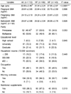
The general characteristics and environmental factors of the study subjects are described in Table 1. The average age of the YP group was 31.54 ± 2.24, and it was 37.05 ± 2.07 for the AP group. About 53.6% of YP and 34.9% of AP were primipara. The education levels of the pregnant women were relatively high: 71% of the YP group and 74% of the AP group had bachelor degrees. The AP group had higher household income than the YP group, but the difference was not considerable. More than 90% of both groups experienced morning sickness. Also, 87% for the YP group and 93% of the AP group took nutritional supplements and there was no remarkable difference between the two groups.
The lifestyle habits of the subjects are summarized in Table 2. For exercise frequency during pregnancy, 27.5% of the YP group exercised once or twice a week and 32.6% of the AP group exercised more than three times a week. The types of exercise performed were mostly walking or doing yoga for both groups. For pre-pregnancy alcohol drinking experience, the YP group had a greater percentage than the AP group, with 72.5% and 51.2%, respectively (P < 0.05). However, the AP group had a higher percentage of women with a longer period of pre-pregnancy alcohol consumption, which was approximately more than 11 years (P < 0.0001). For past smoking, the rate was 13% for the YP group and 7% for the AP group. And regarding a previous smoking period of more than 7 years, AP showed a higher percentage at 7% compared to YP with 5.8%. For current coffee consumption frequency, the groups showed similar results in which 39% of the YP group and 44% of the AP group did not drink coffee.
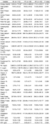
The nutrition intakes of the subjects are shown in Table 3. The energy intakes of the YP group and the AP group were 1,916.2 ± 889.85 kcal and 1,716.2 ± 518.17 kcal, respectively. The total protein intakes of the YP group and the AP group were 76.63 ± 44.62 g and 73.36 ± 58.83 g, respectively. The total fat intake of the YP group was higher at 59.78 ± 40.28 g compared to that of the AP group at 49.74 ± 24.42g. The carbohydrate intakes of the YP group and the AP group were 272.90 ± 103.10 g and 257.00 ± 79.95 g, respectively. The total calcium intakes of both groups were similar, i.e. 599.10 ± 266.40 g for the YP group and 603.20 ± 243.70 g for the AP group. The total Fe intakes from supplemental Fe and food were 80.24 ± 29.87 mg/day and 77.23 ± 28.84 mg/day for YP and AP, respectively. Total folate intakes from supplements and food were 325.5 ± 94.33 µg and 356.2 ± 126.8 µg for YP AP, respectively. The vitamin intakes of the two groups showed no considerable differences. However, a difference was found in niacin intake i.e., 16.83 ± 8.20 mg/day for the YP group and 13.76 ± 5.28 mg/day for the AP group. The ratio of P/M/S was 1/1.3/1.3 for the YP group and 1/1.5/1.4 for the AP group. Both groups had a slightly high intake of saturated fatty acid.
The iron indices of the subjects are indicated in Table 4. The white blood cell (WBC) counts of the YP group and AP group were 9.58 ± 2.70 103/µl and 9.04 ± 3.14 103/µl, respectively, which were within normal range but toward the high end. The red blood cell (RBC) counts of the YP group and AP group presented as similar values, which were 3.73 ± 0.71 106/µl and 3.80 ± 0.47 106/µl, respectively. The hemoglobin (Hg) and hematocrit (HCT) levels of the YP group were 11.63 ± 1.51 g/dl and 34.55 ± 4.16%, and they were 11.60 ± 1.40 g/dl and 34.15 ± 3.96% for the AP group, respectively. The two groups showed similar trends; however values were slightly lower than for non-pregnant women.
The pregnancy outcomes of the pregnant women and their newborn babies are shown in Table 5. The average weight and height of newborns in the YP group were 3.24 ± 0.53 kg and 48.46 ± 1.45 cm, respectively. Those of the AP group were 3.25 ± 0.36 kg and 48.08 ± 1.51 cm, respectively. The Apgar scores of the YP and AP groups were 8.48 ± 1.74 and 8.91 ± 0.28, respectively. Therefore, the average weights and Apgar scores of the two groups showed no considerable differences. The gestational periods of the YP and AP groups were 38.93 ± 3.23 weeks and 38.93 ± 1.36 weeks, respectively. Also, 37.1% of the AP group had a shorter gestational period which was less than 38.7 weeks. Natural spontaneous vaginal delivery was 69% for the YP group and 63% for the AP group. The total maternal weight gain of the YP group was 13.38 ± 3.55 kg and that of the AP group was 13.6 ± 3.93 kg; no significant differences were found between the two groups.
In order to investigate the lifestyle factors influencing infant birth weight, ANOVA was utilized. The considerable factors influencing birth weight are presented in Table 6. Some significant relationships between birth weight and gestational age, maternal weight gain, and exercise frequency during pregnancy were separately identified. For a gestational period less than 38.7 weeks, birth weight was 2.95 ± 0.08 kg, and it was 3.30 ± 0.08 kg for 38.7-40 weeks, and 3.42 ± 0.08 kg for longer than 40 weeks. Birth weight increased as gestational period increased (P < 0.0001). When maternal weight gain was higher than 15 kg, birth weight became remarkably greater compared to that with less than 15 kg of weight gain (P < 0.03). For women who did not exercise and those who exercised 1-3 times in a week, birth weight was remarkably higher compared to babies of women who exercised 1-3 times a month (P < 0.01).
The purpose of this research was to determine the daily lifestyles, nutrient intakes, blood iron indices, and differences in pregnancy outcomes of pregnant women according to age. The only daily lifestyle factor that showed an attentive difference was experience and duration of drinking alcohol prior to pregnancy. Sayal [18] reported that if a pregnant woman drinks more than 4 glasses of alcohol per day, risk to the embryo's behavior and mental health increases, and if a woman drinks every day prior to pregnancy, there is a high risk of presenting hyperactivity and carelessness during pregnancy [18]. Yang et al. [19] observed that drinking alcohol during pregnancy strongly affected abruption of the placenta [19]. Among the research subjects, there were no women who drank while they were pregnant, but some women in the YP group showed high alcohol intakes prior to pregnancy; therefore, active nutrition education and promotion of refraining from drinking during pregnancy are necessary.
The daily average energy intake of the subjects was 1,840.8 ± 774.46 kcal, which was about 80% of the estimated necessary energy intake for pregnant women according to the Dietary Reference Intakes for Koreans 2005 (KDRI) [20]; the YP group showed an energy intake of 83% and the AP group showed an intake of 75% of the DRI, respectively. Protein intake was 142% of the DRI in the YP group and 136% in the AP group. Except for niacin intake, which was considerably higher in the YP group than in the AP group (P < 0.05), no nutrients showed significant intake differences. The nutrients consumed below the average requirements according to the KDRIs were energy (YP 83% : AP 75%), calcium (YP 75% : AP 75%), potassium (YP 60% : AP 58%), and folic acid (YP 62% : AP 69%).
It has been reported that if folate intake is insufficient in the early stage of pregnancy, then the risk of birth defects such as neural tube defects (NTD) will be higher [21]. Kim and Lee [22] reported that Korean women tend to show low intake levels of folate prior to pregnancy and after birth [22]. Folate is the most necessary nutrient in the early stage of pregnancy (21-28 days after pregnancy), and it is strongly recommended that fertile women who are older in age and planning pregnancy regularly consume folate prior to pregnancy.
Guelinckx [23] reported that vegetable intake can be increased simply by reading a brochure for nutrition intervention through nutrition counseling with a nutritionist [23]. Nutrition intervention including nutrition education brochures, video materials, and nutrition counseling, should be reviewed from various standpoints before and during pregnancy.
The iron intakes from food for the YP and AP pregnancy groups were 18% and 19%, respectively, and the women consumed 80% of their iron from supplements. Their total iron intakes were 80.24 ± 29.87 mg and 77.23 ± 28.84 mg, respectively, which were 1.8 and 1.7 times higher than the upper intake of 45 mg in the KDRIs. A similar result was reported by Jang and Ahn [24] in that pregnant women in Korea consumed excess iron.
To prevent an iron deficiency during pregnancy, most pregnant women take iron supplements. Appropriate iron supplement intake increases iron status during pregnancy and after childbirth and also increases hemoglobin content in pregnant women [25] [26]. In addition, it increases birth weight in infants and consequently decreases the risk of delivering a child of low birth weight [27].
However, it was reported that excessive iron intake from supplements may cause harmful effects in the immune response of pregnant women [28]. Therefore, excess iron intake is not desirable for successful pregnancy outcomes.
Moreover, it was reported that excessive iron intake may prevent the absorption of other small amounts of inorganic substances and derive negative effects in oxidization pathways resulting in negative symptoms such as increased oxidative stress, constipation, and impaired judgment. Therefore, the appropriate amount of iron to supplement during pregnancy is still under dispute [29]. When pregnant women consume iron excessively and then stop iron supplements after birth, it can lead to concern, such that a woman who delivers a child can show iron deficiency symptoms even though she is in a normal nutritive state for iron. Therefore, it is essential to create an appropriate dietary strategy to implement proper iron intake from meals during pregnancy and discuss the risk of surplus iron from supplements during pregnancy.
In the case of increased calcium intake during pregnancy, it was previously found that exposed infants had increased bone mass when they reached 9 years old [30]. The result of this study emphasizes the importance of appropriate calcium intake. As a result, it is necessary to plan dietary menus with sufficient calcium and to perform appropriate amounts of outdoor exercise during pregnancy.
By interpreting a table for the iron status of the pregnant women, each separate item was at a normal level and there were no large differences between the YP and AP groups. For Hg and HCT, values were 11.5-16.9 g/dl and 36-46%, respectively, which were lower than for average adult women. However, the standards determined by WHO [31] for anemia in pregnant women are < 11.0 g/dl for Hg and > 33.0% for HCT, which can lead to the conclusion that the pregnant women with 11.5-16.9 g/dl Hg and 36-46% HCT did not have symptoms of anemia. In terms of pregnancy outcomes, there were no differences between the two groups for infant birth weight and height, Apgar score, pregnancy duration, or weight gain of the mother. According to Lee et al. [32], as a mother's age increases, she is likely to have an infant of low birth weight; however, this can be overcome by education, marriage status, and race.
The result of no significant difference in pregnancy outcomes according to mother's age, could lead to two possible conclusions. First, even though a woman may be of advanced age to have a baby, if she is able to maintain healthy habits before and during pregnancy then good pregnancy outcomes can be expected. Second, the result might be due to the small age gap between the two age groups. The average ages of the two groups were only about 6 years apart. Therefore, increasing the age difference between these two groups of pregnant women would present better results.
During pregnancy, woman who exercised 1-3 times a month gave birth to babies of lower weight than woman who exercised 1-3 times a week or did not exercise. Because the number of pregnant women who exercised 1-3 times a month was only 4 persons, we deem that other unobserved personal factors affected infant birth weight. In conclusion, pregnant women, even of advanced age, can obtain desirable pregnancy outcomes if they engage in healthy daily life-style habits, including optimal intakes of folate and calcium.
Figures and Tables
Table 4
Iron indices by the age of pregnant women

1) Mean ± SD
YP: Young age group of pregnant women, AP: Advanced age group of pregnant women, WBC: White Blood Cell, RBC: Red Blood Cell, HG: Hemoglobin, HCT: Hematocrit, MCV: Mean Corpuscular Volume, MCH: Mean Corpuscular Hemoglobin, MCHC: Mean Corpuscular Hemoglobin Concentration, RCDW: Red Cell Distribution Width, PLT: Platelet, PDW: Platelet Distribution Width, MPV: Mean Platelet Volume
References
1. Statistics Korea. Birth data in 2008 [Internet]. cited 2009 August 19. Available from:
http//kostat.go.kr.
2. Hinz S, Rais-Bahrami S, Kempkensteffen C, Weiske WH, Schrader M, Magheli A. Fertility rates following vasectomy reversal: importance of age of the female partner. Urol Int. 2008. 81:416–420.

3. Khoshnood B, Bouvier-Colle MH, Leridon H, Blondel B. Impact of advanced maternal age on fecundity and women's and children's health. J Gynecol Obstet Biol Reprod (Paris). 2008. 37:733–747.
4. Kilic S, Yilmaz N, Zülfikaroglu E, Sarıkaya E, Kose K, Topcu O, Batioglu S. Obesity alters retrieved oocyte count and clinical pregnancy rates in high and poor responder women after in vitro fertilization. Arch Gynecol Obstet. 2010. 282:89–96.

5. Simchen MJ, Shulman A, Wiser A, Zilberberg E, Schiff E. The aged uterus: multifetal pregnancy outcome after ovum donation in older women. Hum Reprod. 2009. 24:2500–2503.

6. Usta IM, Nassar AH. Advanced maternal age. Part I: obstetric complications. Am J Perinatol. 2008. 25:521–534.

7. Salihu HM, Wilson RE, Alio AP, Kirby RS. Advanced maternal age and risk of antepartum and intrapartum stillbirth. J Obstet Gynaecol Res. 2008. 34:843–850.

8. Mbugua Gitau G, Liversedge H, Goffey D, Hawton A, Liversedge N, Taylor M. The influence of maternal age on the outcomes of pregnancies complicated by bleeding at less than 12 weeks. Acta Obstet Gynecol Scand. 2009. 88:116–118.

9. Altman AD, Bentley B, Murray S, Bentley JR. Maternal age-related rates of gestational trophoblastic disease. Obstet Gynecol. 2008. 112:244–250.

10. Kale A, Kuyumcuoğlu U, Güzel A. Is pregnancy over 45 with very high parity related with adverse maternal and fetal outcomes? Clin Exp Obstet Gynecol. 2009. 36:120–122.
11. Heo H, Hwang JY, Kim DG, Lee HJ, Sim JC, Yang HS. A clinical study of pregnancy and delivery in pregnant women 35 years and older. Korean J Obstet Gynecol. 2004. 47:458–463.
12. Yoon SJ, Chang BW, Kim HM, Kim SC, Son YS, Woo BH. A clinical study on the recent tendency of pregnancy and delivery in elderly gravida. Korean J Obstet Gynecol. 1996. 39:476–484.
13. Choi SR, Kim GJ, Lee SP, Kim SY, Yoon SJ, Lee ED. A clinical study of pregnancy and delivery in women aged 40 years and older. Korean J Obstet Gynecol. 2003. 46:612–616.
14. Park HJ, Lee SH, Cha DH, Kim IH, Jun HS, Lee KJ, Song SA, Park HR, Chung CJ, Lee CN. Pregnancy outcomes in women aged 35 and older. Korean J Obstet Gynecol. 2006. 49:2066–2074.
15. Choi JH, Han HJ, Hwang JH, Chung SR, Moon H, Park MI, Cha KJ, Choi HS, Oh JE, Park YS. Meta analysis of clinical studies of pregnancy and delivery in elderly gravida. Korean J Obstet Gynecol. 2006. 49:293–308.
16. Kil K, Lee GSR, Kwon JY, Park IY, Kim SJ, Shin JC, Kim SP. Impact of maternal age of 40 years or older on pregnancy outcomes. Korean J Perinatol. 2007. 18:125–130.
17. Kim TE, Lee SP, Park JM, Whang BC, Kim SY. The effects of maternal age on outcome of pregnancy in healthy elderly primipara. Korean J Perinatol. 2009. 20:146–152.
18. Sayal K, Heron J, Golding J, Alati R, Smith GD, Gray R, Emond A. Binge pattern of alcohol consumption during pregnancy and childhood mental health outcomes: longitudinal population-based study. Pediatrics. 2009. 123:e289–e296.

19. Yang Q, Wen SW, Phillips K, Oppenheimer L, Black D, Walker MC. Comparison of maternal risk factors between placental abruption and placenta previa. Am J Perinatol. 2009. 26:279–286.

20. The Korean Society of nutrition. Dietary reference intakes for Koreans. 2005. Seoul: The Korean Nutrition Society.
21. Black MM. Effects of vitamin B12 and folate deficiency on brain development in children. Food Nutr Bull. 2008. 29:S126–S131.
22. Kim YJ, Lee SS. The relation of maternal stress with nutrients intake and pregnancy outcome in pregnant women. Korean J Nutr. 2008. 41:776–785.
23. Guelinckx I, Devlieger R, Mullie P, Vansant G. Effect of lifestyle intervention on dietary habits, physical activity, and gestational weight gain in obese pregnant women: a randomized controlled trial. Am J Clin Nutr. 2010. 91:373–380.

24. Jang HM, Ahn HS. Serum iron concentration of maternal and umbilical cord blood during pregnancy. Korean J Community Nutr. 2005. 10:860–868.
25. Pena-Rosas JP, Viteri FE. Effects of routine oral iron supplementation with or without folic acid for women during pregnancy. Cochrane Database Syst Rev. 2006. 3:CD004736.
26. Viteri FE, Berger J. Importance of pre-pregnancy and pregnancy iron status: can long-term weekly preventive iron and folic acid supplementation achieve desirable and safe status? Nutr Rev. 2005. 63:S65–S76.

27. Cogswell ME, Parvanta I, Ickes L, Yip R, Brittenham GM. Iron supplementation during pregnancy, anemia, and birth weight: a randomized controlled trial. Am J Clin Nutr. 2003. 78:773–781.

28. Ward RJ, Wilmet S, Legssyer R, Crichton RR. Iron supplementation during pregnancy-a necessary or toxic supplement? Bioinorg Chem Appl. 2003. 1:169–176.
29. Rioux FM, LeBlanc CP. Iron supplementation during pregnancy: what are the risks and benefits of current practices? Appl Physiol Nutr Metab. 2007. 32:282–288.

30. Cole ZA, Gale CR, Javaid MK, Robinson SM, Law C, Boucher BJ, Crozier SR, Godfrey KM, Dennison EM, Cooper C. Maternal dietary patterns during pregnancy and childhood bone mass: a longitudinal study. J Bone Miner Res. 2009. 24:663–668.

31. Yu KH, Yoon JS. Comparison and Evaluation of Hematological Indices for Assessment of Iron Nutritional Status in Korean Pregnant Women(III). Korean J Nutr. 2000. 33:532–539.


 PDF
PDF ePub
ePub Citation
 Print
Print


 XML Download