

 PDF
PDF ePub
ePub Citation
 Print
Print


Introduction
Citrus fruits are among the most abundant fruit crops in the world [1,2]. Generally, the fruit is processed into juice as an ingredient in various value-added products for its special flavor. After juice extraction, the fruit pulp is mostly dumped as waste at large expense. The amount of residue from citrus fruits accounts for 50% of the original amount of the whole fruit [3]. Hence, citrus press cakes (CPCs), which are produced in tons per day, represent a problem for management and can lead to environmental pollution due to microbial spoilage. Manipulation of citrus processing wastes is now becoming a very important environmental issue.
Large amounts of CPCs are generated by juice processing factories annually, and as CPCs mainly consist of citrus peel, they are rich in natural bioactive compounds. Citrus peel contains greater amounts of flavonoids than other parts of the citrus fruit [4]. Flavonoids are a group of phytochemicals that are distributed throughout the plant kingdom and exhibit a wide variety of biological activities [5]. In addition, in a previous study, the phenolic and flavonoid compounds present in CPCs showed strong antioxidant activities in a cell-free system [6]. CPCs have great potential for further use as functional ingredients in many industrial applications. Thus, further commercial exploitation of this by-product has gained increasing interest. However, development of an effective recovery process for CPCs in order to obtain a high quality and commercially attractive product is essential.
Drying is one of the oldest methods of food preservation. One of the ways to shorten the drying time is to supply heat by infrared radiation [7]. Far-infrared radiation (FIR) has been successfully applied for drying of foods and agricultural products. Therefore, FIR is an increasingly popular method for drying food products. In addition, FIR drying has many advantages over the most common convective drying techniques.
It has been reported that oxygen free radicals and other reactive oxygen species (ROS) can cause oxidative injury to living organisms and thus play an important role in many lifestyle-related diseases such as cancer [8,9]. Recent research has focused on the extraction of low-cost antioxidants of natural origin that can replace synthetic alternatives. Many plants and colored fruits are known to contain large amounts of phenolic antioxidants besides the well-known carotenoids, vitamin C, and vitamin E. Phenolic antioxidants in plants and fruits are mainly composed of variety of phenolic acids, flavonoids, and catechins [10-13]. Some phenolic compounds in plants have the potential to suppress lipid peroxidation, prevent DNA oxidative damage, and scavenge ROS [10].
Despite a previous successful CPCs drying process, the aim of the present study is to evaluate the effectiveness of the FIR drying system and to determine the protective effect of methanol extract of FIR-dried CPCs on H2O2-mediated DNA damage in Vero cells. Thus, this work describes the application of the FIR drying technique to CPCs to obtain good quality dried product that could be used as an excellent source of antioxidants.
Materials and methods
Materials
CPCs were obtained from Jeju Special Self Governing Province Development Co., South Korea. The fluorescent probe 2', 7'-dichlorodihydroflurescin diacetate (DCF-DA) and 3-(4-5-dimethyl-2yl)-2-5-diphynyltetrasolium bromide (MTT) were purchased from Sigma Co. (St. Louis, MO, USA). Dulbecco's modified Eagle's medium (DMEM), penicillin / streptomycin were purchased from Gibco BRL Co. (Paisley, UK). All other chemicals used in this study were of analytical grade.
Far-infrared radiation drying of CPCs
CPCs stored at -50℃ were dried in the FIR drying system (Model TOURI-Q, Seoul, Korea). Operating conditions of the FIR drying system were adjusted as previously described [5,14]. Dried CPCs were pulverized into fine powder using a grinder (MF 10 basic mill, GMBH & Co., Staufen, Germany) and sieved through a No. 50 standard testing sieve. Wet CPCs were dried separately using the freeze-drying (FD) method and the freeze-dried CPCs were compared to the FIR-dried CPCs.
Preparation of methanolic extracts from dried CPCs
The proximate chemical composition of FIR-dried CPCs was as follows: Moisture 6.3%, ash 4.07%, protein 9.45%, fat 2.6% and carbohydrate 77.58% [14]. The total phenolic content (TPC) and the total flavonoid content (TFC) of the methanol extract from FIR-dried CPCs were 2,438.6 (mg/100 g) and 1,300.2 (mg/100 g), respectively [5].
Twenty grams of ground CPCs were mixed in absolute methanol and kept in a shaking incubator (120 rpm) for 24 h at room temperature. Then the extracts were filtered and evaporated under vacuum at 40℃ in order to obtain the dry extracts. Samples were dissolved in DMSO prior to use in the cell experiments.
Cell culture
The monkey kidney fibroblast cell line (Vero) was cultured and maintained in DMEM containing 10% heat-inactivated calf serum, streptomycin (100 µg/mL) and penicillin (100 unit/mL) at 37℃ in an incubator, under humidified atmosphere containing 5% CO2.
Determination of intracellular ROS generation (DCFH-DA assay)
Intracellular ROS generation was determined using the oxidation-sensitive dye DCFH-DA according to the method described by Engelmann et al. [15]. The Vero cells were seeded in 96-well plates at a concentration of 1.0 × 10⁵ cells/mL. After 16 h of incubation at 37℃, the cells were treated with 10 µL of sample (25, 50 and 100 µg/mL) and incubated at 37℃ under a humidified atmosphere. After 30 min, H2O2 was added at 1 mM and then the cells were incubated for an additional 30 min under the same conditions. Finally, DCFH-DA (5 µg/mL) was introduced to the cells, and DCFH-DA fluorescence was detected at an excitation wavelength of 485 nm and an emission wavelength of 535 nm, using a Perkin-Elmer LS-5B spectrofluorometer (Walthman, MA, USA).
Determination of cell viability (MTT assay)
Cell viability was estimated using the MTT assay as described by Mosmann [16]. The Vero cells were seeded in 96-well plates at a concentration of 1.0 × 10⁵ cells/mL. After 16 h of incubation at 37℃, the cells were treated with 10 µL of the sample (25, 50 and 100 µg/mL) and incubated at 37℃ under a humidified atmosphere for 1 h. Then, 10 µL of H2O2 (1 mM) was added and incubated for 24 h at 37℃. MTT stock solution (50 µL; 2 mg/mL) was then added to each of the wells, to a total reaction volume of 200 µL. After 4 h of incubation, the plates were centrifuged for 5 min at 800 × g and the supernatant was aspirated. The formazan crystals in each well were dissolved in 150 µL of dimethylsulfoxide (DMSO) and the absorbance was measured using enzyme-linked immunosorbent assay (ELISA) reader (Sunrise; Tecan Co. Ltd., Australia) at 540 nm. Relative cell viability was evaluated in accordance with the quantity of MTT converted to the insoluble formazan salt. The optical density of the formazan generated in the control cells was considered to represent 100% viability. The data are expressed as mean percentages of viable cells versus the respective control.
Lipid peroxidation inhibitory assay
Lipid peroxidation was evaluated by measuring malondialdehyde (MDA) as previously described [17]. Vero cells were seeded in a culture dish at a concentration of 1.0 × 10⁵ cells/mL and incubated at 37℃ in a humidified atmosphere. After 16 h, cells were treated with different concentrations of the extracts from CPCs (50 and 100 µg/mL). One hour later, 0.7 mM of H2O2 was added, and the cells were incubated for an additional hour. The cells were then harvested and washed with phosphate-buffered saline (PBS) and homogenized in ice-cold 1.15% KCl. The extracts containing 100 µL of the cell lysates were combined with 0.2 mL of 8.1% sodium dodecylsulfate (SDS), 1.5 mL of 20% acetic acid (adjusted to pH 3.5), and 1.5 mL of 0.8% thiobarbituric acid (TBA). The mixture was brought to a final volume of 4.0 mL with distilled water and heated at 95℃ for 2 h. After cooling to room temperature, 5.0 mL of a mixture of n-butanol and phyridine (15:1 v/v) was added to each mixture and vortexed thoroughly. After centrifugation at 1500 × g for 10 min, the supernatant was isolated and the absorbance was measured at 532 nm.
Nuclear staining with Hoechst 33342
The nuclear morphology of the cells was evaluated using the cell-permeable DNA dye, Hoechst 33342. Cells with homogeneously stained nuclei were considered viable, whereas the presence of chromatin condensation and/or fragmentation was indicative of apoptosis [18,19]. Vero cells were seeded in 24-well plates at a concentration of 1.0 × 10⁵ cells/mL. Sixteen hours after seeding, the cells were treated with the sample at a concentration of 100 µg/mL and further incubated for 1 h at 37℃ in a humidified atmosphere prior to exposure to H2O2 (1 mM). After 24 h, 1.5 µL of Hoechst 33342 (stock 10 mg/mL), a DNA-specific fluorescent dye, was added to each well, followed by 10-min incubation at 37℃. The stained cells were then observed under a fluorescence microscope equipped with a CoolSNAP-Pro colour digital camera (Media Cybernetics, Carlsbad, CA) in order to examine the degree of nuclear condensation.
Determination of DNA damage (Comet assay)
Each extract was diluted in PBS to make the final concentrations of 25, 50, and 100 µg/mL. Vero cells were treated in three different ways. First, Vero cells (4 × 10⁴ cell/mL) were incubated without the extract for 30 min at 37℃ in a dark incubator (I). Second, Vero cells (4 × 10⁴ cell/mL) were incubated without the extract for 30 min at 37℃ and then oxidatively damaged with 100 µM H2O2 for 5 min on ice (II). Third, Vero cells (4 × 10⁴ cell/mL) were incubated with the extracts for 30 min at 37℃ in a dark incubator and then treated with 100 µM H2O2 for 5 min on ice (III). After each treatment, the cell suspension was centrifuged at 1500 × g for 5 min, and washed with PBS.
The alkaline comet assay was conducted as described by Singh et al. [20] with slight modifications. The cell suspensions made in the previous steps were mixed with 100 µL of 0.7% low melting agarose (LMA), and added to slides precoated with 1.0% normal melting agarose. After solidification of the agarose, slides were covered with another 100 µL of 0.7% LMA and then immersed in lysis buffer (2.5 M NaCl, 500 mM EDTA, 1 M Tris buffer, 1% sodium laurylasarcosine, and 1% Triton X-100) for 90 min. Later, slides were transferred into unwinding buffer for another 20 min for DNA unwinding. The slides were next placed in the electrophoresis tank containing 300 mM NaOH and 1 mM Na2EDTA (pH 13.0). For electrophoresis of the treated DNA, an electric current of 25 V/300 mA was applied for 20 min. The slides were then washed two times with neutralizing buffer (0.4 M Tris, pH 7.5) for 10 min and treated with ethanol for another 5 min before staining with 40 µL of ethidium bromide (20 µL/mL). Measurements were made by image analysis (Komet, Andor Technology, Belfast, U.K.) and fluorescence microscopy (LEICA DMLB, Wetzar, Germany) to determine the percentage of fluorescence in the tail (tail intensity, TI; 50 cells from each of two replicate slides).
Results
Intracellular ROS scavenging activity
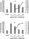
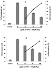
We investigated the scavenging ability of methanol extract of CPCs on intracellular ROS. The DCF fluorescence intensity reached a peak in the cells treated with only H2O2. However, treatment with the extracts from CPCs decreased the fluorescence intensity in Vero cells. In contrast, the fluorescence intensity decreased gradually when the sample concentration increased, indicating that the ROS scavenging activity was dose-dependent. The extract from FIR-dried CPCs showed slightly lower activity than that from freeze-dried CPCs at all concentrations used (Fig. 1A and 1B). However, as the figure shows, methanol extract of FIR-dried CPCs exhibited comparatively good ROS scavenging activity versus the freeze-dried CPCs at the concentration of 100 µg/mL.
Cell viability
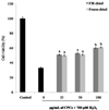
The protective effect of methanol extract from dried CPCs on H2O2-induced cell damage is shown in Fig. 2. Low cell viability was observed in cells treated only with H2O2. However, the dried CPCs prevented H2O2-induced cellular damage, restoring cell survival to significant level. In this study, the protective effect of the extract from FIR-dried CPCs was comparable to that of the extract from freeze-dried CPCs at all three concentrations.
Lipid peroxidation inhibitory activity
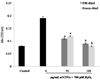
In the present study, when cells were treated only with H2O2, a dramatic increase in MDA concentration was observed, indicating severe peroxidative damage to the cell membrane (Fig. 3). However, pretreatment of the cells with increasing concentrations of the methanol extract from FIR-dried CPCs prevented the phospholipid oxidative alteration in a concentration-dependent manner. In addition, the lipid peroxidation inhibitory activity of the extract of FIR-dried CPCs was comparable with that of the extract of freeze-dried CPCs.
Protective effect against H2O2-mediated DNA damage and cell apoptosis
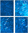
In the present study, the profound antioxidative activity exhibited by dried CPCs was further evaluated with regard to its ability to protect against H2O2-induced cell apoptosis (Fig. 4). Clear image of the negative control showed no cell apoptosis (Fig. 4A), but typical fluorescence photographs revealed shrunken nuclei, chromatic condensation, and apoptotic bodies in the Vero cells after 24 h of H2O2 treatment (Fig. 4B). However, when the cells were treated with methanol extracts from FIR or freeze-dried CPCs at 100 µg /mL for 1 h prior to H2O2 treatment, a considerable reduction in apoptotic bodies was observed (Fig. 4C and 4D). Both samples exhibited similar protective effects against H2O2-induced apoptosis in Vero cells.
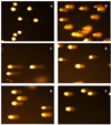
The results of inhibition assays and photomicrographs of DNA damage are shown in Fig. 5 and 6, respectively. Methanol extract of FIR-dried CPCs showed good inhibitory effects against H2O2-mediated DNA damage in a dose-dependent manner. The exhibited inhibitory effects were also comparable to those of the freeze-dried sample. In addition, photomicrographs of different DNA migration profiles were obtained. In the group treated only with hydrogen peroxide, the DNA was completely damaged and the amounts of tail DNA were significantly increased (Fig. 6B). But addition of the methanol extracts from FIR or freeze-dried CPCs with the hydrogen peroxide remarkably reduced the amounts of tail DNA in a dose-dependent manner (Fig. 6C, 6D, 6E and 6F).
Discussion
Disposal of food processing by-products is a major problem for the food industry. Currently, there is a trend in exploiting and utilizing the by-products in the development of functional ingredients such as antioxidants. Bio-waste fractions seem to be particularly useful as a source of biologically important components. Biogenic food processing residues are easily accessible and can be collected under controlled conditions [21]. A more intense use of these residues as a bioactive resource might be a sustainable remedy to reduce the costs of waste disposal. However, in most cases, food-processing residues are wet and it is therefore essential to apply an effective drying process as a preservation step. In the present study, we demonstrated the possibility of using CPCs more efficiently and discuss its potential as a natural antioxidant resource after FIR drying. FIR drying required significantly less drying time (6.5 h) than freeze drying (48 h) to convert the wet CPCs into dried form. Therefore, FIR drying is more effective and economical than freeze drying. In addition, our previous study demonstrated free radical scavenging and antioxidant activity of FIR-dried CPCs by different assays [5]. Moreover, CPCs dried by FIR at 80℃ showed higher antioxidant activities in tested assays than freeze-dried CPCs. Therefore, FIR drying is an effective and economical process that may increase the bioactivities by liberating covalently bound compounds [14,22].
It is well-known that H2O2 is the principal ROS responsible for the oxidation of DCHF to DCF [23]. Our results indicate that methanol extracts from FIR-dried CPCs have efficient antioxidant properties. In addition, one of our previous studies demonstrated the H2O2-scavenging ability of FIR-dried CPCs in a cell-free system [5]. Many researchers have worked with different flavonoid compounds from different origin, and have revealed that such compounds exhibit strong scavenging effects against intracellular ROS formation in the DCFH-DA assay [24]. Hence, the activity shown in this assay may be due to direct scavenging of ROS by active compounds, mainly flavonoids available in the methanol extract of dried CPCs. In our previous study [5], we reported that the TPC and TFC of the methanol extract of FIR-dried CPCs were 2,438.6 (mg/100 g) and 1,300.2 (mg/100 g), respectively. In addition, the previous work revealed that hespiridin and narirutin were the major flavonoid constituents in FIR-dried CPCs.
The level of lipid peroxidation was measured in order to verify the effect of the extracts on the specific molecular alterations that eventually result in oxidative damage in Vero cells. Membrane phospholipids constitute major target sites for the cytotoxic effects of HO· radicals and other ROS, in that polyunsaturated fatty acids are prone to peroxidation, leading to the formation of hydroperoxides, which are subsequently degraded to malondialdehyde (MDA) [25]. Although H2O2 is not highly reactive, the primary mechanism of H2O2 toxicity is the formation of highly reactive radical species such as HO. in the presence of transition metals such as Fe2+ or Cu2+ [26]. Formation of HO. radicals and other ROS may initiate the deleterious reactions on lipids and cause DNA damage.
Oxidative stress leads to oxidative damage to biomolecules and cells. It is well documented that DNA damage leads to mutations, which are responsible for related diseases such as cancer, coronary heart disease, arteriosclerosis and inflammatory disorders. During oxidative stress, large amounts of ROS are produced and may trigger chemical chain reactions such as lipid peroxidation or DNA oxidation, causing cell damage [27,28]. ROS can damage nucleic acids by altering purine and pyrimidine bases and causing DNA strand breakage [29]. Over the years, many scientific investigations have shown that natural antioxidants purified from plant materials have potential inhibitory effects against H2O2-mediated DNA damage and harmful free radicals. Nowadays, interest has increased in the possible health benefits of potent antioxidant and free radical scavenging activities of natural sources. Moreover, natural antioxidants are important components of the human diet, although they are not considered as nutrients.
Previously we reported that methanol extract of FIR-dried CPCs strongly scavenged hydrogen peroxide in vitro [5]. Hence, the extract from FIR-dried CPCs could be used to protect cells, as confirmed by the comet assay. However, to the best of our knowledge, there are no reports on protective effective effects against DNA damage by methanol extract of FIR-dried CPCs. Therefore, in this study we used the comet assay, a sensitive biological assay, to measure the DNA damage in Vero cells. The exhibited protective effect against H2O2-mediated DNA damage and cell apoptosis might be related to the abilities of biologically active components, especially flavonoids, in the extract of dried CPCs.
In conclusion, CPCs can be recovered as a value-added by-product after FIR drying. Antioxidant properties of the methanol extract of FIF-dried CPCs were comparable to those of freeze-dried samples. Since FIR drying significantly reduced the drying time of wet CPCs compared with freeze drying, the FIR drying technique is an effective and economical alternative drying method that can be adopted in many industrial applications. In addition, the present study demonstrated the dried CPCs retained important bioactive components and can be considered as a good source of natural antioxidants. Moreover, these value-added CPCs recovered from citrus by-products are economically attractive and worthy of industrial exploration and application.
 XML Download