

 PDF
PDF ePub
ePub Citation
 Print
Print


Introduction
There have been conflicting reports on the role of ovary (estrogen) and testis (testosterone) in growth and cholesterol metabolism. Our recent study (Lee et al., 2008) demonstrated that a lower growth rate in female rats was due to estrogen, which induced secretion of the stress hormone cortisol. Similarly, other studies (Burgess & Handa, 1992; Lesniewska et al., 1990) also showed gender differences in cortisol secretion, indicating that estrogen elevates basal levels of cortisol, corticosterone and adrenocorticotropin, and their responses to various stimuli in rats. Short-term estradiol treatment to young men increased cortisol and norepinephrine concentrations in the saliva following stress over a placebo control (Kirschbaum et al., 1996).
In the Framingham study, the risk of cardiovascular disease was shown to be significantly increased in women who had taken estrogen (Wilson et al., 1985). Manhem et al. (1996) reported that estrogen administration resulted in an enhanced cardiovascular response to mental stress in young menstruating women. In contrast, estrogen has long been known to exert cardioprotective effect (Barrett-Connor & Bush, 1991), although the precise mechanism underlying its benefits is unknown (Sudhir et al., 1997). Antioxidant activity of estrogen has been suggested as its role in cardioprotection (Walsh et al., 1999), but Santanam et al. (1998) found no inhibitory action of estrogen on LDL oxidation at physiological concentrations.
Our previous studies (Lee et al., 1999; Lee et al., 2008) showed a much higher plasma cholesterol level in female than in male rats when they were fed a hypercholesterolemic diet (CD). Naito et al. (1995) proposed that the effect of sex hormones on lipid metabolism is not likely to account for the sex difference in cardiovascular disease. Furthermore, in the 1995 Ancel Keys lecture, Barrett-Connor (1997) indicated that plasma estrogen levels do not explain coronary heart disease (CHD) risk in either sex.
In the present study, we conducted two experiments to determine the effect of gender and gonadectomy on growth, feed efficiency and plasma cholesterol levels, using pigs as a model animal to confirm the findings of our previous study done with rats (Lee et al., 2008).
Materials and Methods
Animals and diets
In experiment 1 conducted to assess the effect of gonadectomy (GDX) on growth and feed efficiency in female pigs, five sham-operated intact and five GDX Landrace female pigs (26 kg) were allowed to have free access to water and feed until they reached market weight (approximately 100 kg). Body weight and feed consumption were recorded biweekly, and daily body weight gain, daily feed intake and feed efficiency (gain/feed) were calculated during the feeding period.
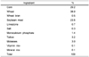
In experiment 2 conducted to determine the effect of gender and GDX on plasma cholesterol levels in pigs, 10 male (26 kg) and 10 female (26 kg) Landrace pigs were used; five male and five female pigs were assigned to sham-operated intact or GDX. Pigs were allowed to have free access to water and diet without added cholesterol until they were 6 months old (male 104 and female 98 kg) and thereafter a hypercholesterolemic diet (Table 1) containing 0.5% cholesterol and 0.1% cholate for 10 days. All animal management and sampling procedures were in accordance with the Guide for the Care and Use of Agricultural Animals in Agricultural Research and Teaching (Consortium, 1988).
Gonadectomy, plasma sample preparation and carcass measurement
Gonadectomy was carried out surgically by removing testicles or ovaries. Testicles were removed through a small incision in the tip of the scrotum after ligation of the spermatic cords. Ovariectomy was done through a dorsal paramedial incision at the level of the lower pole of the kidneys. The surgery was done under light anesthesia and the incision was closed with stitches. The stitches were removed 7 d after surgery. The control intact pigs were sham-operated leaving the organs intact. The surgery on pigs used in both experiments was done when they were 10 days old. At the end of feeding period, pigs were fasted for 16 hr and killed (Exp. 1), and from the pigs used in experiment 2, blood samples were collected into vacutainer tubes containing EDTA before and after a 10-day feeding period of the hypercholesterolemic diet. Plasma was prepared from the blood samples by centrifugation, and stored at -20℃ for later analysis. For pigs used in experiment 1, the average value of measurements at 11th thoracic and 1st lumbar vertebra was used as the back fat thickness. Dressing percentage was calculated from the slaughter weight over the live weight.
Analyses of cholesterol
Total and high-density lipoprotein (HDL) cholesterol concentrations in plasma were determined using commercial assay kits (International Reagent Corp., Tokyo, Japan for the former, and WAKO Pure Chemical Ind., Osaka, Japan for the latter) according to the manufacturer's instruction. Low-density lipoprotein (LDL) plus very-low-density lipoprotein (VLDL) cholesterol concentration was calculated by subtracting HDL cholesterol from the total.
Statistical analysis
The student t-test was used to assess the effect of ovariectomy on pig performance in experiment 1. Data from experiment 2 were analyzed by the two-way analysis of variance (SAS, 1988). In the analysis of variance, the main sources of variation for all variables were gender and gonadectomy. When the F-value in the analysis of variance was significant, the Duncan's multiple range test was used to compare individual means.
Results
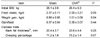
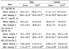
Overiectomy increased average daily gain (P<0.05), but had no effect (P>0.5) on feed efficiency during the growing-finishing period (Table 2). Back fat thickness and dressing percentage in ovariectomized pigs tended to be higher (P>0.05) than those found in sham-operated intact female pigs (Table 2). No differences in the plasma total or HDL cholesterol levels and HDL/LDL+VLDL ratio were found before feeding the hypercholesterolemic diet between sexs or between intact and GDX (Table 3). However, plasma cholesterol levels in pigs fed the hypercholesterolemic diet for 10 days were much higher in females than in males (161 vs 104 mg/100 mL plasma). GDX increased plasma cholesterol levels in male but not in female pigs. HDL/LDL+VLDL ratio appeared to be higher (P<0.01) in male than in female pigs, and was not influenced by GDX in either sex.
Discussion
The present pig study data (Table 2) support our previous findings of the rat study (Lee et al., 2008), in which depressed growth in female rats compared with male rats was mostly due to their ovarian activity. Estrogen secreted in the ovary was responsible for the growth depression because the injection of estradiol markedly depressed growth in GDX rats of either sex, while enhancing plasma cortisol levels. This gender difference was abolished by ovariectomy and was reinstated by estradiol administration (Le Mevel et al., 1979). The pattern of cortisol secretion was also different between sexes in the nonhuman primate macaques, and estradiol implant in castrated male macaques elicited a female pattern of plasma cortisol levels (Norman et al., 1992; Smith & Norman, 1987).
In healthy men, short-term treatment with estradiol led to enhanced hypothalamic-pituitary-adrenal (HPA) and sympathetic responsiveness to psychological stress, resulting in increased adrenocorticotropic hormone (ACTH), cortisol and norepinephrine concentrations in the saliva compared to the placebo (Kirschbaum et al., 1996). Similarly, Burgess and Handa (1992) showed that corticosterone and ACTH levels after a 5-second footshock stress with one mamp were much higher and maintained for a prolonged time in OVX rats when administered with estradiol. In addition, they found that estrogen treatment resulted in a loss of the glucocorticoid receptor's ability of autoregulation.
Interestingly, estrogen has been reported to have both stimulatory and inhibitory effects on HPA functions, depending on the time after ovariectomy and different doses of estradiol (Luber et al., 1991; Redei et al., 1994). Cortisol, as well as catecholaminergic responses to stress, has also been known to vary with estrous cycle in women (Collins et al., 1985) and in rats (Carey et al., 1995).
Taken together, our data obtained from both rat (Lee et al. 2008) and the present pig studies, and others' (Le Mevel et al., 1979; Phillips & Poolsaguan, 1978) clearly indicate that estrogen induces the secretion of hormones that increase with stress, in turn resulting in growth depression. Therefore, ovariectomy may be an economically viable practice to improve growth of female pigs.
Studies have shown marked gender difference in plasma cholesterol levels in Sprague Dawley rats (Lee et al., 1999; Lee et al., 2008) and in guinea pigs (Fernandez et al., 1995), when animals were fed hypercholesterolemic diets. The present study also showed a gender difference in plasma cholesterol levels in pigs, and gonadectomy of male but not female increased plasma total cholesterol and LDL-C concentrations without decreasing HDL-C when pigs were fed the hypercholesterolemic diet (Table 3).
Testosterone replacement therapy in hypogonadal and elderly men showed a beneficial effect on cardiovascular system, decreasing total cholesterol and atherogenic fraction of LDL-C without significant alterations in HDL-C levels or its subfractions HDL2-C and HDL3-C (Zgliczynski et al., 1996). Tchernof et al. (1997) also showed that increased testosterone levels were associated with reduced triacylglycerol and Apo B; total and LDL cholesterol concentrations; and increased HDL/total cholesterol and HDL2-C / HDL3-C ratios. Hypotestosteronemia was observed in Chinese male patients with coronary heart disease (CHD), and positive correlation of plasma testosterone level with plasma HDL-C was found but negative correlation with plasma Lp(a) level, suggesting that testosterone had a protective effect against atherosclerosis (Zhao & Li, 1998).
On the contrary, several studies have shown that testosterone has an adverse effect on cholesterol metabolism by altering lipoprotein profiles. Total testosterone concentrations and sex hormone-binding globulin (SHBG) were significantly associated with LDL size in men (Haffner et al., 1996). Testosterone, per se, when administered at sufficiently high doses and for long durations, can significantly raise serum levels of total cholesterol, triacylglycerol, LDL-C and Apo-B, meanwhile lower HDL-C levels in androgenized women, indicating that male predilection for cardiovascular disease may be due to adverse effects of high androgen levels on lipid and lipoprotein profiles (Goh et al., 1995). In a study done with men, Anderson et al. (1995) also showed that serum HDL-C levels were significantly depressed by intramuscular injection of testosterone enanthate (200 mg per week for 12 months), while plasma total and LDL cholesterol or triacylglycerol levels were not affected.
Handa et al. (1997) reported that the plasma free testosterone level was associated with lower levels of HDL-C, but total estradiol was related to elevated levels of HDL-C in Japanese men in their early fifties, supporting that testosterone may be causally associated with atherosclerosis in men through altered lipoprotein metabolism. Male hamsters fed a hypercholesterolemic diet containing 0.05% cholesterol developed greater elevations in plasma total cholesterol and HDL-C levels, and a higher rate of early aortic atherosclerosis, compared with female hamsters (Wilson et al., 1999). Further studies appear to be inevitable to clarify the involvement of testosterone in cholesterol metabolism, such as cholesterol synthesis (e.g., HMG-CoA reductase gene expression) and absorption, or cholesterol oxidation and excretion (e.g., cholesterol 7α-hydroxylase gene expression).
Estrogen's involvement in lipoprotein metabolism (e.g., an increased ratio of HDL to LDL cholesterol) has been implicated in its role in cardioprotection (Campos et al., 1997; Wagner et al., 1991). If estrogen plays any role at all in reducing cardiovascular disease in females, it may be through actions on other than controlling blood cholesterol levels. Naito et al. (1995) indicated that the effect of sex hormones on lipid metabolism is not likely to account for the sex difference in cardiovascular disease because: 1) men with premature myocardial infarction was shown to have increased estrogen levels (Phillips, 1976), and 2) men who had received high doses of estrogen showed an increased frequency of cardiovascular events (Coronary Drug Project Research Group, 1976; Veterans Administration Cooperative Urological Research Group, 1967).
In conclusion, ovarian activity depresses growth in female pigs and thus ovariectomy (or inhibition of estrogen secretion or its activity) can be an economically viable method to improve growth of female pigs. Testis (testosterone) is responsible for the lower plasma cholesterol level in male pigs than in female pigs when they were fed hypercholesterolemic diets.
 XML Download