Journal List > Nutr Res Pract > v.3(4) > 1051029

Lee, Choi, and Seo: Grape skin improves antioxidant capacity in rats fed a high fat diet

Abstract

This study was conducted to investigate the effect of dietary grape skin on lipid peroxidation and antioxidant defense system in rats fed high fat diet. The Sprague-Dawley rats were fed either control (5% fat) diet or high fat (25% fat) diet which was based on AIN-93 diet for 2 weeks, and then they were grouped as control group (C), control + 5% grape skin group (CS), high-fat group (HF), high fat + 5% grape skin group (HFS) with 10 rats each and fed corresponding diets for 4 weeks. The hepatic thiobarbituric acid reacting substances (TBARS) were increased in high fat group as compared with control group, but reduced by grape skin. The serum total antioxidant status, and activities of hepatic catalase and superoxide dismutase, xanthine oxidase and glucose-6-phosphatase were increased by supplementation of grape skin. Glutathione peroxidase activity was significantly higher in CS group than in C group. Grape skin feeding tended to increase the concentration of total glutathione, especially in control group. The ratio of reduced glutathione to oxidized glutathione was lower in high fat groups than in control groups. The ratio was increased by dietary supplementation of grape skin in control group. These results suggest that dietary supplementation of grape skin would be effective on protection of oxidative damage by lipid peroxidation through improvement of antioxidant defense system in rats fed high fat diet as well as rats with low fat diet.

Introduction

Obesity is generally linked to complications in lipid metabolism and oxidative stress (Oben et al., 2007). Increased oxidative stress in accumulated fat is an important pathogenic mechanism of obesity-associated metabolic syndrome (Furukawa et al., 2004).
France has a lower incidence of coronary heart disease, despite of high consumption of total fat and saturated fat (Belleville, 2002). It was reported that the reason of this effect would be high or regular consumption of red wine (Cui et al., 2002). Antioxidative properties of red wine are believed to be due to polyphenol components, which are mainly derived from grape skin (Falchi et al., 2006; Yunoki et al., 2008).
compounds and grape skin has a particularly high content of resveratrol (Falchi et al., 2006; Frederiksen et al., 2007). Resveratrol is a phytoalexin produced by plants that are under attack. It has been reported to produce a variety of pharmacological effects. These effects contain anti-oxidant, anti-cancer, and anti-inflammatory properties (Aggarwal et al., 2004; Bralley et al., 2007; Delmas et al., 2005; Su et al., 2006; Zhang et al., 2006). Resveratrol has also been shown to modulate lipoprotein metabolism, reduce the synthesis of lipids, inhibit aggregation of platelets and suppress cellular processes associated with tumorigenesis (Fukao et al., 2004; Miura et al., 2003; Schneider et al., 2000; Scarlatti et al., 2003; Zhang et al., 2006).
Also, grape skins contain a large amount of anthocyanins, a phytochemical. Anthocyanins possess various physiological activities such as anti-oxidant activity, anti-obesity effect and inhibition of lipid peroxidation (Acquaviva et al., 2003; Ramirez-Tortosa et al., 2001; Tsuda et al., 2003).
Grape is one of the world's largest fruit crops, which are rich sources of polyphenols (FAO, 2005). However, there is almost no report on the effect of grape skin related to oxidative stress by obesity. Thus, this study was conducted to investigate the effects of dietary supplementation of grape skin on lipid peroxidation and antioxidant defense system in rats fed high fat diet.

Materials and Methods

Preparation and analysis of grape skin

Campbell Early (Vitis Labruscana Bailey) red grapes were obtained from Gyeongsan, Korea. The grape skins were separated from the pulps, freeze-dried and grinded in a mortar for passing through 40 mesh of a sieve.
Total phenol content of grape skin was determined by the Folin-Ciocalteau colorimetric method (Singleton et al., 1965) and expressed as gallic acid equivalents per g dry weight. Total antioxidant capacity of grape skin was measured by the method of Re et al. (1999) and expressed as trolox equivalent antioxidant capacity.
Resveratrol content of grape skin was assayed using the method developed by Cho et al. (2003). Resveratrol was analyzed using HPLC (Shimadzu SCL-10Avp, Japan) equipped with Waters µ Bondapak C18 column (3.9×300 mm). The mobile phase was a mixture of methanol and water (40:60, v/v). Resveratrol was measured with UV-Vis detector at 320 nm with resveratrol (Sigma Chemical Co., USA) as the external standard.

Experimental animals and diets

Forty male Sprague-Dawley rats weighing between 165 and 185 g were purchased from the Orient bio NHP (Seongnam, Korea). The animals were housed in stainless steel cages in a room with 12/12h light/dark cycle at 21 ± 3℃ and a relative humidity of 60 ± 5%. Rats were maintained in accordance with the Guidelines for the Care and Use of Laboratory Animals of Yeungnam University. Rats were fed chow diet (Jinyang Co., Korea) and tap water ad libitum for 1 week before the experiment.
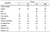
After 1 week of adjustment, the rats were randomly divided into two groups and fed either a normal diet (n=20) or high fat diet (n=20) for 2 weeks. After 2 weeks, they were divided into 4 groups of 10 rats each. The dietary treatment consisted of AIN-93-based diets with or without grape skin powder for 4 weeks. Vitamin and mineral mixtures for diets were purchased from Harlan Terklad Company (Madison, WI, USA). The experimental diets of four groups are described in Table 1.
The animals were given feed and tap water ad libitum during the experimental period. The food consumption and body weight were measured every other day and every week, respectively.

Sample preparation

At the end of the experimental period, the animals were fasted overnight (12 h) and then sacrificed. Blood was drawn from the abdominal aorta, and serum was obtained by sediment for 30 min, centrifugation (Hanil SUPRA 25k, Korea) at 3,000 rpm for 10 min. The liver was rapidly removed and rinsed with physiological saline. The mitochondria, microsomes, and cytosol were separated according to the method of Hogeboom (1955). All samples were stored at -70℃ until analysis.

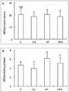
Measurement of thiobarbituric aicd--reactive substances (TBARS) concentrations in serum and liver

As a marker of lipid peroxidation, serum TBARS concentration was measured by the method of Yagi (1976) and the thiobarbituric acid reactive substances (TBARS) assay kit (Zeptometrix Co., NY, USA) was used.
Hepatic concentration of TBARS was measured by the method of Ohkawa et al. (1979). The n-butanol layer was taken for measurement at 532 nm with spectrophotometer. The concentrations of samples were compared with 1,1,3,3-tetraethoxypropane standards (Sigma Chemical Co., USA).

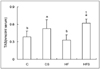
Measurement of serum total antioxidant capacity

The serum total antioxidant capacity was determined as described by Miller et al. (1993) with a slight modification. Total antioxidant status assay kit (ABTS Randox Lab., Crumlin, UK) was used. Data was expressed as nmol of inhibited 2,2'-azinobis-(3-ethyl-benzothiazoline-6-sulfonic acid) radical cation (ABTS·+)/ml of serum.

Determination of antioxidant enzyme activities

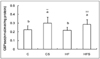
Catalase activity in hepatic mitochondrial fraction was measured using the method of Aebi (1974), by the rate of decomposition for H2O2 at 240 nm. Superoxide dismutase (SOD) activity was assayed in the hepatic cytosol fraction using the method of Marklund and Marklund (1974). The hepatic glutathione peroxidase (GSH-Px) activity was measured by the modified method of Paglia and Valentine (1967). The activity was expressed as nmol of reduced NADPH/min/mg of protein. Glutathione S-transferase (GST) activity was determined in the hepatic cytosol fraction, as developed by Habig et al. (1974). The hepatic xanthine oxidase (XO) activity was measured using the procedure developed by Stripe and della Corte (1969). The glucose-6-phosphatase (G6Pase) activity in the hepatic microsome fraction was determined using the method of Baginski et al. (1983) with a slight modification. Protein concentration was determined by the method of Lowry et al. (1951) with bovine serum albumin as the standard.

Measurement of total glutathione and GSH/GSSG

Total glutathione and oxidized glutathione (GSSG) contents were determined in liver according to the method of Anderson (1985) with a slight modification. Total glutathione content was measured using the solution (0.1 M potassium phosphate buffer with 1 mM EDTA (pH 7.0), 6 mM DTNB, 6 unit glutathione reductase and 1 mM NADPH) for 5 min at 25℃ based on an increase in the absorbance at 412 nm.
GSSG was assayed using 0.1 M potassium phosphate buffer (pH 7.0) containing 0.1 M EDTA, 1 mM NADPH and 20 unit glutathione reductase at 340 nm.
The amount of reduced glutathione (GSH) was calculated by subtraction of GSSG from total glutathione.

Statistical analysis

Results were presented as means ± standard deviations. For statistical analysis, a two-way analysis of variance (ANOVA) followed by Duncan's multiple-range test was first carried out to test for any difference between groups. If differences were established, the values were compared using Student's t-test.

Results

Total phenol, total antioxidant capacity and resveratrol contents of grape skin

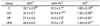
Total phenol, total antioxidant capacity and resveratrol contents of grape skin used in this experiment are presented in Table 2. Total phenol content of the grape skin was 42.9 mg/g. Total antioxidant status of the grape skin was 2.26 trolox equivalents. Resveratrol content of the grape skin was 46.5 µg/g.

Growth performance of rats

The final body weight of HF and HFS groups was higher compared to C and CS groups. During the experimental period, the food intake of C and CS groups was higher than that of HF and HFS groups. Food efficiency ratio of HF and HFS groups was higher than that of C and CS groups. However, there were no significant differences in body weight, food intake and food efficiency ratio between groups with and without grape skin (Table 3).

Thiobarbituric aicd-reactive substances (TBARS) concentrations in serum and liver

Although serum level of TBARS was not different among the experimental groups (Fig. 1A), the hepatic level of TBARS was increased in HF group as compared with C group. In the present study, dietary supplementation of grape skin decreased the hepatic concentration of TBARS (Fig. 1B).

Serum total antioxidant capacity and antioxidant enzyme and glucose-6-phosphatase activities

As shown in Fig. 2, serum total antioxidant status of HF group was not different from that of C group. But grape skin feeding increased the serum total antioxidant capacity significantly in C and HF groups.
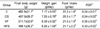
The hepatic enzyme activities of rats are presented in Tables 4 and 5. Catalase activity was decreased by high fat diet as compared with control diet. However, supplementation of grape skin increased catalase activity in both control and high fat-fed groups. While there is no difference in SOD activity between C and HF groups, dietary supplementation of grape skin increased the activity in C and HF groups. Although no significant differences were found in GST activity among the groups, GSH-Px activity of CS group was significantly higher than that of C group. Hepatic XO activity of HF group was increased compared with C group, but the activity was lower in HFS group than in HF group. The hepatic G6Pase activity did not change by high fat diet, but dietary supplementation of grape skin increased the activity in C and HF groups (Fig. 3).

Concentration of total glutathione and GSH/GSSG

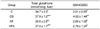
There was no significant difference in the total glutathione concentration between C and HF groups. However, grape skin feeding tended to increase the concentration of total glutathione, especially in C group. The ratio of GSH and GSSG was lower in HF groups than in C groups. The ratio of GSH to GSSG was increased in C groups by dietary supplementation of grape skin, but did not differ in HF groups (Table 6).

Discussion

Obese condition has a more elevated degree of oxidative stress than normal condition. Increased body fat stimulates excessive reactive oxygen species (ROS) production by NADPH oxidase activation (Furukawa et al., 2004). ROS, especially superoxide anion and hydrogen peroxide, can cause oxidative damage to lipids, proteins and DNA. These ROS are known to be participated in various chronic diseases including diabetes (Akhileshwar et al., 2007), cancer (Pelicano et al., 2004.), heart failure (Campolo et al., 2006) and hypertension (Vasdev & Gill, 2005).
Grape skin has been postulated to possess antioxidative activity in vitro and in vivo conditions (Falchi et al., 2006; Yassa et al., 2008; Young et al., 2000). In this study, we investigated the effect of antioxidant capacity by dietary grape skin of Campbell Early cultivated in Korea.
Many studies (Bray et al., 2004; Decorde et al., 2009; West & York, 1998) showed that high fat diet can lead to visceral obesity in rodent animal models. It is usually assumed that high-calorie and/or high-fat diets can lead to obesity (Caballero, 2007). In this study, dietary supplementation of high fat increased the final body weight compared with control diet.
Lipid peroxidation and antioxidant enzyme activities are important markers of oxidative stress. In the present study, the serum level of TBARS was not different among the experimental groups. However, the hepatic level of TBARS was increased in high fat group compared with control group. Dietary supplementation of grape skin decreased the hepatic concentration of TBARS in control and high fat groups.
Several studies have also shown elevated lipid peroxidation products in obesity (Amirkhizi et al., 2007; Beltowski et al., 2000; Feillet-Coudray et al., 2009; Olusi, 2002). High intake of dietary fat directly enhanced ROS overproduction which increased lipid peroxidation (Zhang et al., 2005). On the other hand, dietary supplementation of grape skin lowered the hepatic level of lipid peroxidation in this experiment. These results suggest that the polyphenol components such as resveratrol in grape skin might have antioxidant capacity. Previous studies have reported that resveratrol inhibited experimental lipid peroxidation in rats (Kasdallah-Grissa et al., 2006; Sener et al., 2007).
The removal of ROS is accomplished by enzymatic and non-enzymatic reactions in biological systems. In enzymatic reactions, SOD converts superoxide anions to hydrogen peroxide (H2O2), and H2O2 can be rapidly degraded by catalase and GSH-Px to H2O (Evans & Halliwell, 2001).
The present study revealed that grape skin might cause the induction of enzyme activities of catalase, SOD, G6Pase and GSH-Px in liver. In addition, we observed significant increase in serum total antioxidants capacity by feeding grape skin. A possible explanation of this effect is that grape skin contains high concentrations of anthocyanidins as well as various polyphenolic compounds (Makris et al., 2007; Negro et al., 2003). Bae (2008) reported that grape skin contains a high content of dietary fiber besides phenolic compounds as physiological active compounds. Therefore, dietary fiber may be used as a potential ingredient for physiological activities of grape skin. All of these active compounds have been shown to function as antioxidants by scavenging various free radicals.
Ramirez-Tortosa et al. (2001) showed that anthocyanin-rich extracts decreased vitamin E deficiency-induced hydroperoxides in liver and increased plasma antioxidant capacity. Also, Cui et al. (2002) suggested that dietary supplementation of grape polyphenols might prevent cardiac disease by inducing the antioxidant activity. The grape-derived polyphenols have been associated with a beneficial effect on the vascular system. Health benefits of grape polyphenols may enhance the redox-sensitive formation of vasoprotective factors and prevent oxidative stress (Madeira et al., 2009).
Reduced glutathione (GSH) has many different functions such as protection against ROS. During the reduction of hydrogen peroxide, GSH is converted into an oxidized form, glutathione disulfide (GSSG), and GSSG is reduced back to GSH by NADPH-dependant glutathione reductase (Fang et al., 2002; Sies, 1999).
In this study, high-fat diet might lead to depletion of liver content of reduced glutathione because the ratio of GSH and GSSG was lower in high fat groups than in control groups. It might be due to the increased generation of free radical species. Rocha et al. (2009) showed that hepatic GSH tended to be higher in high fat-fed rats than in standard diet-fed rats. But hepatic concentration of GSH was significantly increased in high fat diet-fed rats receiving 6 mg/l resveratrol in its drinking water compared with standard diet or high fat diet-fed rats without resveratrol. In the present study, grape skin feeding tended to increase the concentration of total glutathione, especially in control group. Hsu and Yen (2007) showed that GSH content was decreased and GSSG content increased in obese rats induced by a high fat diet. In this study, GSH/GSSG tended to be lower by a high fat diet, but not significantly. We also found that dietary supplementation of grape skin increased the GSH/GSSG ratio compared to control group without grape skin. GSH was the first line of antioxidant defense system by scavenging free radicals (Yassa et al., 2008). In conformity with the increase of ROS and lipid peroxides, the GSH level was decreased and the GSSG level was increased.
In conclusion, the present study demonstrated that grape skin has antioxidant effects by improving oxidative stress markers such as lipid peroxidation, catalase, SOD, GSH-Px activities. There results suggest that dietary supplementation of grape skin may alleviate the damage of oxidative stress induced by obesity through modulating antioxidant defense system and possibly reduce the risk of obesity-associated diseases.

Figures and Tables

Fig. 1
Effect of dietary supplementation of grape skin on serum (A) and hepatic (B) concentrations of TBARS in rats. Mean ± S.D. Values with the same superscript letter are not significantly different at P < 0.05. *Significant at P < 0.05 as compared with control group (C). ††Significant at P < 0.01 as compared with high fat group (HF).
nrp-3-279-g001
Fig. 2
Effect of dietary supplementation of grape skin on serum total antioxidant status in rats. Mean ± S.D. Values with the same superscript letter are not significantly different at P < 0.05. Significant at P < 0.05 as compared with high fat group (HF). Data were expressed as nmol of inhibited ABTS[2,2'-azinobis-(3-ethyl-benzothiazoline-6-sulfonic acid)] radical cation/ml of serum.
nrp-3-279-g002
Fig. 3
Effect of dietary supplementation of grape skin on hepatic activity of G6Pase in rats. Mean ± S.D. Values with the same superscript letter are not significantly different at P < 0.05. G6Pase : Glucose-6-phosphatase. **Significant at P < 0.01 as compared with control group (C). ††Significant at P < 0.01 as compared with high fat group (HF).
nrp-3-279-g003
Table 1
Composition of experimental diets (%)
nrp-3-279-i001

aMineral mixture according to AIN-93 (Teklad, USA)

bVitamin mixture according to AIN-93 (Teklad, USA)

Table 2
Total phenol, total antioxidant capacity and resveratrol content of grape skin
nrp-3-279-i002

1)TAS: Total Antioxidant Status

Table 3
Effect of dietary supplementation of grape skin on body weight gain, food intake and food efficiency ratio in rats
nrp-3-279-i003

Mean ± S.D.

Values with the same superscript letter within the column are not significantly different at P < 0.05.

1)FER: Food Efficiency Ratio

Table 4
Effect of dietary supplementation of grape skin on hepatic activities of catalase and SOD in rats (unit/min/mg protein)
nrp-3-279-i004

Mean ± S.D.

Values with the same superscript letter within the column are not significantly different at P < 0.05.

1)Superoxide dismutase

*Significant at P < 0.05 as compared with control group (C).

**Significant at P < 0.01 as compared with control group (C).

Significant at P < 0.05 as compared with high fat group (HF).

††Significant at P < 0.01 as compared with high fat group (HF).

Table 5
Effect of dietary supplementation of grape skin on hepatic activities of GST, GSH-Px and XO in rats (nmol/min/mg protein)
nrp-3-279-i005

Mean ± S.D.

Values with the same superscript letter within the column are not significantly different at P < 0.05.

1)Glutathione-S-transferase

2)Glutathione peroxidase

3)Xanthine oxidase

*Significant at P < 0.05 as compared with control group (C).

**Significant at P < 0.01 as compared with control group (C).

††Significant at P < 0.01 as compared with high fat group (HF).

Table 6
Effect of dietary supplementation of grape skin on hepatic levels of total glutathione and GSH/GSSG in rats
nrp-3-279-i006

Mean ± S.D.

Values with the same superscript letter within the column are not significantly different at P < 0.05.

*Significant at P < 0.05 as compared with control group (C).

**Significant at P < 0.01 as compared with control group (C).

Significant at P < 0.01 as compared with high fat group (HF).

Notes

This research was supported by the Yeungnam University research grants in 2007.

References

1. Acquaviva R, Russo A, Galvano F, Galvano G, Barcellona ML, Bolti GL, Vanella A. Cyanidin and cyanidin 3-O-β-D-g lucoside as DNA cleavage protectors and antioxidants. Cell Biol Toxicol. 2003. 19:243–252.
crossref
2. Aebi H. Bergmeyer HU, editor. Catalase. Methods of Enzymatic Analysis. 1974. 11. New York. USA: Academic Press;673–684.
crossref
3. Aggarwal BB, Bhardwaj A, Aggarwal RS, Seeram NP, Shishodia S, Takada Y. Role of resveratrol in prevention and therapy of cancer: Preclinical and clinical studies. Anticancer Res. 2004. 24:2783–2840.
4. Akhileshwar V, Patel SP, Katyare SS. Diabetic cardiomyopathy and reactive oxygen species related parameters in male and female rats: A comparative study. Indian J Clin Biochem. 2007. 22:84–90.
crossref
5. Amirkhizi F, Siassi F, Minaie S, Djalali M, Rahimi A, Chamari M. Is obesity associated with increased plasma lipid peroxidation and oxidative stress in women? ARYA Atherosclerosis Journal. 2007. 2:189–192.
6. Anderson ME. Determination of glutathione and glutathione disulfide in biological samples. Methods Enzymol. 1985. 113:548–555.
7. Bae HS. Effect of grape skin with resveratrol emplification on lipid metabolism and antioxidative system in rats fed high cholesterol diet. 2008. Gyeongbuk. Republic of Korea: Yeungnam University;MS thesis.
8. Baginski ES, Foa PP, Zak B. Bergmeyer HU, editor. Glucose-6-phosphatase. Methods of Enzymatic Analysis. 1983. 2. New York. USA: Academic Press;876–880.
crossref
9. Belleville J. The Franch paradox: Possible involvemenet of ethanol in the protective effect against cardiovascular disease. Nutrition. 2002. 18:173–177.
crossref
10. Beltowski J, Wojcicka G, Gorny D, Marciniak A. The effect of dietary-induced obesity on lipid peroxidation, antioxidant enzymes and total plasma antioxidant capacity. J Physiol Pharmacol. 2000. 51:883–896.
11. Bralley EE, Hargrove JL, Greenspan P, Hartle DK. Topical anti-inflammatory activities of Vitis rotundifolia(Muscadine grape) extracts in the tetradecanoylphorbol acetate model of ear inflammation. J Med Food. 2007. 10:636–642.
crossref
12. Bray GA, Paeratakul S, Popkin BM. Dietary fat and obetisy: a review of animal, clinical and epidemiological studies. Physiology & Behavior. 2004. 83:549–555.
crossref
13. Caballero B. The global epidemic of obesity: An overview. Epidemiol Rev. 2007. 29:1–5.
crossref
14. Campolo J, Maria RD, Caruso R, Accinni R, Turazza F, Parolini M, Roubina E, Chiara BD, Cighetti G, Frigerio M, Vitali E, Parodi O. Blood glutathone as independent marker of lipid peroxidation in heart failure. Int J Cardiol. 2006. 117:45–50.
crossref
15. Cho YJ, Kim JE, Chun HS, Kim CT, Kim SS, Kim CJ. Contents of resveratrol in different parts of grapes. Korean Journal of Food Science and Technoloy. 2003. 35:306–308.
16. Cui J, Juhasz B, Tosaki A, Maulik N, Das DK. Cardioprotection with grape. J Cardiovasc Pharmacol. 2002. 40:762–769.
17. Decorde K, Teissedre PL, Sutra T, Ventura E, Cristol JP, Rouanet JM. Chardonnay grape seed procyanidin extract supplementation prevents high-fat diet-induced obesity in hamsters by improving adipokine imbalance and oxidative stress markers. Mol Nutr Food Res. 2009. 53:659–666.
crossref
18. Delmas D, Jannin B, Latruffe N. Resveratrol: Preventing properties against vascular alterations and ageing. Mol Nutr Food Res. 2005. 49:377–395.
crossref
19. Evans P, Halliwell B. Micronutrients: Oxidabt/antioxidant status. Br J Nutr. 2001. 85:S67–S74.
20. Falchi M, Bertelli A, Scalzo R, Morassut M, Morelli R, Das S, Cui J, Das DK. Comparison of cardioprotective abilities between the flesh and skin of grapes. J Agric Food Chem. 2006. 54:6613–6622.
crossref
21. Fang YZ, Yang S, Wu G. Free radicals, antioxidants and nutrition. Nutrition. 2002. 18:872–879.
crossref
22. STAT database. FAO. 2005. Access on 7/20/2007. www.Fao.org.
23. Feillet-Coudray C, Sutra T, Fouret G, Ramos J, Wrutniak-Cabello C, Cabello G, Cristol JP, Coudray C. Oxidative stress in rats fed a high-fat high-sucrose diet and preventive effect of polyphenols: Involvement of mitochondrial and NAD(P)H oxidase systems. Free Radic Biol Med. 2009. 46:624–632.
crossref
24. Frederiksen H, Mortensen A, Schroder M, Frandsen H, Bysted A, Kunthsen P, Rasmussen SE. Effects of red grape skin and seed extract supplementation on atherosclerosis in watanabe heritable hyperlipidemic rabbits. Mol Nutr Food Res. 2007. 51:564–571.
crossref
25. Fukao H, Ijiri Y, Miura M, Hashimoto M, Yamashita T, Fukunaga C, Oiwa K, Kawai Y, Suwa M, Yamamoto J. Effect of trans-resveratrol on the thrombogenicity and atherogenicity in apolipoprotein E-deficient and low-density lipoprotein receptor-deficient mice. Blood Coagul Fibrinolysis. 2004. 15:441–446.
crossref
26. Furukawa S, Fujita T, Shimabukuro M, Lwaki M, Yamada Y, Nakajima Y, Nakayama O, Makishima M, Matsuda M, Shimomura I. Increased oxidative stress in obesity and its impact on metabolic syndrome. J Clin Invest. 2004. 114:1752–1761.
crossref
27. Habig WH, Pabst MJ, Jakoby WB. Glutathione-S-transferase. The first enzymatic step in mercapturic acid formation. J Biol Chem. 1974. 249(22):7130–7139.
28. Hogeboom GH. Fractionation of cell components of animal tissues. Meth Enzymol. 1955. 1:16–19.
29. Hsu CL, Yen GC. Effect of gallic acid on high fat diet-induced dyslipidaemia, hepatosteatosis and oxidative stress in rats. Br J Nutr. 2007. 98:727–735.
crossref
30. Kasdallah-Grissa A, Mornagui B, Aouani E, Hammami M, Gharbi N, Kamoun A, El-Fazaa S. Protective effect of resveratrol on ethanol-induced lipid peroxidation in rats. Alcohol Alcohol. 2006. 41:236–239.
crossref
31. Lowry OH, Rosebrough NJ, Farr AL, Randall RJ. Protein measurement with the Folin phenol reagent. J Biol Chem. 1951. 193:265–275.
crossref
32. Madeira SV, Auger C, Anselm E, Chataigneau M, Chataigneau T, Moura RS, Schini-Kerth VB. eNOS activation induced by a polyphenol-rich grape skin extract in porcine coronary arteries. J Vasc Res. 2009. 46:406–416.
crossref
33. Makris DP, Boskou G, Andrikopoulos NK. Polyphenolic content and in vitro antioxidant characteristics of wine industry and other agri-food solid waste extracts. J Food Compost Anal. 2007. 20:125–132.
crossref
34. Marklund S, Marklund G. Involvement of the superoxide anion radical in the autoxidation of pyrogallol and a convenient assay for superoxide dismutase. Eur J Biochem. 1974. 47:469–474.
crossref
35. Miller NJ, Rice0Ebans C, Davies MJ, Gopinathan V, Milner A. A novel method for measuring antioxidant capacity and its application to monitoring the antioxidant status in premature neonates. Clin Sci. 1993. 84:407–412.
crossref
36. Miura D, Miura Y, Yagasaki K. Hypolipidemic action of dietary resveratrol, a phytoalexin in grapes and red wine, in hepatoma-bearing rats. Life Sci. 2003. 73:1393–1400.
crossref
37. Negro C, Tommasi L, Miceli A. Phenolic compounds and antioxidant activity from red grape marc extracts. Bioresour Technol. 2003. 87:41–44.
crossref
38. Oben JE, Enyegue DM, Fomekong GI, Soukontoua YB, Agbor GA. The effect of Cissus quadrangularis (GQR-300) and a Cissus formulation (CORE) on obesity and obesity-induced oxidative stress. Lipids Health Dis. 2007. 6:4–12.
39. Ohkawa H, Ohishi N, Yagi K. Assay for lipid peroxides in animal tissues by thiobarbituric acid reaction. Anal Biochem. 1979. 95:351–358.
crossref
40. Olusi SO. Obesity is an independent risk factor for plasma lipid peroxidation and depletion of erythrocyte cytoprotectic enzymes in humans. Int J Obes. 2002. 26:1159–1164.
crossref
41. Paglia DE, Valentine WN. Studies on the quantitative and qualitative characterization of erythrocyte glutathione peroxidase. J Lab Clin Med. 1967. 70:158–169.
42. Pelicano H, Carney D, Huang P. ROS stress in cancer cells and therapeutic implications. Drug Resist Updat. 2004. 7:97–110.
crossref
43. Ramirez-Tortosa C, Andersen OM, Gardner PT, Morrice PC, Wood SG, Duthie SJ, Collins AR, Duthie GG. Anthocyanin-rich extract decreases indices of lipid peroxidation and DNA damage in vitamin E-depleted rats. Free Radic Biol Med. 2001. 31:1033–1037.
crossref
44. Re R, Pellegrini N, Proteggente A, Pannala A, Yang M, Rice-Evans C. Antioxidant activity applying an improved ABTS radical cation decolorization assay. Free Radic Biol Med. 1999. 26:1231–1237.
crossref
45. Rocha KK, Souza GA, Ebaid GX, Seiva FRF, Cataneo AC, Novelli ELB. Resveratrol toxicity: Effects on risk factors for atherosclerosis and hepatic oxidatice stress in standard an high-fat diets. Food Chem Toxicol. 2009. 47:1362–1367.
crossref
46. Scarlatti F, Sala G, Somenzi G, Signorelli P, Sacchi N, Ghidoni R. Resveratrol induces growth inhibition and apoptosis in metastatic breast cancer cells via de novo ceramide signaling. FASEB J. 2003. 17:2339–2341.
crossref
47. Schneider Y, Vincent F, Duranton B, Badolo L, Gosse F, Bergmann C, Seiler N, Raul F. Anti-proliferative effect of resveratrol, a natural component of grapes and wine, on human colonic cancer cells. Cancer Lett. 2000. 158:85–91.
crossref
48. Sener G, Topaloglu N, Sehirli O, Ercan F, Gedik N. Resveratrol alleviates bleomycin-induced lung injury in rats. Pulm Pharmacol Ther. 2007. 20:642–649.
crossref
49. Sies H. Glutathione and its role in cellular functions. Free Radic Biol Med. 1999. 27:916–921.
crossref
50. Singleton VL, Joseph A, Possi JR. Colorimetry of total phenolics with phosphomolybdic-phosphotungstic acid reagents. Am J Enol Vitic. 1965. 16:144–158.
51. Strip F, della Corte E. The regulation of rat liver xanthine oxidase; Conversion in vitro of the enzyme activity from dehydrogenase (Type D) to oxidase (Type O). J Biol Chem. 1969. 244:3855–3863.
52. Su H, Hung L, Chen J. Resveratrol, a red wine antioxidant, possesses an insulin-like effect in streptozotocin-induced diabetic rats. Am J Physiol Endocrinol Metab. 2006. 290:E1339–E1346.
crossref
53. Tsuda T, Horio F, Uchida K, Aoki H, Osawa T. Dietary cyanidin 3-O-β-D-glucoside-rich purple corn color prevents obesity and ameliorates hyperglycemia in mice. J Nutr. 2003. 133:2125–2130.
crossref
54. Vasdev S, Gill V. Antioxidants in the treatment of hypertension. Int J Angiol. 2005. 14:60–73.
crossref
55. West DB, York B. Dietary fat, genetic predisposition, and obesity: lessons from animal models. Am J Clin Nutr. 1998. 67:505S–512S.
crossref
56. Yagi K. A simple fluorometric assay for lipoperoxide in blood plasma. Biochem Med. 1976. 15:212–216.
crossref
57. Yassa N, Razavi BH, Hadjiakhoondi A. Free radical scavenging and lipid peroxidation activity of the shahani black grape. Pak J Biol Sci. 2008. 11:2513–2516.
crossref
58. Young JF, Dragsted LO, Daneshvar B, Lauridsen ST, Hansen M, Sandstrom B. The effect of grape skin extract on oxidative status. Br J Nutr. 2000. 84:505–513.
crossref
59. Yunoki K, Sasaki G, Tokuji Y, Kinoshita M, Naito A, Aida K, Ohnishi M. Effect of dietary wine pomace eztract and oleanolic acid on plasma lipids in rats fed high-fat diet and its DNA microarray analysis. J Agric Food Chem. 2008. 56:12052–12058.
crossref
60. Zhang X, Dong F, Ren J, Driscoll MJ, Culver B. High dietary fat induces NADPH oxidase-associated oxidative stress and inflammation in rat cerebral cortex. Exp Neurol. 2005. 191:318–325.
crossref
61. Zhang Y, Liu Y, Wang T, Li B, Li H, Wang Z, Yang B. Resveratrol, a natural ingredient of grape skin: Antiarrhythmic efficacy and ionic mechanisms. Biochem Biophys Res Commun. 2006. 340:1192–1199.
crossref
TOOLS
Similar articles