

 PDF
PDF ePub
ePub Citation
 Print
Print


Introduction
Adequate amounts of calcium are essential throughout the life cycle to promote bone and overall health, and to help reduce the risk of osteoporosis (Nieves, 2005). Osteoporosis is considered a major public health threat for an estimated 44 million U.S. women and men 50 y old, causing an annual health care cost of $17 billion in 2001 (McCarron & Heaney, 2004). Unfortunately, children, adolescents, especially girls, do not consume the recommended 2-3 servings of milk and milk products each day (Fiorito et al., 2006). As a result of declining milk consumption in recent years, 70% of girls aged 6-11 y does not meet current calcium recommendations (Fiorito et al., 2006).
Osteoporosis and osteoporotic fractures have substantial clinical and health effects. An estimated 75 million persons in Europe, Japan, and the United States are affected by osteoporosis (Report of a WHO Scientific Group, 2003). Osteoporosis is characterized by low bone density and microarchitectural deterioration of bone tissue, and there is increasing interest in environmental factors that may influence bone density. Nutrition clearly plays a role; nutrient inadequacies, eg, calcium and vitamin D, have received the most emphasis (Gourlay & Brown, 2004; Heaney et al., 2002; Ilich & Kerstetter, 2000). Dairy consumption has been postulated to reduce the risk of osteoporosis and metabolic disturbances. Several observational studies have found associations between calcium intake or intake of dairy products and bone mineral density. The recommended calcium intake changes with age (Institute of Medicine, 1997). One of the highest daily intakes is required after age 50. Important dietary sources of calcium are dairy products (milk, yogurt, and cheese), dark green vegetables; canned fish with bones (but not fish fillets); nuts; and more recently, fortified foods (including juices, waffles, cereals, crackers, and snack foods). A large proportion of the worlds' population consumes low-calcium diets. Women in developed nations, such as the United States and northern Europe, where the incidence of osteoporosis is high, generally have inadequate calcium status.
Of the nutrients, calcium has been the most extensively studied to determine its role in osteoporosis. Approximately, 99% of the body's calcium is stored in the bone as hydroxyapatite, which provides structural integrity for the skeleton (Broadus, 1993). Vitamin D may play a role in the development of osteoporosis due to its effect on calcium metabolism. Plasma calcium homeostasis is maintained by vitamin D in conjunction with PTH. Low blood calcium level activates PTH, which in turn stimulates the production of vitamin D in the kidney. In response, vitamin D enhances the absorption of calcium, decreases the excretion of calcium and stimulates calcium resorption from bone (Linder, 1991).
Human growth hormone (hGH) has been shown in some studies to have anabolic effects on bone but taurine effects on bone are unknown. The major hormones that regulate tissue growth and metabolism all have a major influence on skeletal growth and remodeling, including the growth hormone-insulin-like growth factor (GH-IGH) system. The GH-IGH system determines body size and regulates the distribution of body fat, lean body mass, and bone modeling and remodeling after epiphyseal closures (Sjogren et al., 1999). GH can stimulate IGF production not only in the liver but also in other target organs, including bone. The GH-IGF system stimulates both resorption and formation (Yakar et al., 2002).
Taurine could act either directly or indirectly by enhancing growth factor production (Boujendar et al., 2002). However, no significant association between taurine intake and spine or femur bone mineral density was observed. An association between taurine intake and bone turnover markers was also not found. The present study is unique in that it investigated the relations of taurine and bone. Influence of taurine on early postmenopausal bone loss is not well understood. Dietary supplement with excess taurine could yield increased sulfuric acid production in the body. Under those circumstances, the skeleton may be called on to act as a buffer to neutralize acid generated from high-sulfuric diets.
On the other hand, interest has been expressed in the relation of skeletal maintenance to age-related decreases in IGF-I concentrations (Jensen et al., 2002). Recombinant IGF-I increases bone formation activity in postmenopausal women (Ebeling et al., 1993; Ghiron et al., 1995; Grinspoon et al., 1995), suggesting that increasing IGF-I concentrations may help restore bone mass. IGF-I production is markedly affected by nutrient intake (Jensen et al., 2002). The paper recently demonstrated that taurine supplementation of the maternal diet restored normal IGF-II expression in islet cells of fetuses of low protein-fed rats (Boujendar et al., 2002). Our understanding of how taurine affects calcium homeostasis and skeletal metabolism is limited.
Low calcium diets can produce rachitic lesions in young growing animals, but in adults the condition becomes osteoporosis. It has been reported that calcium deficient, ovariectomized rats develop bone changes (especially decrease in bone density) similar to estrogen deficient osteoporotic women (Donahue et al., 1988).
Therefore, we examined in ovariectomized rats whether the preventive effect of taurine on ovarian hormone deficiency-induced bone loss. Taurine supplementation has been shown to have an effect on femur bone mineral content in ovariectomized rats in our research (Choi & DiMarco, 2009). It therefore seemed desirable to find out whether the beneficial effect of taurine on OVX rats fed calcium deficient diet could also be reproduced.
Materials and Methods
Animals and diet
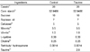
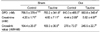
Forty female Sprague-Dawley rats (body weight 200 ± 5 g, 9 weeks old) were randomly divided into two groups. One group was ovariectomized (OVX) and the other group received sham operation (SHAM), and then each rat group was further divided into control diet and taurine supplemented (2.0%) diet group for 6 weeks. All rats were fed on experimental diet and deionized water ad libitum for 6 weeks. The rats were weighed weekly. The dietary supply of vitamins, minerals (except Ca : 50% of recommended level ), and protein was in accordance with the recommended dietary allowances for rats from the American Institute of Nutrition (AIN-93G; Reeves et al., 1993) and shown in Table 1.
Bone and bone markers determination
Bone mineral density (BMD) and bone mineral content (BMC) were measured using PIXImus (GE Lunar Co, Wisconsin, USA) in spine and femur on 6 weeks after feeding. Bone formation was measured by serum osteocalcin and alkaline phosphatase (ALP) concentrations. Bone resorption was measured by deoxypyridinoline (DPD) crosslinks immunoassay and corrected for creatinine. Serum osteocalcin, growth hormone, insulin-like growth factor-1 (IGF-1), parathyroid hormone (PTH) and calcitonin were analyzed using radioimmunoassay kits.
Statistical analysis
The statistical significance of differences among the groups was evaluated by two-way ANOVA, using a computer software package (version 9.13, SAS Institute Inc, Cary, NC). Individual comparisons were made by Duncan's multiple range test using the ANOVA. A P value <0.05 was considered statistically significant. Data are expressed as means ± SD.
Results
Weight gain and FER
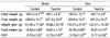
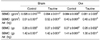
As expected, weight gain for the OVX rats was significantly greater than that of SHAM rats (p<0.05). The results of this study indicate that body weight gain was higher in OVX groups than in SHAM groups regardless of diets. And the weight gain for the OVX rats were not lower than that for rats fed 100% calcium level as recommended (Table 2). Mean food intake was 14.52 ± 3.85 g/day and 13.81 ± 1.73 g/day for the control and taurine groups within SHAM rats, respectively. No difference was found in food intake between control diet and taurine diet (Table 2).
Urine Ca, P, deoxypyridinoline, creatinine and crosslink value
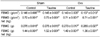
Urinary calcium excretion between SHAM and OVX groups was not significantly different (Table 5). Crosslink values were 164.4 ± 26.8 and 169.3 ± 36.9 (nM/mM) for the control and taurine groups within SHAM group, respectively. Crosslink value was increased in ovariectomized groups. Although urinary crosslink value was generally higher in the control group than in the taurine group (Table 6), the difference was not statistically significant.
Spine and Femur BMD, BMC, BMD per weight and BMC per weight
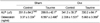
The bone mineral density and bone mineral content data are shown in Table 7 and Table 8. The spine BMC and BMD were significantly less in rats fed 50% of recommended calcium diet than in SHAM and OVX rats fed 100% of recommended calcium diet (Choi & DiMarco, 2009). In the control diet, spine BMD was significantly lower in OVX rats than in SHAM rats (0.095 ± 0.01 and 0.084 ± 0.08 g/cm2, respectively p<0.05), and in the taurine group, spine BMD was significantly lower in OVX rats than in SHAM rats. They were not significantly different from each other within SHAM and OVX groups by taurine supplementation (Table 7).
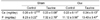
Mean femur BMD and BMC in the taurine diet group did not differ significantly from that in the control diet group in OVX rats fed 50% of recommended Ca diet. Also femur BMD/weight and BMC/weight of taurine group was not significantly higher than that of control group in OVX rats. (Table 8).
Discussion
Osteoporosis is a metabolic bone disease characterized by a defect in bone remodeling and the loss of normally mineralized bone. Maximum skeletal mass is achieved in young adults at 18-25 y of age. After age 40, the slow phase of bone loss begins in both sexes and continues at a rate of 0.5-1%/y until late in life. In women after menopause, there is an additional rate of bone loss of 2-3%/y because of the decreasing estrogen concentrations associated with aging. Osteoporosis is responsible for 1.2 million fractures in the United States annually. The most common fractures are of the vertebrae, distal radius, and hip (Riggs & Melton, 1986). Bone mineral density (BMD) and bone metabolism are affected by genetic, endocrine, mechanical, and nutritional factors, with extensive interactions between the different factors. Calcium has been reported as the most important nutrient associated with peak bone mass (Recker et al., 1992) and may be the only one for which there is epidemiological evidence of a relation to fracture rate. Low calcium intake is particularly common in many countries.
There is a general consensus that dietary calcium moderately reduces the rate of cortical bone loss in late menopause (Reid et al., 1995; Soroko et al., 1994). A positive association between bone density and BMI or body weight has been well documented in many large epidemiologic studies (Ravn et al., 1999). Bone-protective effects of BMI may increase weight bearing (Slemenda, 1995), increase aromatization of androgen to estrogen in adipose tissue (Frumar et al., 1980), lower concentrations of sex hormone-binding globulin (van Hemert et al., 1989), or directly increase bone formation induced by high circulating concentrations of insulin (Reid et al., 1995) and other hormones secreted by the cells of the pancreatic islets (Reid, 2002).
In recent years, a growing body of observational and clinical studies supports a role for dietary calcium and dairy foods in controlling body weight and excess adiposity. The investigation has shown an inverse relation between calcium intake and body weight in older women (Zemel et al., 2000).
Davies et al. (2000) and Heaney et al. (2002) reported a negative association between calcium intake and body weight, body fat, and weight gain in young, middle-aged, and older women and calculated that a 300 mg increase in daily calcium intake was associated with a 3 kg difference in body weight.
As expected, weight gain for the OVX rats was significantly greater than those for SHAM rats (p<0.05). The results of this study indicate that body weight gain was higher in OVX groups than in SHAM groups regardless of diets. And the weight gain for the OVX rats were not lower than that for rats fed 100% calcium level as recommended. In recent years, several clinical and epidemiologic studies have reported a consistent inverse association between calcium intake and body weight (Lin et al., 2000; Loos et al., 2004). A possible physiologic mechanism explaining this relation was recently proposed by Zemel et al. (2000), who showed that an increase in intracellular calcium concentrations in human adipocytes after stimulation with parathyroid hormone (PTH) and 1,25-dihydroxyvitamin D [1,25(OH)2D] is able to switch lipid metabolism from lipolysis to lipogenesis, which results in an increase in triglycerol storage. Consistently, 1,25(OH)2D and PTH have been found to be positively associated with body mass index (BMI) (Bell et al., 1985). Because serum PTH and 1,25(OH)2D concentrations are regulated by calcium intake, this metabolic pathway would be responsible for the higher risk of overweight and obesity in subjects with a low calcium intake and for the weight loss after increases in dietary calcium intake.
Bone development requires sufficient amounts of many nutrients, but calcium has received the greatest attention because of its great mass in the skeleton. Low calcium diet, decreased calcium absorption, and increased loss are probably some of the most important mechanisms involved in bone loss in aging humans. When OVX rats were fed low calcium diet, the decrease in calcium absorption due to OVX became significant (Kalu, 1991) and bone loss was enhanced (Nordin et al., 1979). Low calcium diet and OVX have a great potential for weakening the bone quality of cortical bone and trabecular bone, respectively, and have an additive effect when combined.
This experiment was originally designed to test the ability of a taurine supplement to minimize bone loss during postmenopausal model with low calcium intake. Sulfur is predominantly responsible for determining the net endogenous acid production from protein because it is the acid precursor that is oxidized to sulfuric acid (Frassetto et al., 1998). It would therefore make sense that a dietary supplement with excess sulfur-containing amino acids (taurine) could yield increased sulfuric acid production in the body. However, these data suggest that bone mineral density per weight was increased, without changes in bone resorption and bone formation, in the taurine supplemented group, and therefore have the potential to increase bone mineral density if either the study is extended or more taurine is supplemented. The beneficial effect of taurine on ovariectomized rats fed calcium deficient diet was not reproduced. Because in this study, the control and taurine groups consumed identical diets as in the previous study (besides the calcium content), and we do not know whether intestinal calcium absorption will be increased by more taurine supplement. These results indicate that no significant differences in spine and femur BMD were found due to 2% taurine diet in OVX rats fed calcium deficient diet for 6 weeks. No positive effects of taurine on bone mineral density were found in the present study. Our failure to see an association between taurine supplementation and bone mineral density may have been related to the fact that we used calcium deficient diet and the same amount of taurine. Further investigations of the relation between taurine and calcium intake level for bone mineral density are warranted.
The results of this study indicate that body weight gain and food intake were higher in OVX groups than in SHAM groups regardless of diets. There were no differences in weight gain between the control and taurine groups in both OVX and SHAM groups. Within the OVX groups, serum calcium concentration was lower in rats fed taurine than in rats fed the control diet. Serum ALP, osteocalcin, urinary calcium, and phosphate were not different in each group. Within the OVX group, there were no differences in spine BMD and BMC and femur BMD and BMC. The beneficial effect of taurine on ovariectomized rats fed calcium deficient diet was not reproduced. So the beneficial effect of taurine on bone seems different by calcium intake levels.

 XML Download