

 PDF
PDF ePub
ePub Citation
 Print
Print


Introduction
DHEA and DHEA-sulfate are the precursors of androgens and estrogens produced from adrenal glands (Roberge et al., 2007). Their levels start to decrease after middle age and animal and human studies have reported that age-associated chronic diseases involving dysregulation of metabolism may be due to the decline in the levels of these compounds (Labrie, 2007). Although there have been in vivo studies about the protective effects of DHEA against cancer, obesity, diabetes, postmenopausal osteoporosis, sexual dysfunction and cardiovascular disease as modulators of adrenocortical steroid synthesis (Allolio et al., 2007; Kim and Choi, 2005), its mechanism of action needs to be studied more in detail before safely used as supplements in humans for different diseases.
Reactive oxygen species are continuously formed as by-products of aerobic metabolism and from reactions of drugs and environmental toxins such as carcinogens (Cerutti, 1985). These play important roles in tumor progression by damaging DNA, proteins and unsaturated lipids (Champe & Harvey, 1987). The cells have protective mechanisms that can minimize the toxic effects from these compounds. These mechanisms include antioxidant chemicals (e.g. vitamin E), enzymes that catalyze antioxidant reactions using NADPH as a source of reducing electrons and a liver microsomal cytochrome P-450 monooxygenase system, which also utilizes NADPH to convert steroids and drugs to soluble forms (Wu et al., 1989).
We have previously shown that DHEA decreases glutathione S-transferase placental from (GST-p) foci in the liver and lipid peroxdation using a hepatocellular carcinoma model in rats (Kim & Choi, 2005). Here, we aimed to determine the mechanism of protective effects of DHEA supplement during chemical hepatocellular carcinogenesis in vivo in comparsion to those with vitamin E supplement, an effective intracellular reducing agent.
Materials and Methods
Animals
Six-week-old male Sprague Dawley rats were purchased from Seould National University and housed at the animal care facility at Seoul National University (Seoul, Korea). All rats were kept under standard temperature, humidity, and timed lighting conditions and were provided with rat chow and water ad libitum. Animals were induced with hepatocellular carcinoma by i.p. injection of diethylnitrosamine (200 mg/kg body weight) followed by administration of diets containing 0.01% of 2-acetylaminofluorene for 6 weeks. Partial hepatectomy surgeries were performed 1 week after the start of 2-acetylaminofluorene diet to effectively induce progression of tumor as previously published (Ito et al., 1988).
Diets and materials
Rat chow was purchased from Purina (purified rodent diet 5053, St. Louis, Missouri) and was supplemented with 1.5% vitamin E (Sigma T-3376, DL-α tocopheryl acetate, St. Louis, MO) and 0.5% DHEA (Sigma D-400, DHEA 3-acetate, St. Louis, MO) as described previously (Kim & Choi, 2005).
Biochemical assays
Frech or frozen livers were weighed and five volumes of ice-cold homogenization buffer [154 mM KCl, 50 mM Tris-HCl, 1mM EDTA, pH 7.5] were added. The tissue was homogenized and was fractionated by spinning at 1,000 g for 13 min at 4℃. The middle layer was centrifugated at 10,000 g for 13 min at 4℃. Then the supernatant was centrifugated at 100,000 g for 65 min at 4℃ to obtain cytosolic and microsomal fractions as described in detail previously (Kim & Choi, 2005).
Malate dehydrogenase activities
Malate dehydrogenase activities were measured in the cytosolic fraction using modified methods of Ochoa (1969). Briefly, 500 µl of triethanolamine buffer [0.4 M, pH 7.4], 50µl of L-malate [30 mM], 100 µl of MnCl2, 4H2O [0.12 M] and 200µl of NADP [3.4 mM] were mixed with 1,400 µl of cytosolic fraction using a vortex. Absorption at 270 nm during 1 min at 26℃ was measured every 6 seconds. One unit is increased in absoption by 0.01 during 1 min.
Glucose 6-phophate dehydrogenase activities
Glucose 6-phophate dehydrogenase activities were measured in the cytosolic fraction using modified methods of Lohr and Waller (1974). Briefly, 2.4 ml of triethanolamine buffer [50 mM, pH 7.5] and 0.5 ml of cytosolic fraction (~0.8 mg/ml protein) were mixed using a vortex. Then, 50 µl of NADP solution was added and was kept at 25℃ for 5 minutes. Next, 50 µl of glucose 6-phosphate was added and increases in absorption was immediately measured at 340 nm for every 2 minutes up to 6 minutes. Specific activity was calculated as µmole of substrate converted per minute per mg protein.
Se-glutathione peroxidase
Cytosolic Se-glutathione peroxidase activites were measured using a modified method of Tappel (1978) as previously described.
Microsomal cytochrome P-450
Hepatic microsomal fraciton was used to measure cytochrome P-450 content (Omura & Sato, 1964). Fresh microsomal fraction was diluted with phosphate buffer (pH 7.4) to conatin ~ 1 mg/ml protein and sodium dithionate was added and was saturated with CO gas for 1 minute. Then, absorptions of reduced carbon monoxide at 450 and 490 nm were determined spectrophotometrically. Molar extinction coefficient used was 91/mM/cm.
Results
DHEA treament increased cytosolic malate dehydrogenase activities
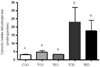
We previously showed (Kim & Choi, 2005) that DHEA treatment decreased NADPH-utilizing antioxidant enzymes in this model so we first determined if activities of enzymes that regulate NADPH synthesis were altered. Interestingly, DHEA treatment significantly increased malate dehydrogenase activities by almost 5 fold vs. carcinogen-treated controls (Fig. 1). This effect was not seen in vitamin E-treated animals. Malate dehydrogenase activities were not different between non-carcninogen treated and carcinogen-treatd groups (Fig. 1, COO vs. TOO). Based on these results, we focused our study on determining the activities of another NADPH producing enzyme, glucose 6-phosphate dehydrogenase. a and b; one-way ANOVA, p<0.05
Activities of cytosolic glucose 6-phosphate dehydrogenase had a tendency to decrease with DHEA treatment
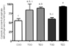
Although DHEA treatment increased malate dehydrogenase activities, it had a strong tendency to decrease glucose 6-phosphate dehydrogenase activities (Fig. 2). Glucose 6-phosphate dehydrogenase activity was increased by almost 1.5 fold with the carcinogen treatment and DHEA supplement decreased it to the level of those of non-carcinogen treated group (COO vs. TOD). However, in the combination group (TED), the enzyme activity was not suppressed as seen in DHEA-only treated animals. a, b, c; one-way ANOVA, p<0.05.
DHEA treatment decreased hepatic cytosolic Se-glutathione peroxidase activities.
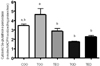
Activities of cytosolic Se-glutathione peroxidase activities tended to increase with carcinogen treatment (COO vs. TOO). The enzyme activities were inhibited almost 50-60% with DHEA treamtment comapred to controls (COO and TOO vs. TOD). Vitamin E treatment also decreased the activities significantly and a synergistic inhibitory effects were seen in combination treatment group (TOO vs. TED).
Hepatic microsomal cytochrome P-450 content was not altered with DHEA treatment.
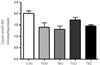
To examine the mechanism of action of DHEA on hepatic microsomal cytochrome P-450 mono-oxygenase system, cytochrome P-450 content was measured. DHEA treatment did not affect cytochrome P-450 content (Fig. 4). We also measured microsomal NADPH cytochrome P-450 reductase activites but they were also not affected with DHEA treatment (data not shown).
Discussion
Here we studied the mechanism of action of DHEA during hepatocellular carcinogenesis in vivo. We found that DHEA significantly increased activities of malate dehydrogenase, an enzyme important for supplying biochemical reductant NADPH in the cytosol. On the other hand, glucose 6-phosphate dehydrogenase activites were reduced by ~25% with DHEA treatment possibly due to the high level of NADPH/NADP+ ratio in the cytosol, provided by increased malate dehydrogenase activity. Reports showed that DHEA treatment decreased mRNA levels of hepatic malate dehydrogenase in broiler chickens (Zhao et al., 2007) and increased the activity in non-insulin dependent diabetic rats (Ladriere et al., 1997). However, mRNA levels do not always correlate with levels of enzyme activity and the dose, duration and the disease model used in these studies were different from the current study. Marrero, et al. showed that DHEA treatment in female mice increased hepatic malate dehydrogenase activites by 2-3 fold (Marrero et al., 1990) but did not alter glucose 6-phosphate dehydrogenase activity, similar to the data from this study. Further studies using different organ systems in various disases models are needed to clarify effect of DHEA on these NADP-dependent enzymes.
Under most metabolic conditions, the ratio of NADPH/NADP+ is high enough to inhibit glucose 6-phosphate dehydrogenase activity. However, with increased demand for NADPH in the cells (e.g. toxic and oxidative stress), the ratio of NADPH/NADP+ decreases and flux through the hexose monophosphate pathway is enhanced by increasing the activity of glucose 6-phosphate dehydrogenase (Champe & Harvey, 1987). Glucose 6-phosphate dehydrogenase is the key enzyme regulating the first and irreversible step of the pentose phsophate pathway. This pathway can then provide ribose-5-phosphates, a precursor for DNA synthesis in cell proliferation. During promotional phase of carcinogenesis, the demand for ribose-5-phosphate for de novo synthesis of DNA may be increased and it is thus particularly important that DHEA treatment suppressed this rate limiting enzyme of the pentose phosphate pathway, glucose 6-phosphate dehydrogenase. This may be one of the protective mechanisms of DHEA during carcinogenesis as reported previously (Kim & Choi, 2005). Although malate dehydrogenase activites increased with DHEA treamtment in this study, it has been reported that its activity is ~15% of that of glucose 6-phosphate dehydrogenase in rat adrenal cortex (Frederiks et al., 1990). If this is the case in rat liver, the decrease in glucose 6-phosphate dehydrogenase would surpass the relative change in the malate dehydrogenase activity in the inhibition of pentose phosphate pathway.
Increases in malate dehydrogenase activities by DHEA treatment may have also provided NADPH to reduce glutathione efficiently. This antioxidant effects of DHEA was seen in decreased Se-dependent glutathione peroxidase activities (Fig. 3), confirming less oxidative damage with DHEA treatment as previously reported (Kim & Choi, 2005). Reduced glutathione (a tripeptide-thiol) can detoxify hydrogen peroxide in most cells in a NADPH-dependent manner (Frederiks et al., 1990) catalyzed by glutathione reductase. Although the cells were producing higher amounts of NADPH through malate dehydrogenase, it may be that the cells were not triggered to increase NADPH-dependent antioxidant enzymes due to already decreasd oxidative stress level in the cell by direct or indirect antioxidant effect of DHEA. The possible mechanism of the non-enzymatic antioxidative activity of DHEA needs further studies in the future. Furthermore, the effects of DHEA may depend on the phase in which these enzymatic activities were measured during the progression of hepatocellular carcinoma.
Vitamin E has been known to be a strong nonenzymic antioxidant, preventing oxidative damage in the cell components by reducing free radicals as reported (Muller et al., 2003). Here, vitamin E decreased glutathione peroxidase activities (Fig. 3) probably through different mechanism than that from DHEA; vitamin E did not alter malate dehydrogenase or glucose 6-phosphate dehydrogenase activities in this study.
Lastly, the content of cytochrome P-450 (Fig. 4) and NADPH-dependent cytochrome P-450 reductase activities (data not shown) were not affected by DHEA treatment. These data suggest that the mechanism of actions of DHEA is independent from these liver microsomal cytochrome P-450 mono-oxygenase system, a major pathway for the hydroxylation of aromatic and aliphatic compounds sush as stroids and alcohols and carcinogens (Wu et al., 1989) which also uses NADPH to to convert them into soluble forms. This is different from previous reports that showed that DHEA decreased cytochrome P-450 enzymes, especially P4501A and 3A (Fitzpatrick et al., 2001). It is possible that because we measured total levels of cytochrome P-450 in this study, we may not have found any difference in the overall levels and may have missed isozyme-specific effects of DHEA.
Our data suggest that DHEA has protective effects during hepatocellular carcinoma by upregulating NADPH-producing malate dehydrogenase and may thus spare cells from reactive oxygen species-induced damages during carcinogenesis. In the future, more studies with different concentration of DHEA in various stages of cancer progression will be required to find out further details of its mechanism of action in vivo.
 XML Download