

 PDF
PDF ePub
ePub Citation
 Print
Print


Introduction
Atherosclerosis is a chronic inflammatory disease of arterial blood vessels and the development of atherosclerosis is influenced by genetic, lifestyle and nutritional factors (Beattie & Kwun, 2004; Henriksen et al., 2008). At present time, atherosclerosis and its complications remain a major cause of morbidity and mortality worldwide. Cardiovascular and cerebrovascular diseases cause over 15 million deaths every year - one third of the global total and coronary heart disease and stroke accounts for 7.2 million and 4.6 million, respectively (Kharbanda & MacAllister, 2005).
Initiation of an atherosclerotic lesion involves an endothelial cell pro-inflammatory state by the oxidative stress that recruits leukocytes and promotes their movement across the endothelium. These processes require endothelial expression of adhesion molecules such as vascular cell adhesion molecule-1 (VCAM-1), intercellular adhesion molecule-1 (ICAM-1) and E-selectin. Proinflammatory cytokines, such as TNF-a, IL-1b, IL-4, accelerate atherosclerosis by inducing adhesion molecules of endothelium, such as intercellular cell adhesion molecule-1 (ICAM-1) and vascular cell adhesion molecule-1 (VCAM-1) (Zheng et al., 2008). ICAM-1 and VACM-1 are two members of the Ig-like supergene family of adhesion molecules that are normally expressed by endothelial cells (Papagianni et al., 2003) and also in the aortic cells (Desai et al., 2007).
Several studies indicate that zinc is vital to vascular endothelial cell integrity and for preventing severe impairment of the endothelial barrier function (Beattie & Kwun, 2004; Clair et al., 1995; Hennig et al., 1992; McClain et al.,1995), since the endothelial dysfunction is considered as a surrogate marker of vascular pathology leading to atherosclerosis (Minqin et al., 2007). It is also reported that zinc is inversely correlated with the atherosclerotic lesion formation (Ren et al., 2003), which may be through the anti-atherogenic properties by inhibiting activation of oxidative stress-responsive transcription factors that are activated during an inflammatory response in atherosclerosis (Watt et al., 2006). Therefore, zinc can slow down the progression of atherosclerosis, perhaps through its anti-inflammatory properties and its ability to suppress the proliferation of the smooth muscle cells adding to intimal thickening (Berger et al., 2004; Reiterer et al., 2004). Moreover, recently from the pooled microarray data, it is reported that genes involved in cellular zinc homeostatis are modulated in atherosclerotic patients under the state of treatment for decreasing cholesterol synthesis, which implies zinc involvement in atherosclerosis incidence (Costarelli et al., 2008).
In the present study, we evaluated whether zinc deficiency decreased the viability of endothelial cells and mouse aortic ring cell ex vivo model. Also, gene expression of VCAM-1 and ICAM-1 in endothelial cells was assessed to evaluate zinc modulation of CAMs. The study results could imply the potential role of zinc for preventing atherosclerosis.
Materials and Methods
Reagents
Chemicals were obtained from Sigma (St Louis, MO), unless otherwise noted. Cell culture reagents (DMEM, penicillin, HAT, streptomycin and fetal bovine serum) were obtained from Gibco Laboratories (Grand Island, NY, USA). All consumables and plastic wares were used with trace element free products or were soaked in 10% nitric acid solution to prevent zinc and other trace element contamination.
Study 1: In vitro study (EA.hy926 cell line)
Cell culture and zinc treatment
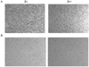
The human endothelial cell line (EA.hy926; ATCC: CRL-2922) was originally derived from human umbilical vein and showed vascular endothelial cell characteristics. EA.hy926 cells were cultured in DMEM culture medium supplemented with 10% FBS, 2% HAT medium supplement (the final concentration of hypoxanthine, aminopterin, and thymidine was 100, 0.4 and 16 mM, respectively) and 1% penicillin/streptomycin in a humidified atmosphere of 5% CO2 at 37℃. The cells were treated just post-confluence because the differentiation marker for endothelial cell, von willebrand factor, was optimally expressed just post-confluence in this EA.hy926 cell line (Fig. 1 A-B). Cellular zinc was depleted using intracellular zinc chelator, N, N, N', N', -tetrakis (2-pyridylmethyl) ethylenediamine (TPEN). Just post-confluence, the cells were treated with either Zn- (0 µM as ZnCl2) or Zn+ (15 µM as ZnCl2) with TPEN (5 or 10 µM) for 6 hours. After then, the cells were exposed to TNFα (10 ng/ml) for 5 hours for inducing cellular oxidative stress. Stock ZnCl2 solution (10 mM) was prepared in appropriate concentration. The media were changed in every 2-3 days.
Cell morphology
The cells were incubated with zinc, either in Zn- (0 µM) or Zn+ (15 µM) with two different levels of intracellular zinc chelator TPEN (5 or 10 µM TPEN) for 6 hours and then, were treated with TNF-α (10 ng/ml) for another 5 hours. The morphological features of EA.hy926 cell line were assessed by inverted light microscope (Leica DMIL, Leica Microsystems, WetZlar, Germany) to observe the bulk morphology for apoptosis for both at 5 µM and 10 µM TPEN treatment.
Trypan blue test
The cells were incubated with zinc either in Zn- (0 µM Zn+ 10 µM TPEN) or Zn+(15 µM Zn + 10 µM TPEN) for 6 hours and then, were treated with TNF-α (10 ng/ml) for another 5 hours. The cell viability of EA.hy926 cell was determined by the trypan blue exclusion assay, in which if the dead cell membrane is leaking and then dead cells uptake trypan blue dye while viable cells don't.
RT- PCR for von Willebrand factor and cellular adhesion molecules (VCAM-1 and ICAM-1)
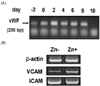
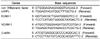
mRNA expression of von Willebrand factor as endothelial cell differentiation and vascular marker, and cellular adhesion molecules (VCAM-1 and ICAM-1) was measured by RT-PCR. For RNA extraction from EA.hy926 cells, Trizol reagent (Invitrogen, USA) was used as commercial instruction. For the synthesis of first-strand cDNA, 100 ng of RNA from each sample were reverse transcribed using 20 U of AMV reverse transcriptase and oligo-p (dT) 1X random primers. The resulting cDNAs were PCR-amplified by using a mixture of the corresponding sense (forward) and antisense (reverse) primers (Table 1). Primers for target and housekeeping genes were obtained from Bioneer Corporation (Daedeok-gu, Daejeon 306-220, Korea). The PCR conditions were 95℃ for 2 min and then 36 cycles at 95℃ for 30s, 55℃ for 45s and 70℃ for 1 min and a final extension at 72℃ for 5 min. The PCR products were separated on 1.8% agarose gel.
Study 2: Ex vivo study (mouse aortic ring culture study)
Mouse aortic ring culture
Mouse aorta from C57BL/6 strain was removed and all adipose tissues were removed. The aorta was dissected in three major parts through the entire aorta for random sampling and then, the aorta was dissected 0.5 mm in thickness for the aortic ring samples to culture in a culture dish, as ex vivo. Mouse aortic ring was cultured in a 96-well plate as one ring per well in DMEM culture medium supplemented with 30% FBS and 1% penicillin/streptomycin in a humidified atmosphere of 5% CO2 at 37℃. The aortic ring culture was carefully examined overnight and cultured for another two to three days, until the aortic ring culture being confirmed viable. After confirming the aortic ring being viable, then MTT assay was done for evaluating aortic ring cell viability.
MTT assay for viability of mouse aortic ring cells
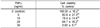
Mouse aortic ring cell viability was determined by the MTT assay on the metabolic reduction of 3-[4, 5-dimethylthiazol-2-y]-2, 5-diphenyltetrazolium bromide (MTT). Aortic ring was plated in a 96-well plate and maintained in growth media for 2-3 days at 5% CO2 at 37℃. After then, the aortic ring was treated with TNFα with the range of 0-30 ng/ml to evaluate aortic cell viability by oxidative stress or TPEN with the range of 0-30 mM to evaluate if cellular zinc depletion by TPEN decreased aortic ring cell viability for 2 days. Ten micorliter of MTT solution was added in each well and incubated at 37℃ for 3 hours to allow for the formation of formazan crystals. After formation of formazan crystals, the MTT medium was then aspirated and replaced with solubilization solution (DMSO) for dissolving the formazan crystals. The plates were read on Micro Elisa reader (Asys Hitee, Expert 96, Asys Co., Austria) at 570nm.
Results
In vitro study
Cell morphology
The cells were shown to be less viable morphologically in zinc- and the viability was much less in higher Zinc chelator treatment group (10 µM TPEN), where less free zinc was available by cells than in lower Zinc chelator group (5 µM TPEN) (Fig. 1). The cells in Zn- showed fewer adherents to the cell dish and visually apoptotic. zinc deficiency decreased endothelial cell viability even within 6 hours, and the less viable morphology was observed in higher zinc chelator level at 10 µM TPEN.
Trypan blue test
The cell viability was decreased by zinc deficiency (0 µM Zn) for 6 hours. Seventy five percent cell viability was observed at the Zn- (0 µM Zn + 10 µM TPEN), while Zn- (15 µM Zn + 10 µM TPEN) showed 94% of cell viability of 6 hours zinc treatment, followed by 10 ng/ml TNFα treatment (data not shown).
mRNA expression for von Willebrand factor, VCAM-1 & ICAM-1
To confirm if this particular cell line was appropriate for testing endothelial characteristics, von Willebrand factor was assessed, since von Willebrand factor is related to the formation of clots and also can be a characteristic of endotherial cells. von Willebrand factor on endothelial cells was expressed from the just pre-confluence and was maximally from the post-confluence 2 days and maintained up to 6 days (Fig. 2A). Therefore, we decided to treat zinc just post-confluence on 2 days on this cell model. The cells were treated for 6 hours with the level of Zn- (1µM) and Zn+ (15 µM), and after then, the cells were exposed with TNFα for 5 hours. mRNA expression of adhesion molecules, VCAM-1 and ICAM-1, were measured by RT-PCR. The mRNA expression of VCAM-1 and ICAM-1 mRNA was higher in Zn+ than Zn-, unexpectedly, under this experimental condition. Even ICAM-1 mRNA expression showed a bit less discrepancy between Zn- and Zn+ (Fig. 2B, data are presented as the representative of three replicates). For the atherosclerotic incidence, once inflammatory stimuli signal to the vascular endothelium, which then in turn mediates leukocyte recruitment and adherence to the endothelium at the early stages of vascular inflammation, ultimately leading to the progression of numerous vascular diseases. VCAM-1 and ICAM-1 are responsible for the process of formation of endothelium adherence.
Ex vivo study
MTT viability for mouse aorta by TNFα & TPEN
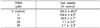
The functional integrity of the aortic ring, under zinc deficiency, was assessed using the MTT viability assay for mouse aortic ring culture. At first, we evaluated if cell viability affected in this particular ex vivo mouse aortic ring culture sample which was treated by TNFα, the inducer of oxidative stress. The viability of mouse aorta cells was inversely proportional to the TNFα concentration (0-30 ng/mL, Table 2), which confirmed the TNFα-induced oxidative stress, and again which decreased mouse aortic ring viability. On considering the 0 ng/mL of TNFα is 100% cell viability, 10 ng/ml TNFα treatment showed marginal viability level (78.4%), and then we decided to treat with this level for consecutive zinc chelator experiment for mouse aortic ring viability by cellular zinc deficiency. Then, we evaluated whether less cellular zinc availability could decrease mouse aortic ring viability. Cellular zinc depletion was induced by increasing the addition of intracellular zinc chelator, TPEN (0-30 µM). The aortic ring cell viability showed that as the added TPEN level increased, which means less cellular zinc is available, the aortic ring cell viability was decreased as shown in Table 3. The results showed that as cellular zinc was less available, then aortic cell viability decreased.
Discussion
The present study evaluated the potential role of zinc on vascular disease induced by oxidative stress, in that whether cellular zinc deficiency decreased the viability of both the endothelial cell in EA.hy926 cell line and the mouse aortic cells ex vivo model. Also it was examined whether cellular zinc deficiency modulated the mRNA expression of VCAM-1 and ICAM-1 in endothelial cells. To confirm beforehand if this particular endothelial cell line, EA.hy926 cells, shows the appropriate condition for vascular and inflammatory morphology, von Willebrand factor (vWF) gene which is considered as the endothelial cell differentiation was measured and confirmed vWF gene expression in the cells under the present study condition. vWF mRNA was expressed from 0 to 6 days of EA.hy 926 cell culture after TNFα treatment. vWF is normally synthesized by the endothelial cells and stored in the Weibel-Palade bodies and it is secreted on endothelial activation (Collins et al., 1993). Normally, the increased plasma vWF levels are associated with cardiovascular disease (Theilmeier et al., 2008).
In the present study, the results showed that zinc could protect endothelial cell viability both in endothelial in vitro and mouse aortic ex vivo model under TNFα-induced oxidative stress condition, which causes cardiovascular abnormality such as atherosclerosis. TNFα induce the inflammatory signal for increasing the oxidative stress and thus increase the cell apoptosis through stimulating the oxidative stress in endothelial cells (Ling et al., 2007). Ex vivo study showed that as more cellular zinc chelator TPEN was added, which means less cellular zinc is available, then the mouse aortic ring culture viability was decreased under TNFα treatment (Table 3). In vitro study results also showed as being consistent with the results of ex vivo, in which zinc deficiency (Zn-) followed by TNFα treatment showed more apoptotic and lower cell viability than zinc adequacy (Zn+) (Fig. 1 and trypan blue test results).
In fact, it is reported that zinc deficiency induces cell death in various cellular models (Clegg et al., 2005), however, not much confirmed in endothelial cell models and nor in mouse aortic ring culture model, ex vivo. The endothelial cell model from porcine pulmonary arteries showed lower cell viability under zinc deficiency induced by TPEN zinc chelator (Hennig et al., 1999; Meerarani et al., 2000). Therefore, the findings in the current study confirmed that zinc could protect cell death in human origin endothelial EA.hy926 cells, which has not been reported before, in addition to mouse aortic ring culture condition. The preventive role of zinc for the decreased cell viability in endothelial cells and mouse aortic tissue culture in the current study results can be interpreted that zinc can increase endothelial cell viability under oxidative stress, which can be protective for atherosclerosis in the endothelium of blood vessels.
In our in vitro model, Zn- showed low cell viability than Zn+ in human origin endothelial EA.hy926 cells at 6-hour zinc treatment prior to TNFα treatment, which causes intracellular oxidative signal. Under TNFα exposure, the high cell viability in Zn+ might result from the anti-oxidative property of zinc, since zinc has an antioxidant role as a component of systemic antioxidant enzyme such as Cu/Zn-SOD (Formigari et al., 2007). The intracellular oxidative signals may provide a molecular mechanism linking the oxidative signal which is one of risk factors for the early pathogenesis of atherosclerosis through the expression of some vascular inflammatory genes, such as cell adhesion molecules VCAM-1 and ICAM-1 (Alexander et al., 1995; Kunsch & Medford, 1999). It is reported that the cell adhesion molecules VCAM-1 and ICAM-1 are expressed in endothelial cells by oxidative stress, such as TNFa and other inflammatory molecules (Choi et al., 2004; Reiterer et al., 2004). In the present study, mRNA expression of VCAM-1 and ICAM-1 under zinc treatment in EA.hy926 cells was evaluated and unexpectedly, VCAM-1 and ICAM-1 mRNA expression was highly expressed in Zn+ under the current experimental condition of 6-hour zinc treatment (1 µM or 15 µM) prior to TNFα exposure (10 ng/ml). The potential explanation of the current contradicting results for VCAM-1 and ICAM-1 mRNA expression might be due to shorter zinc treatment time period (6 hours with 1 or 15 µM Zn) and stronger TNFα exposure condition (10 ng/ml for 5 hours), compared to longer Zinc treatment (20 µM for 24 hours) and weaker TNFα exposure (10 ng/L for 2 hours) (Reiterer et al., 2004). Further study for VACM-1 and ICAM-1 expression under optimal experimental condition would be needed for getting final conclusion.
Normally, more endothelial adhesion molecules, VCAM-1 and ICAM-1, induce more oxidative stress, which may cause the progression of atherosclerosis. Even under the current particular experimental condition, a potential explanation for higher mRNA expression VCAM-1 and ICAM-1 in Zn+ would be suggested: One possibility would be that the VCAM-1 and ICAM-1 were up-regulators of atherosclerosis, which means these were expressed normally before the progression of atherosclerosis. In our study, Zn+ showed more VCAM-1 and ICAM-1 expression which biologically induces more expression of anti-oxidative enzymes. Recent study showed that the antioxidant defense system against lipid peroxidation was increased, which can propose to retard the development of atherosclerosis. Also, the increase in Cu/Zn-superoxide dismutase (Cu/Zn-SOD) activity may reduce vascular cell-mediated oxidation of low-density lipoprotein in mouse aorta (Yang et al., 2004).
Taken all together, zinc adequate can protect endothelial damage which is induced by oxidative stress through increasing endothelial cell viability and aortic ring culture cell viability. Also, zinc modulates cellular adhesion molecules (VDAM-1 and ICAM-1) mRNA expression in endothelial cells. These findings are cautiously interpreted that zinc can be a factor for preventing atherosclerosis incidence. Further investigation will undertake for clarifying the mechanism by zinc for anti-atherosclerosis.
 XML Download