

 PDF
PDF ePub
ePub Citation
 Print
Print


Introduction
Recent studies have demonstrated the potent antioxidant activity of green tea polyphenolics, leading to suggestions that green tea drinking reduces the risk for cancer (Ke et al., 2002; McKay & Blumberg, 2002; Su & Arab, 2002). Green tea and its constituents have been extensively studied both in vitro and in animal models of carcinogenesis (Yang et al., 2002). Although these compounds have been shown to be efficacious in a number of models of carcinogenesis, the epidemiological data on cancer prevention remain inconclusive (Arab & Il'yasova, 2003; Huang & Xu, 2004; Yang et al., 2002). Numerous potential mechanisms have been proposed for the cancer-preventive activity of green tea based on the strong antioxidative activity of tea polyphenols (Ahmad et al., 2001; Dreosti, 1996; Feng et al., 2001). Many studies have focused on epigallocatechin gallate and theaflavins as chemopreventive agents that inhibit the growth and induce cell cycle arrest and apoptosis in various cancer cell lines (Takada et al. 2002; Yang et al., 1998; Yang et al., 2002). The relative importance of any of these mechanisms in vivo remains to be determined since most in vitro studies have employed concentrations of tea compounds that far exceed those found in animal plasma or tissue following reasonable tea consumption (Lambert & Yang 2003). To overcome this shortcoming, we designed experiments involving green tea supplementation as the sole source of fluid at human-achievable doses.
Chemoprevention consists of the pharmacological use of one or more substances with the purpose of delaying, or even reverting, the carcinogenic process before the emergence of malignancy (Gescher et al., 2001). Despite the great advances in diagnosis and treatment, cancer mortality rates (especially of liver cancer) remain high (Guyton & Kensler, 1997; Korea National Statistical Office, 2008), mainly in developing countries (Barret, 2002). Although in vitro and in vivo animal studies have provided evidence of the beneficial effects of green tea polyphenols at most stages of cancer development and have identified many possible sites of action in cancer prevention (Chen et al., 2000; Lin, 2002) the relevance of these studies is uncertain due to the experimental polyphenol concentrations being higher than those attainable via normal dietary intake of green tea (Henning et al., 2005; Yang et al., 2001).
Therefore, the present study evaluated the chemopreventive activities of green tea ingestion both prior to the initiation stage (pre-initiation) and during the promotion stage (post-initiation) of hepatocarcinogenesis. We intended to clarify the effects of the duration and timing of green tea ingestion on hepatocarcinogenesis induced by diethylnitrosamine (DEN) and 2-acetylaminofluorene (2-AAF) in male rats. We conducted a study on the hematocarcinogenesis since liver cancer is still a leading cause of cancer mortality in Korea, which is the highest liver cancer mortality among OECD countries (Korea National Statistical Office, 2008). The percentage area of foci that were positive for placental glutathione S-transferase (GST-P) was measured as a marker of preneoplastic lesions, since the degree of induction of GST-P positive foci is directly correlated with the incidence of hepatocellular carcinomas in long-term in vivo systems (Tatematsu et al., 1988). Because DEN and 2-AAF are metabolized through phase I and phase II drug-metabolizing enzymes, we hypothesized that green tea modulates these enzymes, and thereby affects hepatocarcinogenesis. Therefore, we measured cytochrome P450 (CYP) content and glutathione S-transferase (GST) activities. We also measured the contents of thiobarbituric acid reactive substances (TBARS) in liver microsomes as an index of lipid peroxidation. Two sets of experiments were conducted with different durations to examine the extents to which green tea modulates the chemical carcinogenic effects before and after initiation.
Materials and Methods
Preparation and analysis of green tea
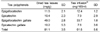
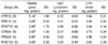
Twenty-five grams of green tea (Sullok Green Tea, Pacific Corporation, Seoul, Korea) were added to 1 L of 80℃ boiled, distilled and deionized water and left to stand at room temperature (25℃) for 5 min. The brewed tea was filtered through several layers of cheese cloth. The contents of several phenolic compounds were analyzed by reverse-phase high performance liquid chromatography (TOSOH with a UV detector at 280nm, Tokyo, Japan; C18 Shiseido Cap Cell Pack, 5µm particle size, 250×4.6mm, Shiseido, Tokyo, Japan). The mobile phase contained 250 mL of 25% 1, 2, 3, 4-tetrahydro-9-fluorenone and 750mL of 1% phosphoric acid. The column was maintained at room temperature (25℃), and the flow rate of the mobile phase was 0.8mL/min. Table 1 lists the polyphenols composition of brewed green tea used in the experiment.
Animals and diets
Study protocols were approved by the Institutional Animal Care and Use Committee of Seoul National University.
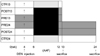
There were two experimental periods, of 13 and 24 weeks, each with pre-initiation and post-initiation and two control groups. Male Sprague-Dawley rats (80~90 g) were supplied by the Seoul National University Animal Care Facility. The rats were housed at two per suspended stainless steel cage with wire mesh bottoms that were kept in a humidity- and temperature-controlled room (55 ± 1% and 20 ± 1℃, respectively) with a 12-h light:dark cycle. The rats had free access of deionized water and a commercial diet (Rat chow, SamYang Animal Chow Co., Seoul, Korea) for one week before they were divided into six experimental groups, with six rats allocated to each group. The experimental design is shown in Fig. 1. Rats in two control groups of 13 weeks (CTR13) and 24 weeks (CTR24) consumed a diet formulated to meet recommended nutrient levels for rats (AIN76A) (AIN76A, American Institute of Nutrition, 1980), and distilled water. Rats in the tea groups consumed an AIN76A diet, with green tea being the sole source of fluid for the entire 13- or 24-week experimental period (PRE13, PRE24, POST13 and POST24). Rats were injected with DEN intraperitoneally as a single dose of 200 mg/kg body weight after 4 weeks of feeding. From 2 to 12 weeks after the DEN injection, the rats consumed a diet that included 0.02% (W/W) 2-AAF. We sacrificed rats at weeks 13 and 24.
Placental glutathione S-transferase positive foci
Upon killing the animals by decapitation after 12 h of fasting, the livers were immediately excised and rinsed with saline solution. Blot-dried livers were weighed and cut into 2- to 3-mm-thick sections with a blade. These liver slices were fixed in ice-cold acetone for immunohistochemical examination of GST-P positive foci. The avidin-biotin-peroxidase-complex method was used to demonstrate GST-P positive liver foci. Immunohistochemical analysis was carried out with sequential treatments of rabbit anti-rat placental glutathione S-transferase (BMI Inc., Tokyo, Japan) as a primary antibody, swine anti-rabbit IgG antibody (BMI Inc.) as a secondary antibody and peroxidase-antiperoxidase complex (Vectastain Elite ABC kit, Vecta Laboratories, Burlingame, USA). Final visualization of GST-P positive foci was enzymatically activated by 3,3-diaminobezidine (Sigma Chemical Co., St. Louis, USA) and H2O2 as substrate. The total areas of GST-P positive foci with diameters larger 0.2mm in the liver sections were measured using an image analyzer program (Bummi Universe, Ansan-si, Korea), and expressed as a percentage of the total area.
Biochemical assays
Microsomal and cytosolic fractions were prepared according to a modified version of the methods of Sohn et al. (1994). A portion of the liver was finely minced in three weight volumes of ice-cold 50 mM Tris-HCl buffer, pH 7.4, containing 1mM EDTA, and then homogenized. Homogenates were centrifuged at 12,000×g for 20min and, microsomes were obtained by centrifuging the resulting supernatant at 105,000×g for 1h. After collecting the cytosolic fraction, microsomes were resuspended in three weight volumes of 50 mM Tris-HCl buffer, pH 7.4, containing 1mM EDTA. Cytosol and microsomal suspensions were frozen by liquid nitrogen and stored at -70℃ until assayed.
Lipid peroxides of the hepatic microsomes were determined by measuring the formation of TBARS (Buege & Aust, 1978). Malondialdehyde as the product of lipid peroxidation was reacted with thiobarbituric acid, and the absorbance of the resulting chromophore was measured at 535nm. GST activity was assayed in the hepatic cytosolic fraction using the method of Habig et al. (1974). Total CYP content was determined according to the method of Omura and Sato (1964). Fresh liver microsomes were added to sodium dithionate, and the reduced hemoprotein was combined with carbon monoxide by bubbling CO gas through the solution. The characteristic absorbance at 450 and 490nm was measured by dual-beam spectroscopy. The protein amounts of hepatic microsomes and cytosolic fractions were determined by Lowry's method (Lowry et al., 1951).
Results
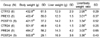
Green tea ingestion significantly reduced the final body weight when introduced from the beginning of the experiment (p<0.05 for PRE13 vs. CTR13 and PRE24 vs. CTR24, Table 2). However, when tea was introduced from the post-initiation stage (POST13 and POST 24), final body weights were not different from corresponding control groups. Although tea did not affect liver weights (Table 2), the relative liver weight was significantly increased with tea ingestion from post-initiation up to week 24 (3.36 ± 0.43 g of POST24 vs 2.56 ± 0.25 g of CTR24, p<0.05).
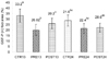
Fig. 2 presents the area of GST-P positive foci, the preneoplastic lesions. Suppressive effects of green tea ingestion on the induction of GST-P positive foci were observed both at weeks 13 and 24 (p<0.05) compared to the corresponding control groups. However, at week 13, green tea ingestion from prior to the introduction of initiator reduced lesion further than green tea ingestion started from post-intiation (p<0.05, PRE13 vs POST13) while no difference was observed between green tea ingestion from pre- and post-intiation at week 24. The areas of GST-P positive foci at week 13 were similar to those at week 24 (33.2 ± 5.8% of CTR13 vs. 28.6 ± 5.1% of CTR24).
TBARS content, GST activity and CYP content are given at Table 3. Liver microsomal lipid peroxidations determined by TBARS of rats ingested tea were significantly lower than those of control groups (p<0.05). The average of TBARS content at week 13 was higher than at week 24 (p<0.05). No significant differences were observed among groups of GST activity and CYP content.
Discussion
In the present study, we observed anticarcinogenic effects of green tea with different duration and timing of ingestion on rat hepatochemical carcinogenesis induced by DEN and 2-AAF. We sacrificed rats at weeks 13 and 24 from the initiation of the experiment because we would like to examine if green tea modulated the metabolism in rat liver induced by chemical carcinogens at different periods of time and if it prolonged the effects after finishing administration of chemical carcinogens. As an index of preneoplastic lesion, we measured the area of GST-P positive foci which shows a good correlation with the incidence of hepatocellular carcinomas revealed in long term in vivo systems (Tatematsu et al., 1988). When we terminated the experiment at week 13, green tea ingestion from the post-initiation stage decreased the area of GST-P positive foci by about 22% while about 32% reduction was observed when tea was introduced from the beginning of the experiment. However, further reduction of green tea ingestion before the initiator was not observed when the experiment was continued until week 24. The results support the suppressive effect of green tea on hepatocarcinogenesis in rats with a human-achievable dose, which is a relatively low dose compared to in vitro or single dose in vivo studies. These results suggest that green tea reduced carcinogenesis during drugs (initiator or promoter) administration, but it might not affect on hepatocarcinogenesis after administering carcinogens because no further reduction of GST-P lesion up to week 24 compared to week 13 was observed. It seemed to be more effective to drink tea before the administration of carcinogens than during drug administration for a short-term period, but if tea ingestion is prolonged for a longer period, the same effect of tea ingestion was observed whether tea drinking was started from pre- or post-intiation. Yang et al. (2001) previously reported that polyphenols, which are effective when given during the post-initiation period by inhibiting tumor promotion and progression, are believed to be more useful in preventing cancer in humans than are polyphenols, which are effective only when given before and during the carcinogen treatment. Our study showed no difference in cancer preventive effects of tea between pre- and post-initiation ingestion if tea was ingested for an extended period up to 24 weeks. Regarding the results of 24-week experiment of no difference between PRE24 and POST24 in GST-P positive foci areas, tea may have inhibitory activity against hepatocarcinogenesis when administered before and during the initiation or promotion stages. The broad inhibitory activity of tea has been reported on lung cancer as well as skin cancer (Conney et al., 1997). Our results can add another support of broad inhibition activity of tea on hepatocarcinogenesis. The results of 13-week experiment, however, showed differences between PRE13 and POST13 in GST-P lesion. With a relatively low dose, duration of tea ingestion seems to be important. This result may explain the controversial results found from epidemiology studies. Some cross-sectional studies without information on the duration of tea drinking may result in biased effects of tea drinking. Even if we used a relatively low dose of tea, using tea as a sole source of drinking liquid seemed to be stressful to the growing rats in this experiment. Rats in PRE 13 and 24 groups did not show full growth as the control groups and we lost one animal each from PRE13 and 24 groups in the middle of the experimental period. However rats in POST initiation groups grew as well as the control groups although one more animal in POST24 could not survive up to the termination of the experiment. For the fast growing young animals, initial growth retardations due to drinking tea instead of water could be somewhat stressful to fully recover their physiological functions.
We measured TBARS contents to determine the level of lipid peroxidation in liver microsome. TBARS contents were higher at week 13 than week 24 because TBARS induced by chemical carcinogens (Tian et al., 2001). Farombi et al. (2000) reported that TBARS contents of rat fed 0.02% 2-AAF in diet induced 2.5 times higher than those of rats fed control diet. In the present study, the contents of TBARS in the control group at week 13 (one week after 2-AAF administration) were 1.75 times higher than those at week 24 (ten weeks after 2-AAF administration, CTR13 vs CTR24). On the other hand, for the tea groups, TBARS contents at week 13 were about 5.5 times higher than those at week 24. At week 13, chemical carcinogen induced lipid peroxidation effects would be higher than at week 24 due to a shorter time period since 2-AAF administration. This may be confirmed by the previous study, which indicated TBARS induction was decreased as time passed by (Farombi et al., 2000; Tian et al., 2001). TBARS contents of rats in the tea groups were lower than those of rats in the control groups. The reduction of TBARS by green tea was more significant with longer ingestion of tea. According to the investigation with breast cancer patients, plasma malondiadehyde level may decrease in advanced stages of cancer (Alagol et al., 1999). Therefore, the big difference of TBARS levels between week 13 and 24 in the tea groups may appear from the combination effects of the carcinogens, tea and the metabolism in late stage of cancer.
GST activities of rats sacrificed at week 13 were not changed by green tea. It has been reported that lipid peroxidation reduced glutathione levels, and then GST activity decreased (Ozdemirler et al., 1999). However, we could not observe negative correlation between TBARS and GST activity. CYP contents of the green tea groups at both week 13 and 24 were not different from that of the corresponding control groups. CYP is a main component of phase I drug metabolism, which activates carcinogenesis. Previously, DNA damage can be reduced through decreasing Phase I drug metabolites, CYP3A, by grapefruit juice (Miyata et al., 2004) and we expected the same effects with green tea. However, such effect was not observed in our study. Green tea ingestion did not modify Phase I and II drug metabolism during hepatocarcinogenesis in the present study. The anticarcinogenic effects, therefore, may be mainly attributable to the antioxidants effects in green tea.
In conclusion, this study proved that green tea has anticarcinogenic effects through reduction of preneoplastic lesions, GST-P positive foci. The effect was more significant when tea was ingested before the drug administration for a short term, but for a long term period, green tea ingestion was beneficial regardless of ingestion timing.
 XML Download