Journal List > Nutr Res Pract > v.2(4) > 1050970

Shim, Kim, and Lee: Effects of soybean isoflavone extract on the plasma lipid profiles and antioxidant enzyme activity in streptozotocin-induced diabetic rats

Abstract

The present study evaluated the effects of various dosages of soybean isoflavone extract on lipid profiles, lipid peroxidation and antioxidant activities in streptozotocin-induced diabetic rats. The one normal control group was fed an AIN-76-based experimental diet and four diabetic groups were fed the same diet, supplemented with four different levels of soybean isoflavone extract for seven weeks. The daily dosages of pure isoflavone for four diabetic groups were set to be 0 mg (diabetic control), 0.5 mg (ISO-I), 3.0 mg (ISO-II) and 30.0 mg (ISO-III) per kilogram of body weight, respectively. The plasma total cholesterol levels and the TBA-reactive substances contents in the liver and kidney were significantly lowered in ISO-II and ISO-III groups compared to those in the diabetic control group. The levels of plasma HDL-cholesterol, plasma vitamin A and hepatic superoxide dismutase were significantly increased in those two groups compared with the diabetic control group. The present study demonstrated the possibility that the diets supplemented with 3.0 mg and 30.0 mg of soybean isoflavone extract may have beneficial effects on the plasma lipids, tissue lipid peroxidation and partly on antioxidant system in diabetic animals and there were no significant differences between the ISO-II and ISO-III groups. The results suggest that the effective daily dosage level of isoflavone for improving lipid metabolism in diabetic rats may be above 3.0 mg per kilogram body weight.

Introduction

Isoflavones, one of the phytoestrogens have structural and functional similarity to human estrogen with affinity to the estrogen receptor (Kudou et al., 1991). They contain three main compounds, in four chemical forms; aglycones (daidzein, genistein and glycitein), glucosides (daidzin, genistin and glycitin), their malonyl-glycoside and acetyl-glycoside forms (Ohta et al., 1980). Recent epidemiological evidences and experimental data from animal studies strongly support the beneficial effects of isoflavones in preventing various chronic diseases (Anderson et al., 1999; Setchell & Cassidy, 1999). The most abundant food sources of isoflavones are soybeans and their products (Wang & Murphy, 1994). Soybeans contain 12 types of isoflavones (Anderson et al., 1998; Kuiper et al., 1997) and the contents of soy isoflavones can vary from 50 to 300 mg/100 g (Eldridge & Kwolek, 1983). Soy isoflavones have been reported to attenuate bone loss from the lumbar spine in perimenopausal women (Alekel et al., 2000), to lower blood cholesterol levels (Jenkins et al., 2002; Kirk et al., 1998; Raines & Ross, 1995), and to have antioxidant properties in vivo and in vitro experiments (Wei et al., 1993; Wei et al., 1996). Due to these abilities of soybean isoflavones, they have shown the beneficial effects on prevention and attenuation in chronic diseases including cancers (Zhang et al., 2004), cardiovascular diseases (Teede, 2001), osteoporosis (Arjmandi et al., 1996) and menopausal symptoms (Somekawa et al., 2001).
Recently, it has been reported that soy isoflavones may also have favorable effects on diabetes mellitus in animals and humans. Genistein was reported to increase glucose-stimulated insulin secretion in both insulin-secreting cell lines and mouse pancreatic islets (Liu et al., 2006) and effectively prevent the glucose autoxidation mediated LDL oxidation which results in atherosclerosis (Exner et al., 2001). Jayagopal et al. (2002) demonstrated that supplementation with phytoestrogens had the beneficial effects on insulin resistance, glycemic control and lipid profiles in postmenopausal women with type 2 diabetes. Ali et al. (2005) showed that isoflavones had hypoglycemic and hypolipidemic effects in lean SHR/N-cp rats but not in obese rats. Lee (2002) exhibited that soybean isoflavone extract had positive effects on the elevation of plasma vitamin A and vitamin E in diabetic animals.
However, to our knowledge, the studies investigating the effects of soybean isoflavone supplementation on lipid metabolism associated with diabetes mellitus are in short. Also, recently, inconsistent results have been reported for the effects of isoflavones in diabetic rats. Lee (2006) investigated the effects of genistein (60 mg/100 g diet) and soy protein (20 g/100 g diet)-supplemented diets administered to streptozotocin-induced diabetic rats for three weeks and reported the beneficial effects on the levels of lipids in the serum and liver as well as the activities of antioxidant enzymes in the liver. On the other hand, Hsu et al. (2003) found no favorable effect on the plasma lipid levels and the antioxidant enzyme activities in diabetic rats fed a diet supplemented with isoflavones (240-1920 mg/100 g diet) for 24 days. Additionally, we judged that the dosages of isoflavones administered to diabetic rats in these studies were enormously high. Kim and Kwon (2001) reported that total isoflavone intake per capita in Korean population was estimated as 14.88 ± 6.26 mg/d (genistein : 7.32 ± 3.24 mg/d , daidzein 5.81 ± 2.88 mg/d, and glycitein 1.75 ± 0.52 mg/d) and Lee et al. (2000) presented that the mean intake of Korean middle-aged women was assessed to be 24.11 mg/d (0.0 mg-144.30 mg). We already reported that 2.5 mg/100 g diet of soybean isoflavone had the beneficial effects on glucose tolerance and suppression of weight loss in streptozotocin-induced diabetic rats (Shim et al., 2007).
Therefore, the purpose of this study was to examine the effects of supplementation of soybean isoflavone on the lipid profiles as well as the antioxidant activities in streptozotocin-induced diabetic rats at the lower dosage than those used in the previous studies (Hsu et al., 2003; Lee, 2006). Additionally, we investigated the adequate level of supplementation of soybean isoflavone extract which may exhibit the beneficial effects on the lipid profiles and antioxidant system in diabetic animal.

Materials and Methods

Experimental animals and diet

We used the same experimental animals and diets as described in our previous paper (Shim et al., 2007). Briefly, eighty weanling male Sprague-Dawley rats (Biogenomics Co., Korea) were divided into five groups; normal control group, diabetic control group, and three diabetic experimental groups supplemented with three different levels of isoflavone; ISO-I (0.5 mg/kg BW/day; 0.4 mg/100 g diet), ISO-II (3.0 mg/kg BW/day; 2.5 mg/100 g diet) and ISO-III (30.0 mg/kg BW/day; 25.4 mg/100 g diet). The amounts of isoflavone were calculated based on approximate average daily feed intake (30 g) of mature rats and the initial mean body weight (257 g) of experimental rats. All diets contained an identical concentration of energy and nutrients (Table 1) and the animals were fed experimental diets for seven weeks.

Induction of diabetes

previous paper (Shim et al., 2007). Briefly, diabetes was induced by an injection of streptozotocin (Sigma Chemical, St Louis, MO, USA) at a dose of 50 mg/kg BW. The rats were considered to be diabetic only if their fasting blood glucose levels exceeded 180 mg/dL. The maintenance of the diabetic state was confirmed by measuring the fasting blood glucose level at the 5th week of the experimental period.

Blood and tissue preparation

The blood and tissue preparations were described in our previous paper (Shim et al., 2007). Briefly, after fasting for 12 hours, the blood was drawn from the inferior vena cava of the rats with a syringe treated with heparin (100 units/mL). The plasma was separated by centrifuging at 3,000 rpm for 20 minutes and stored frozen at -70℃. The liver, kidney and heart tissues were excised, rinsed, weighed and stored in a liquid nitrogen tank.

Assessment of concentrations of plasma and tissue lipids

The concentrations of triglyceride (Bucolo & David, 1973), total cholesterol (Allain et al., 1974) and HDL-cholesterol (Finley et al., 1978) in the plasma were measured using enzymatic analysis kits (Asan pharmaceuticals, Hwasung, Korea). Total lipids in liver tissue were extracted by the Folch method (Folch et al., 1957), and then triglyceride (Sidney & Bernard, 1973) and total cholesterol (Sale et al., 1984) concentrations were measured using commercial enzymatic kits (Asan Pharmaceuticals).

Assessment of lipid peroxides in plasma and urine

We assessed the lipid peroxides as thiobarbituric acid-reactive substances (TBARS) by measuring malondialdehyde (MDA) levels in the plasma and tissues. TBARS in the plasma and urine were measured according to the Taladgis method (Taladgis et al., 1964). An aliquot (0.5 mL) of plasma or urine sample was mixed with 3 mL of 5% trichloroacetic acid and 1 mL of 0.06 M TBA, then incubated in 80℃ water bath for 90 minutes. The mixtures were cooled to room temperature, centrifuged at 2,000 rpm for 15 minutes and the absorbance of the supernatants was then measured at 535 nm. Nelson et al. (1993) have reported that TBARS in urine presented in association with the amount of creatinine excreted are statistically more significant; we presented the data as nmol MDA/µmoles creatinine. Creatinine in urine was measured according to the Jaffé reaction method (The Korean Society of Food Science and Nutrition, 2000) by using a kit (Asan pharmaceuticals).

Assessment of lipid peroxides in tissues

TBARS in the liver, kidney and heart tissues were measured according to the Uchiyama and Mihara method (Uchiyama & Mihara, 1978). After mixing 0.5 g of tissue and 9 times volume of 0.01 M sodium phosphate buffer (pH 7.0), the mixture (0.5 mL) was homogenized and added to the solution composed of 3 mL of 1% phosphoric acid and 1 mL of 0.06 M TBA, and then boiled for 45 minutes. After cooling, the mixture was added to 4 mL of n-butanol and centrifuged at 3,000 rpm for 10 minutes. The supernatant absorbance was measured at 520 and 535 nm, and the difference of absorbance was used to calculate the peroxide level.

Preparation of hepatic antioxidant enzymes source

A total of 2 g of liver tissue was homogenized with 5 times volume of 0.25 M sucrose solution at 4℃ using a glass teflon homogenizer (Glascol, 099C K44, USD). In order to discard cell debris, the homogenates were centrifuged at 600 ×g for 10 minutes. Then, 3 mL of the supernatant was centrifuged at 10,000 ×g for 20 minutes at 4℃ to obtain the mitochondria supernatant and then 5 mL of the supernatant was centrifuged at 105,000 ×g for 1 hour at 4℃ using an ultracentrifuge (Beckman, Optima TLX-120) to obtain the cytosol supernatant. The protein contents in the cytosolic fraction were determined by the Bradford method (Bradford, 1976) with bovine serum albumin as the standard.

Assay of catalase activity

The activity of catalase was measured by the Abei method (Abei, 1974). A total of 0.1 mL of 30 mM H2O2 was added to 2.89 mL of 50 mM potassium phosphate buffer (KH2PO4: NaH2PO4, 1:1.5, pH 7.0). The mixture was incubated for 5 minutes at 25℃ and then the absorbance was measured at 240 nm. Then, 10 ㎕ of liver tissue preparations was added to the mixture and incubated for 5 minutes at 25℃. The absorbance was then measured at 240 nm for 5 minutes. The H2O2 decomposition rate was calculated and the activity was expressed as H2O2 µmol/min/mg protein.

Assay of glutathione peroxidase activity

Glutathione peroxidase (GPx) was determined by the Paglia and Valentine method (Paglia & Valentine, 1967). One tenth mL of 30 mM glutathione reductase, 0.1 mL of 6 mM NADPH and 0.1 mL of 25 µM H2O2 were subsequently added to 2.6 mL of 0.1 M Tris-HCl buffer (pH 7.2) and incubated for 5 minutes at 25℃. Then, 0.1 mL of liver tissue preparations was added to this mixture and incubated at 25℃ for 5 minutes. The absorbance was then measured at 340 nm. A molar extinction coefficient of 6.22 mM-1cm-1 was used and the activity was expressed as oxidized NADPH nmol/min/mg protein.

Assay of superoxide dismutase activity

Superoxide dismutase (SOD) was assayed by the Marklund and Marklund method (Marklund & Marklund, 1974). SOD was detected based on its ability to inhibit the superoxide-mediated reduction. One tenth mL of liver tissue preparations and 0.1 mL of 15 mM pyrogallol were added to 2.8 mL of 50 mM Tris-HCl buffer (pH 8.6) containing 10 mM EDTA. After incubation for 10 minutes at 25℃, the reaction was terminated by adding 0.1 mL of 1 N HCl and the absorbance was then measured at 420 nm. The enzyme unit was defined as the amount inhibiting the autoxidation of pyrogallol by 50%.

Assessment of antioxidant nutrients (vitamin A & E) concentration in plasma

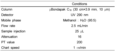
The concentrations of vitamin A and vitamin E in the plasma were measured using the Bieri HPLC method (Bieri et al., 1979). Table 2 shows the HPLC conditions for the determination of vitamin A and E. One hundred ㎕ of α-tocopheryl acetate and 100 ㎕ of retinyl acetate were added to 200 ㎕ of plasma as an internal standard and the solutions were mixed. A total of 200 ㎕ of heptane for HPLC analysis was added to the solution and the mixture was centrifuged at 1500 rpm for 5 minutes. The supernatant was then collected in a brown vial of interception light. This supernatant was filtered using a 0.45 µM membrane filter (Millipore Corporation, Bedford, MA 01730) and dried under N2 gas. This extract was dissolved in a diethyl ether: methanol (1:3, v/v) solution and was then analyzed by HPLC (Waters 500).

Statistical analysis

Data were presented as means and standard errors of the mean. Group means were compared by an Analysis of Variance using Duncan's multiple range test and differences were considered to be statistically significant at p value of less than 0.05. All statistical tests were performed by using the Windows SPSS program (SPSS, Chicago, IL, USA: Version 10.0).

Results

Effect on plasma and hepatic lipids

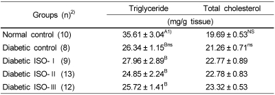
The plasma triglyceride and total cholesterol levels in the diabetic control group were significantly lower (p<0.05) than those in the normal control group (Table 3). Among diabetic rats, there were no significant differences (p<0.05) in the triglyceride levels. However, the levels of plasma total cholesterol in ISO-II and ISO-III groups were significantly lower (p<0.05) than those in the diabetic control and ISO-I groups. The HDL-cholesterol level and the ratio of HDL-cholesterol/total cholesterol in the diabetic control group were significantly lower (p<0.05) than those in the normal control group. Among diabetic groups, the ISO-II and ISO-III groups showed significantly higher (p<0.05) HDL-cholesterol levels and ratios of HDL-cholesterol/total cholesterol than diabetic control and ISO-I groups. The atherogenic index (AI) of the diabetic control group was significantly higher (p<0.05) than that of the normal control group. Among the diabetic groups, the ISO-II and ISO-III groups exhibited significantly lower (p<0.05) AI than the diabetic control and ISO-I groups.
The hepatic triglyceride level in the diabetic control group was significantly lower (p<0.05) than that in the normal control group and there were no significant differences among the diabetic groups (Table 4). There were no significant differences (p<0.05) in the level of hepatic total cholesterol among all groups including the normal control, diabetic control and three isoflavone-supplemented groups.

Effect on the lipid peroxides levels in plasma, urine and tissues

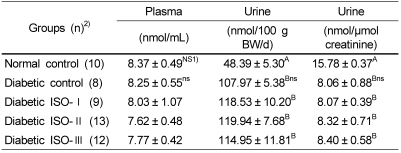
We expressed the lipid peroxides as TBARS by measuring the MDA levels in plasma, urine and tissues. MDA is the final product of the degradation of lipid peroxides, which has been used as a biochemical index of lipid peroxide-mediated tissue damages in vivo (Pierro et al., 1992). The plasma TBARS levels showed no significant differences (p<0.05) in all groups, including normal control, diabetic control and three isoflavone-supplemented groups (Table 5). When the TBARS content in the urine was calculated per 100 g body weight per day, it was found that the levels of all of the diabetic groups were significantly higher (p<0.05) than that of the normal control group. However, based on the amount of creatinine excreted, the urinary TBARS levels in all of the diabetic groups were significantly lower (p<0.05) than that of the normal control group.
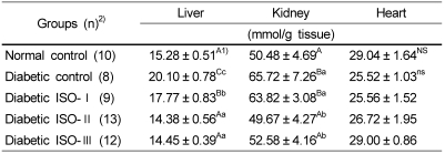
The hepatic TBARS level in the diabetic control group was significantly higher (p<0.05) than that in the normal control group (Table 6). The hepatic levels of all the diabetic isoflavone-supplemented groups were significantly lower (p<0.05) than that of the diabetic control group. The levels of the ISO-II and ISO-III groups were significantly lower (p<0.05) than that of ISO-I group and were not significantly different in comparison to that of the normal control group. The TBARS level in renal tissue of the diabetic control group was significantly higher (p<0.05) than that of the normal control group. Among the diabetic groups, the levels of the ISO-II and ISO-III groups were significantly lower (p<0.05) than those of the diabetic control and ISO-I groups. There were no significant differences in regard to the TBARS levels in the heart tissue among all the groups, including the normal control, diabetic control and three isoflavone-supplemented groups.

Effect on antioxidant enzyme activity in liver tissue

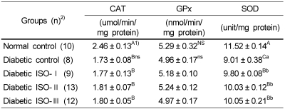
In comparison of the activities of antioxidant enzymes between the diabetic control and normal control groups, the catalase and SOD activities were significantly lower (p<0.05) in the diabetic control group, but there was no significant difference (p<0.05) in the activity of GPx (Table 7). Among the diabetic groups, there were no significant differences (p<0.05) in the activities of catalase and GPx. However, the SOD activities in all of the isoflavone-supplemented groups were significantly higher (p<0.05) than that in the diabetic control group.

Effect on plasma antioxidant vitamins

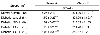
The concentration of the plasma vitamin A in the diabetic control group was significantly lower (p<0.05) than that of the normal control group (Table 8). Vitamin A concentrations in the ISO-II and ISO-III groups were significantly higher (p<0.05) than those in the diabetic control and ISO-I groups. There were no significant differences (p<0.05) in the concentrations of vitamin E among all the experimental groups.

Discussion

This study examined the effects of diets containing various levels of soy isoflavone extract on lipid profiles in the plasma and tissues, lipid peroxidation and antioxidant enzymes and vitamins in streptozotocin-induced diabetic rats. The supplementation of soybean isoflavone to diabetic rats significantly lowered the plasma cholesterol level, however, there was no effect on the plasma triglyceride level. Lee (2006) demonstrated that the concentrations of serum triglyceride and cholesterol were significantly reduced in diabetic groups fed a diet supplemented with genistein for 3 weeks. Ali et al. (2005) exhibited that soy isoflavones did not reduce triglyceride level in obese SHR/N-cp rats which are a genetic animal model of obesity and type 2 diabetes. Hsu et al. (2003) reported that soy isoflavone supplementation had no significant effect on the reduction of triglyceride and cholesterol levels in fasting plasma, in which soy isoflavones at the three levels (240, 480, 1920 mg/100 g diet) were administrated to streptozotocin-induced diabetic rats for 24 days. From the previous studies conducted by other investigators, the effects of isoflavones on blood lipids in diabetic animals appeared inconsistent.
The increases of total triglyceride and total cholesterol in plasma were frequently shown in diabetes mellitus states (Kaleem et al., 2006; Kesavulu et al., 2001; Lee, 2006). Insulin has a role in inhibiting the hormone sensitive lipase and activating lipoprotein lipase (LPL). Therefore, in diabetes state, the mobilization of free fatty acids from peripheral deposits is increased and serum total triglyceride levels are elevated (Al-Shamaony et al., 1994; Taskinen, 1987). However, in this study, the plasma triglyceride and cholesterol concentrations were significantly reduced in the diabetic control group in comparison to that in the normal control group. It may be inferred that the decreased levels of these lipids in the present study might have been caused by the markedly reduced body weight in the diabetic rats, which was reported in the previous paper by Shim et al. (2007). At the end of 7 weeks of experiment period, the mean final body weight of the normal control and the diabetic control groups was 522.50 g and 277.36 g, respectively. We had previously obtained similar results from several studies using diabetic animals (Lee et al., 1996; Park & Lee, 2003).
It has been observed in this study that the supplementation of soybean isoflavone at the levels of 3.0 mg (ISO-II) and 30 mg (ISO-III) per kilogram of body weight per day in diabetic rats resulted in the increases of plasma HDL-cholesterol, the ratio of HDL-cholesterol/total cholesterol in plasma and remarkable improvement of atherogenic index. Anthony et al. (1996) reported that soy protein including isoflavone significantly increased the concentration of HDL-cholesterol and the ratio of HDL-cholesterol/total cholesterol and also significantly reduced the total plasma cholesterol concentrations in rhesus monkeys. Lee (2006) demonstrated that a 60 mg/100 g diet of genistein increased the serum HDL-cholesterol level in diabetic rats. Jayagopal et al. (2002) presented that a diet supplemented with phytoestrogens (soy protein 30 g/d, isoflavones 132 mg/d) for 12 week significantly lowered total cholesterol, LDL cholesterol and cholesterol/HDL cholesterol ratio in postmenopausal women with type 2 diabetes mellitus. The results of these studies support the conclusion that soybean isoflavones may be effective in improving the plasma lipid profiles in diabetes mellitus.
It is known that the quantity of triglycerides in the liver is significantly increased in uncontrolled diabetes mellitus, which results from raised plasma free fatty acids levels. Since the antilipogenic effect of isoflavone has been reported (Duncan et al., 1999; Naaz et al., 2003), we hypothesized that hepatic lipid content in diabetic rats might be reduced by isoflavone supplementation. However, in this study, the concentrations of hepatic triglyceride and cholesterol were not significantly reduced in the isoflavone-supplemented groups in comparison to that in the diabetic control group. Lee (2006) reported a significant reduction of hepatic fat contents in diabetic rats fed a diet supplemented with genistein at the intake level of 60 mg/100 g diet for 3 weeks. Compared to the above mentioned study, we supplemented less amounts of isoflavones (0.42-25.40 mg/100 g diet) for longer period (7 weeks). It could be assessed that the reduction of hepatic lipid levels by isoflavone might be impacted by the amount of isoflavone supplement and the duration of diabetic state. On the other hand, the significant reduction of hepatic triglyceride in the diabetic control group in comparison to that in the normal control group might be due to the severe loss of body weight in diabetic state.
It has been reported that lipid peroxides levels have been elevated in humans with diabetes (Griesmacher et al., 1995; Turk et al., 2002) as well as diabetic rats (Lee, 2006; Morel & Chisolm, 1989). In diabetic state, hyperglycemia may induce a formation of reducing sugars through glycolysis and polyol pathway. These reducing sugars can easily react with lipids and proteins (nonenzymatic glycation reaction), increasing the production of reactive oxygen species (Kaneto et al., 1996; Palmeira et al., 2001). In the study of Griesmacher et al. (1995), the serum TBARS levels were significantly increased in all patients suffering from diabetes mellitus, suggesting that the increased formation of free radicals was responsible for the enhanced lipid peroxidation. Morel and Chisolm (1989) reported that lipid oxidation had been increased in a lipoprotein fraction containing VLDL and LDL of diabetic rats. They also demonstrated the relationship between the in vivo oxidation of lipoproteins in diabetes and the potential for tissue damage as monitored by in vitro cytotoxicity. Whereas, in the study of Seven et al. (2004), plasma lipid peroxide levels between the normal control and the diabetic control groups were not significantly different, which were consistent with the results of this study. The supplementation of soybean isoflavone had no significant effect on the reduction of plasma TBARS in diabetic rats in our study. However, urinary excretion of TBARS per 100 g body weight per day was significantly increased in all diabetic groups over that in the normal control group. It seemed to be due to the kidney's compensatory mechanism for the elimination of the increased lipid peroxides in vivo. The increased urinary TBARS has been considered as a biomarker for the increased lipid peroxidation in vivo under diabetic conditions. Gallaher et al. (1993) reported that the urinary excretion of TBARS in diabetic rats was increased 5-fold compared to that in normal rats. When the amounts of urinary TBARS were calculated based on the amount of creatinine excreted, the TBARS levels of the diabetic groups were significantly lower than that of the normal control group. This result might be explained from the increased excretion of creatinine induced by the accelerated degradation of lean body mass in the diabetic state, compared to the normal condition.
The TBARS levels of the liver and kidney tissues in the diabetic control group were significantly higher than that in the normal control group, which were consistent with the results of previous studies (Kaleem et al., 2006; Lee, 2006; Seven et al., 2004). The supplementations of both 3.0 mg and 30.0 mg soybean isoflavone extract per kilogram of body weight per day significantly lowered the levels of TBARS in the liver and kidney to almost that of the normal control group. Lee (2006) reported that the administration of genistein significantly decreased the level of hepatic TBARS, which was significantly elevated in streptozotocin-induced diabetic rats. These results indicate that isoflavones may have a beneficial effect on the reduction of lipid peroxidation in the liver and kidney.
The effects of soybean isoflavone extract on antioxidant system in diabetic rats were investigated. Antioxidant enzymes and antioxidant vitamins play major roles in eliminating lipid peroxides produced in in vivo oxidative reactions. The activities of hepatic catalase and superoxide dismutase (SOD) in the diabetic control group were significantly reduced in comparison to those in the normal control group, which were consistent with the results of previous studies (Kaleem et al., 2006; Lee, 2006). Kaleem et al. (2006) stated that the decreased activities of SOD and catalase in both liver and kidney during diabetic state may be due to the production of reactive oxygen free radicals that can themselves reduce the activity of these enzymes. However, all diabetic groups fed the soybean isoflavone-supplemented diets (0.4-25 mg/100 g diet) significantly increased the activities of SOD. In the study of Hsu et al. (2003), a 240-1920 mg/100 g diet of soy isoflavone had no effect on the increases of SOD and GPx activities in diabetic rats. Lee (2006) reported that 60 mg/100 g diet of genistein significantly increased the activities of catalase, SOD and GPx in diabetic rats. Therefore, the relationship between the supplementation level of isoflavones and the activities of antioxidant enzymes in diabetic animals need to be further elucidated.
The SOD converts superoxide anions (O2-) into hydrogen peroxide (H2O2), which, in turn, is degraded to H2O or GSSG by catalase or GPx in the biological antioxidant defense system. Based on the results of this study, the hydrogen peroxides produced by hepatic SOD in diabetic groups supplemented with soybean isoflavone seem to be insufficiently eliminated by catalase and GPx in comparison to the normal control group. Therefore, it is difficult to fully ascertain that the administration of soybean isoflavone to diabetic rats may strengthen the antioxidant enzyme systems to scavenge lipid peroxides. The significant reduction of TBARS in the liver and kidney tissues, observed in this study, appeared not to be related to antioxidant enzyme system, but to the antioxidative property of isoflavone itself. Therefore, we suggest that soybean isoflavone might have a major role in the suppression of lipid peroxidation as a powerful antioxidant in the liver and kidney of diabetic rats. The antioxidative effect of isoflavones had been also reported in previous studies (Pratt et al., 1981; Wei et al., 1993; Wei et al., 1996).
The plasma vitamin A level in the diabetic control was significantly decreased in comparison to that in the normal control group. This result was in agreement with that of the previous study (Tuitoek et al., 1996) in which plasma retinol-binding proteins and retinol were measured in streptozotoc-ininduced diabetic rats. They demonstrated that decreased plasma retinol concentration in diabetic rats may be induced, at least in part, by impaired metabolic transport from the liver. It is also hypothesized that the decreased plasma vitamin A level in diabetic state may be induced by the increased utilization of vitamin A in eliminating or suppressing excess lipid peroxides. The supplementation of soy isoflavone in diabetic rats shows a remarkable effect on the elevation of vitamin A concentration in plasma, especially at the dosage level of 3.0 mg per kilogram of body weight. The level of plasma vitamin A was the highest among diabetic groups and similar to that of normal control group. We could not explain the mechanisms which induced the increases of plasma vitamin A in isoflavone-supplemented diabetic rats, considered to be beyond the scope of this study. To our knowledge, the effects of soybean isoflavones on the transport of vitamin A from the liver to plasma in diabetic animals have not yet been reported. The results of plasma vitamin E levels of diabetic rats compared to normal rats were inconsistent with the results of previous studies (Alper et al., 2006; Ravi et al., 2004). In this study, there were no significant differences between the normal control and the diabetic control groups in plasma vitamin E level. It might be partially explained due to the antioxidant property of vitamin C. When vitamin E interacts with lipid radicals, the vitamin E itself forms α-tocopheroxyl radicals, which can be regenerated to vitamin E by vitamin C in biological system (Alper et al., 2006; Packer 1993). Although the experimental diets used in this study did not contain vitamin C (Table 1), it is possible because rats are able to biosynthesize vitamin C by itself.
In conclusion, the present study has demonstrated that the dosage levels of both 3.0 mg and 30.0 mg soybean isoflavone extract per kilogram of body weight may have favorable effects on the lipid profiles and antioxidant activities, in part, by decreasing the plasma total cholesterol level as well as TBARS levels in the liver and kidney, simultaneously, and by increasing the levels of plasma HDL-cholesterol and vitamin A as well as the activity of hepatic superoxide dismutase. As no significant differences were found between the ISO-II and ISO-III groups, we suggest that 3.0 mg of soy isoflavone per kilogram of body weight might be a suitable daily dosage to alleviate lipid disturbances in diabetic rats.

References

1. Abei H. Catalase in the Method of Enzymatic Analysis. 1974. Vol. 2. New York. USA: Academic Press;p. 673–684.
2. Alekel DL, Germain A, Peterson CT, Hanson HB, Stewart JW, Toda T. Isoflavone-rich soy protein attenuates bone loss in the lumber spine of premenopausal women. Am J Clin Nutr. 2000; 72:844–852. PMID: 10966908.
3. Ali AA, Velasquez MT, Hansen CT, Mohamed AI, Bhathena SJ. Modulation of carbohydrate metabolism and peptide hormones by soybean isoflavones and probiotics in obesity and diabetes. J Nutr Biochem. 2005; 16:693–699. PMID: 16081264.
crossref
4. Allain CC, Poon LS, Chen CS, Richmond W. Enzymatic determination of total serum cholesterol. Clin Chem. 1974; 20:470–475. PMID: 4818200.
crossref
5. Alper G, Olukman M, İrer S, Cağlayan O, Duman E, Yılmaz C, Ülker S. Effect of vitamin E and C supplementation combined with oral antidiabetic therapy on the endothelial dysfunction in the neonatally streptozotocin injected diabetic rat. Diabetes Metab Res Rev. 2006; 22:190–197. PMID: 16216038.
crossref
6. Al-Shamaony L, Al-Khazraji SM, Twailiv IA. Hyperglycemic effect of Artemisia herba alba. II. Effect of a valuable extact on some blood parameters in diabetic animals. J Ethnopharmacol. 1994; 43:167–171. PMID: 7990489.
7. Anderson JJ, Anthony MS, Cline JM, Washburn SA, Garner SC. Health potential of soy isoflavones for menopausal women. Public Hearth Nutr. 1998; 2:489–504.
crossref
8. Anderson JW, Smith BM, Washnock CS. Cardiovascular and renal benefits of dry bean and soybean intake. Am J Clin Nutr. 1999; 70:464S–474S. PMID: 10479219.
crossref
9. Anthony MS, Clarkson TB, Hughes CL, Morgan TM, Burke GL. Soybean isoflavones improve cardiovascular risk factors without affecting the reproductive system of peripubertal rhesus monkeys. J Nutr. 1996; 126:43–50. PMID: 8558324.
crossref
10. Arjmandi BH, Alekel L, Hollis BW, Amin D, Stacewicz-Sapuntzakis M, Guo P, Kukreja SC. Dietary soybean protein prevents bone loss in an ovariectomized rat model of osteoporosis. J Nutr. 1996; 126:161–167. PMID: 8558297.
crossref
11. Bieri G, Toliver JJ, Catignani GL. Simultaneous determination of alpha-tocopherol and retinol in plasma or red blood cells by high pressure liquid chromatography. Am J Clin Nutr. 1979; 32:2143–2149. PMID: 484533.
12. Bradford MM. A rapid and sensitive method for the quantitation of microgram quantities of protein utilizing the principle of protein-dye binding. Anal Biochem. 1976; 72:248–254. PMID: 942051.
crossref
13. Bucolo G, David H. Quantitative determination of serum triglycerides by use of exzymes. Clin Chem. 1973; 19:476–482. PMID: 4703655.
14. Duncan AM, Underhill KE, Xu X, Lavalleur J, Phipps WR, Kurzer MS. Modest hormonal effects of soy isoflavones in postmenopausal women. J Clin Endocirnol Metab. 1999; 84:3479–3484.
crossref
15. Eldridge AC, Kwolek WF. Soybean isoflavones: Effect of environment and variety on composition. J Agric Food Chem. 1983; 31:394–396. PMID: 6682871.
crossref
16. Exner M, Hermann M, Hofbauer R, Kapiotis S, Quehenberger P, Speiser W, Held I, Gmeiner BM. Genistein prevents the glucose autoxidation mediated atherogenic modification of low density lipoprotein. Free Radic Res. 2001; 34:101–112. PMID: 11234992.
crossref
17. Finley PR, Schifman RB, Williams RJ, Luchti DA. Cholesterol in high-density lipoprotein : Use of mg2+/dextran sulfate in its measurement. Clin Chem. 1978; 24:931–933. PMID: 207463.
18. Folch JM, Lees M, Stanley GHS. A simple method for the isolation and purification of total lipids from animal tissue. J Bio Chem. 1957; 226:497–509. PMID: 13428781.
19. Gallaher DD, Csallany AS, Shoeman DW, Olson JM. Diabetes increases excretion of urinary malonaldehyde conjugates in rats. Lipids. 1993; 28:663–666. PMID: 8355596.
crossref
20. Griesmacher A, Kindhauser M, Andert SE, Schreiner W, Toma C, Knoebl P, Pietschmann P, Prager R, Schnack C, Schernthaner G, Mueller MM. Enhanced serum level of thiobarbituricacid-reactive substances in diabetic mellitus. Am J Med. 1995; 98:469–475. PMID: 7733126.
21. Hsu CS, Chiu WC, Yeh SH. Effects of soy isoflavone supplementation on plasma glucose, lipids and antioxidant enzyme activities in streptozotocin-induced diabetic rats. Nutr Res. 2003; 23:67–75.
crossref
22. Jayagopal V, Albertazzi P, Kilpatrick ES, Howarth EM, Jennings PE, Hepburn DA, Atkin SL. Beneficial effects of soy phytoestrogen intake in postmenopausal women with type 2 diabetes. Diabetes Care. 2002; 25:1709–1714. PMID: 12351466.
crossref
23. Jenkins DJ, Kendall CW, Jacson CJ, Connelly PW, Parker T, Faulkner D, Vidgen E, Cunnane SC, Leiter LA, Josse RG. Effects of high- and low-isoflavone soyfoods on blood lipids, oxidized LDL, homocysteine, and blood pressure in hyperlipidemic men and women. Am J Clin Nutr. 2002; 76:365–372. PMID: 12145008.
crossref
24. Kaleem M, Asif M, Ahmed QU, Bano B. Antidiatic and antioxidant activity of Annona squamosa extract in streptozotoc-ininduced diabetic rats. Singapore Med J. 2006; 47:670–675. PMID: 16865205.
25. Kaneto H, Fujii J, Myint T, Miyazawa N, Islam KN, Kawasaki Y, Suzuki K, Nakamura M, Tatsumi H, Yamasaki Y, Taniguchi N. Reducing sugars trigger oxidative modification and apoptosis in pancreatic β-cells by provoking oxidative stress through the glycation reaction. Biochem J. 1996; 320:855–863. PMID: 9003372.
crossref
26. Kesavulu MM, Rao BK, Giri R, Vijaya J, Subramanyam G, Apparao C. Lipid peroxidation and antioxidant enzyme status in Type 2 diabetics with coronary heart disease. Diabetes Res Clin Pract. 2001; 53:33–39. PMID: 11378211.
crossref
27. Kim JS, Kwon CS. Estimated dietary isoflavone intake of Korean population based on National Nutrition Survey. Nutr Res. 2001; 21:947–953. PMID: 11446978.
crossref
28. Kirk EA, Sutherland P, Wang SA, Chait A, LeBoeuf RC. Dietary isoflavones erduce plasma cholesterol and atherosclerosis in C57BL/6 mice but not LDL receptor-deficient mice. J Nutr. 1998; 128:954–959. PMID: 9614153.
29. Kudou S, Fluery Y, Welti D, Magnolato D, Uchida T, Kitamura K, Okaubo K. Malonyl isoflavone glycosides in soybean seeds (Glycine max Merrill). Agric Biol Chem. 1991; 55:2227–2233.
30. Kuiper JM, Carrison B, Grandien K, Enmark E, Haggblad J, Nilsson S, Gustafsson JA. Comparison of the ligand binding specificity and transcript tissue distribution of estrogen receptors alpha and beta. Endocrinology. 1997; 139:4252–4257. PMID: 9751507.
31. Lee HS, Choi MS, Lee YK, Park SH, Kim YJ. Astudy on the development of high-fiber supplements for the diabetic patients-Effect of seaweed supplementation on the lipid and glucose metabolism in streptozotocin-induced diabetic rats. The Korean Journal of Nutrition. 1996; 29:296–306.
32. Lee JS. Effects of soy protein and genistein on blood glucose, antioxidant enzyme activities and lipid profile in streptozitocin-induced diabetic rats. Life Sci. 2006; 79:1578–1584. PMID: 16831449.
33. Lee KA. Effects of isoflavone-rich bean sprout on the metabolism of diabetic rats. 2002. Kyungpook National University of Korea;MS thesis.
34. Lee SK, Lee MJ, Yoon S, Kwon DJ. Estimated isoflavone intake from soy products in Korean middle-aged women. Journal of the Korean Society of Food Science and Nutrition. 2000; 29:948–956.
35. Liu D, Zhen W, Yang Z, Carter JD, Si H, Reynolds KA. Genistein acutely stimulates insulin secretion in pancreatic β-cells through a Camp-dependent protein kinase pathway. Diabetes. 2006; 55:1043–1050. PMID: 16567527.
crossref
36. Marklund S, Marklund G. Involvement of the superoxide anion radical in the autoxidation of pyrogallol and a covenient assay for superoxide dismutase. Eur J Bio Chem. 1974; 47:469–474.
37. Morel DW, Chisolm GM. Antioxidant treatment of diabetic rats inhibits lipoprotein oxidation and cytotoxicity. J Lipid Res. 1989; 30:1827–1834. PMID: 2621411.
crossref
38. Naaz A, Yellayi S, Zakroczymski MA. The soy isoflavone genistein decreaseds adipose deposition in mice. Endocrinology. 2003; 144:3315–3320. PMID: 12865308.
39. Nelson GJ, Morris VC, Schmidt PC, Levander O. The urinary excretion of thiobarbituric acid reactive substances and malondialdehyde by normal adult males after consuming a diet containing solmon. Lipids. 1993; 28:757–761. PMID: 8377591.
40. Ohta n, Kuwata G, Akahori H, Watanabe T. Isolation of a new isoflavone acely glucoside, 6"-O-acetylgenistin, from soybeans. Agric Biol Chem. 1980; 44:469–470.
41. Packer L. The role of anti-oxidative treatment in diabetes mellitus. Diabetologia. 1993; 36:1212–1213. PMID: 8270140.
crossref
42. Paglia PE, Valentine WN. Studies on quantitative and qualitative characterization of erythrocyte glutathione peroxidase. J Lab Clin Med. 1967; 70:158–169. PMID: 6066618.
43. Palmeira CM, Santos DL, Seica R, Moreno AJ, Santos MS. Enhanced mitochondrial testicular antioxidant capacity in Goto-Kakizaki diabetic rats: role of coenzyme Q. Am J Physiol Cell Physiol. 2001; 281:C1023–C1028. PMID: 11502580.
crossref
44. Park SH, Lee HS. Effects of legume supplementation on the glucose and lipid metabolism and lipid peroxidation in streptosotoc-ininduced diabetic rats. The Korean Journal of Nutrition. 2003; 36:425–436.
45. Pratt D, Pietro CD, Porter W, Giffee JW. Phenolic antioxidants of soy protein hydrolyzates. J Food Sci. 1981; 47:24–27.
crossref
46. Raines EW, Ross R. Biology of atherosclerotic plaque formation possible role of growth factors in lesion development and the potential impact of soy. J Nutr. 1995; 125:624S–630S. PMID: 7884544.
47. Ravi K, Ramachandran B, Subramanian S. Effect of Eugenia jambolana seed kernel on antioxidant defense system in streptozotocin-induced diabetes in rats. Life Sci. 2004; 75:2717–2731. PMID: 15369706.
crossref
48. Sale FD, Marchesini S, Fishman PH, Berra B. A sensitive enzymatic assay for determination of cholesterol in lipid extracts. 1984. New York. USA: Academic Press Inc.;p. 347–350.
49. Setchell KDR, Cassidy A. Dietary isoflavones: Biological effects and relevance to human health. J Nutr. 1999; 129:758S–767S. PMID: 10082786.
crossref
50. Seven A, Güzel S, Scymen O, Civelek S, Bolayırl M, Uncu M, Gurçak B. Effects of vitamin E supplementation on oxidative stress in streptozotocin induced diabetic rats: Investigation of liver and plasma. Yonsei Med J. 2004; 45:703–710. PMID: 15344213.
crossref
51. Shim JY, Kim KO, Seo BH, Lee HS. Soybean isoflavone extract improves glucose tolerance and raises the survival rate in streptozotocin-induced diabetic rats. Nutrition Research and Practice. 2007; 1:266–272.
crossref
52. Sidney PG, Bernald R. Improved menual Spectrometric procedure for determination of serum triglyceride. Clin Chem. 1973; 19:1077–1078. PMID: 4744812.
53. Somekawa Y, Chiguchi M, Ishibashi T, Aso T. Soy intake related to menopausal symptoms, serum lipids, and bone mineral density in postmenopausal Japanese women. Obst Gynecol. 2001; 97:109–115. PMID: 11152918.
crossref
54. Taladgis BG, Pearson AM, Duan LR. Chemistry of the 2-thiobarbituric acid test for determination of oxidation of oxidative rancidity in foods. J Sci Food Agric. 1964; 15:602–607.
55. Taskinen MR. Lipoprotein lipase in diabetes. Diabetes Metab Rev. 1987; 3:551–570. PMID: 3552532.
crossref
56. Teede HJ, Dalais FS, Kotsopoulos D, Liang YL, Davis S, McGrath BP. Dietary soy has both beneficial and potentially adverse cardiovascular effects: a placebo-controlled study in men and postmenopausal women. J Clin Endo Metab. 2001; 86:3053–3060.
crossref
57. The Korean Society of Food Science and Nutrition. Handbook of experiments in food science and nutrition. 2000. Seoul. Republic of Korea: Hyoil Publishing Co.;p. 480–483.
58. Tuitoek PJ, Ritter SJ, Smith JE, Basu TK. Streptozotoc-ininduced diabetes lowers trtinol-binding protein and transthyretin concentrations in rats. Br J Nutr. 1996; 76:891–897. PMID: 9014657.
59. Turk HM, Sevinc A, Camci C, Cigli A, Buyukberber S, Savi H, Bayraktar N. Plasma lipid peroxidation products and antioxidant enzyme activities in patients with type 2 diabetes mellitus. Acta Diabetol. 2002; 39:17–122.
crossref
60. Uchiyama M, Mihara M. Determination of malondialdehyde precursor in tissue by TBA test. Anal Biochem. 1978; 86:271–278. PMID: 655387.
61. Wang HJ, Murphy PA. Isoflavone content in commercial soybean foods. J Agric Food Chem. 1994; 42:1666–1673.
crossref
62. Wei H, Cai Q, Rahn RO. Inhibition of UV light- and Fenton reaction-induced oxidative DNA damage by the soybean isoflavone genistein. Carcinogenesis. 1996; 17:73–77. PMID: 8565140.
63. Wei H, Wei L, Frenkel K, Bowen R, Barnes S. Inhibition of tumor promoter-induced hydrogen peroxide formation in vitro and in vivo by genistein. Nutr Cancer. 1993; 20:1–12. PMID: 8415125.
64. Zhang M, Xie X, Lee AH, Binns CW. Soy isoflavone intake are associated with reduced risk of ovarian cancer in southeast China. Nutr Cancer. 2004; 49:125–130. PMID: 15489204.
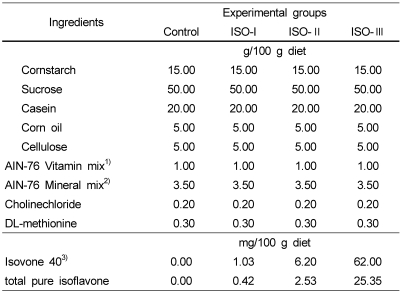
Table 1
The composition of the experimental diets
nrp-2-218-i001

1)AIN-76 vitamin mix (g/kg mix) : thiamin HCl 0.6, riboflavin 0.6, pyridoxine HCl 0.7, nicotinic acid 3, D-calcium pantothenate 1.6, folic acid 0.2, cyanocobalamin 0.001, retinyl palmitate 0.8 (500,000 IU/g), DL-α-tocopherol acetate 20 (2501 IU/g), cholecalciferol 0.0025, menaquinone 0.005, sucrose to make 1 kg

2)AIN-76 mineral mix (g/kg mix) : calcium phosphate dibasic 500, sodium chloride 74, potassium citrate monohydrate 220, potassium sulfate 52, magnesiumoxide 24, manganous carbohydrate 3.5, ferric citrate 6, zinc carbonate 1.6, cupric carbonate 0.3, potassium iodate 0.01, sodium selenite 0.01, chromium potassium sulfate 0.55, sucrose to make 1 kg

3)Isovone 40 (Product No. HF-2002-038, Bioland Inc. Seoul, Korea) contains 40.9% of total pure isoflavone which consists of 36.9% total aglycone type (22% daidzein, 11.7% glycitein, 6.39% genistein) and 4% total glycoside type

Table 2
HPLC conditions for the determination of plasma vitamin A and E
nrp-2-218-i002
Table 3
Effects of soybean isoflavones extract supplementation on plasma lipids concentrations and atherogenic index in STZ-induced diabetic rats
nrp-2-218-i003

ISO: soybean isoflavone extract, STZ: streptozotocin, TG: triglyceride, TC: total cholesterol, HDL: high density lipoprotein, C: cholesterol, AI=atherogenic index

1)Mean ± S.E.

2)Number of animals

3)AI=(TC-HDL-C)/HDL-C

Different capital superscripts in the same column indicate significant difference (p<0.05) among 5 groups by Duncan's multiple comparison test.

Different small superscripts in the same column indicate significant difference (p<0.05) among 4 diabetic groups by Duncan's multiple comparison test.

nsNot significantly different among 4 diabetic groups (p<0.05)

Table 4
Effects of soybean isoflavone extract supplementation on hepatic lipid concentrations in STZ-induced diabetic rats
nrp-2-218-i004

ISO: soybean isoflavone extract, STZ : streptozotocin

1)Mean ± S.E.

2)Number of animals

Different capital superscripts in the same column indicate significant difference (p<0.05) among 5 groups by Duncan's multiple comparison test.

Different small superscripts in the same column indicate significant difference (p<0.05) among 4 diabetic groups by Duncan's multiple comparison test.

NSNot significantly different among 5 groups (p<0.05)

nsNot significantly different among 4 diabetic groups (p<0.05)

Table 5
Effects of soybean isoflavone extract supplementation on the levels of TBA-reactive substances in plasma and urine of STZ-induced diabetic rats
nrp-2-218-i005

ISO: soybean isoflavone extract, STZ: streptozotocin

1)Mean ± S.E.

2)Number of animals

Different capital superscripts in the same column indicate significant difference (p<0.05) among 5 groups by Duncan's multiple comparison test.

Different small superscripts in the same column indicate significant difference (p<0.05) among 4 diabetic groups by Duncan's multiple comparison test.

NSNot significantly different among 5 groups (p<0.05)

nsNot significantly different among 4 diabetic groups (p<0.05)

Table 6
Effects of soybean isoflavone extract supplementation on the levels of TBA-reactive substances in liver, kidney and heart tissues of STZ-induced diabetic rats
nrp-2-218-i006

ISO: soybean isoflavone extract, TBA: thiobarbituricacid, STZ: streptozotocin

1)Mean ± S.E.

2)Number of animals

Different capital superscripts in the same column indicate significant difference (p<0.05) among 5 groups by Duncan's multiple comparison test.

Different small superscripts in the same column indicate significant difference (p<0.05) among 4 diabetic groups by Duncan's multiple comparison test.

NSNot significantly different among 5 groups (p<0.05)

nsNot significantly different among 4 diabetic groups (p<0.05)

Table 7
Effects of soybean isoflavone extract supplementation on hepatic antioxidant enzyme activities in STZ-induced diabetic rats
nrp-2-218-i007

ISO: soybean isoflavone extract, STZ: streptozotocin, GPx: glutathione peroxidase, SOD: superoxide dismutase

1)Mean ± S.E.

2)Number of animals

Different capital superscripts in the same column indicate significant difference (p<0.05) among 5 groups by Duncan's multiple comparison test.

Different small superscripts in the same column indicate significant difference (p<0.05) among 4 diabetic groups by Duncan's multiple comparison test.

NSNot significantly different among 5 groups (p<0.05)

nsNot significantly different among 4 diabetic groups (p<0.05)

Table 8
Effects of soybean isoflavone extract supplementation on plasma levels of vitamin A and E in STZ-induced diabetic rats
nrp-2-218-i008

ISO: soybean isoflavone extract, STZ: streptozotocin

1)Mean ± S.E.

2)Number of animals

Different capital superscripts in the same column indicate significant difference (p<0.05) among 5 groups by Duncan's multiple comparison test.

Different small superscripts in the same column indicate significant difference (p<0.05) among 4 diabetic groups by Duncan's multiple comparison test.

NSNot significantly different among 5 groups (p<0.05)

nsNot significantly different among 4 diabetic groups (p<0.05)

TOOLS
Similar articles