

 PDF
PDF ePub
ePub Citation
 Print
Print


INTRODUCTION
Vascular dementia (VaD), which impairs cognitive abilities, is the second most common type of dementia, accounting for approximately 15–20% of all dementia patients [12]. VaD results from inadequate blood supply to the brain caused by occlusion or rupture of cerebral arteries [1]. Because of its diverse etiologies, VaD can be classified into various subtypes, including hypoperfusion dementia, subcortical VaD, multi-infarct dementia, and strategic infarct dementia [23].
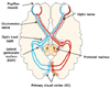
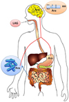
Several animal models that mimic VaD have been developed to study VaD [14]. Of these, the bilateral common carotid artery occlusion (BCCAO) model in rats is the most commonly used. In this model, white matter lesions manifest axonal and myelin injury, vacuolization, and glial cell activation [14]. Thus, the rat BCCAO model mimics hypoperfusion dementia and subcortical VaD in that white matter injury is consistently observed in both subtypes [34]. In hypoperfusion dementia, one etiology is caused by partial or complete blockage of the carotid arteries [34]. In humans, carotid artery stenosis or occlusion was found to be associated with white matter injury [3], which is observed in the regions including the corpus callosum (cc) [56] and optic nerve [7], a bundle of which forms the optic tract (opt) in the visual pathway (Fig. 1). In the rat BCCAO model, optic nerve injury correlates with loss of the pupillary light reflex (PLR), a reflex that controls the pupil diameter in response to light intensity [8]. Conversely, in the subcortical VaD, the most common form of VaD, one etiology is caused by partial blockage of small vessels, which also leads to white matter injury [39].
As there are no treatments approved by the FDA, it is imperative to develop agents to treat VaD [12]. We previously showed that hot water extract of ground wheat [10] and wheat bran (WBE) [11] reduced brain injury in a rat BCCAO model, demonstrating that processed wheat can be developed as a functional food for prevention of VaD. We also found that arabinoxylan, a cell wall polysaccharide consisting of arabinose and xylose, and arabinose itself were the active components responsible for the efficacy [10]. These findings suggest that any polysaccharides containing arabinose might show similar efficacies. Of the cell wall polysaccharides constituting wheat cell walls, arabinogalactan-peptide is also a candidate in addition to arabinoxylan, because arabinogalactan-peptide is composed of 92% polysaccharide arabinogalactan and 8% peptides [1213], and the arabinogalactan domain in arabinogalactan-peptide consists of arabinose and galactose [12]. To test the hypothesis that arabinogalactan-peptide in wheat is also an active component, we selected commercially available LAG as a model polysaccharide representing arabinogalactan-peptide. We then investigated whether larch arabinogalactan (LAG) supplementation could reduce white matter injury and improve PLR in a rat BCCAO model.
MATERIALS AND METHODS
Materials
LAG (82 wt % galactose and 13 wt % arabinose, along with several unidentified, minor compounds) was purchased from Sigma-Aldrich (St. Louis, MO, USA).
Animals
Eight-week-old male Sprague Dawley (SD) rats (280–300 g) were purchased from Samtaco Inc. (Osan, Gyeonggi-do, Republic of Korea). Animals were housed with diet and water available ad libitum before the start of the experiment, and diurnal lighting conditions and temperature-controlled environments were maintained throughout the experiment [1011]. Experiments were conducted according to the guidelines for animal care and protocols on laboratory animal use approved by the Institutional Animal Care and Research Advisory Committee of Catholic University, Daegu, Republic of Korea (Approval No. 2013-0820-CU-AEC-10-Y).
Diet preparation
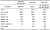
Diets containing LAG were prepared as previously described [14]. For the LAG diet (1 kg), a mixture of LAG (2 g) and corn starch (48 g) was added to 950 g of a modified AIN-93G diet [15] purchased from Unifaith Inc. (Seoul, Republic of Korea) (Table 1). For the basal diet (1 kg), 50 g of corn starch was added to 950 g of the modified AIN-93G diet.
Diet administration [11]
Rats were randomly divided into three groups: (1) sham (n = 6), (2) control (n = 6), and (3) LAG-treated group (100 mg/kg/day) (n = 6). The 100 mg/kg/day dose of LAG was selected based on our previous findings that WBE consisting of 2.4 wt% arabinose significantly reduced white matter injury at a dose of 400 mg/kg/day in the same rat BCCAO model [10]. Therefore, the arabinose dose that is an active component in the same rat BCCAO model [11] is equivalent to approximately 10 mg/kg/day in 400 mg/kg/day WBE. To match 10 mg/kg/day arabinose, we selected a dose of LAG at 100 mg/kg/day because LAG consists of approximately 10% arabinose. In the LAG-treated group (100 mg/kg/day), rats received LAG diet for 5 days before and 4 weeks after bilateral common carotid artery ligation. The amount of LAG diet fed to each rat at the beginning of the experiment was 15 g to give a LAG dose of 100 mg/kg/day based on the assumption that the rats weighed 300 g (Table 1). The amount of LAG diet fed to the rats then increased in proportion to the rise in rat weight because the rats gained weight as the experiment progressed. In the sham and control groups, rats received the basal diet only. Once the rats consumed all of the LAG or basal diet, more basal diet was provided ad libitum.
Bilateral common carotid arteries occlusion (BCAAO)
The rat bilateral common carotid arteries were ligated to mimic vascular dementia as previously described [1011]. The male SD rats were anesthetized through isoflurane inhalation (Hana Pharmaceutical Inc., Seoul, Republic of Korea) throughout the surgical procedure. The bilateral common carotid arteries in the control and LAG-treated groups were exposed and ligated permanently with silk sutures. The rats in the sham group underwent the same experimental procedure without ligation. Four weeks after BCCAO, the PLR was examined, after which the rats were anesthetized again through isoflurane inhalation, and their brains were harvested for further study.
Pupillary light reflex (PLR) assessment
The PLR, which reflects the injury in the visual pathway, was assessed as previously described [11]. Briefly, the PLR of each rat was examined before and after the BCCAO experiments. In the PLR examination, each rat was first adapted to darkness for at least 5 min. One eye was then exposed to a beam of light from a clinical pen light (3M Korea, Ltd., Seoul, Republic of Korea). Then, after the rat was re-adapted to darkness for 1 min, the other eye was also exposed to the light. Loss of PLR in each eye was defined as failure of the pupil to constrict after exposure to a beam of light for 10 seconds.
Luxol fast blue staining
Luxol fast blue staining was performed to assess white matter injury as previously described [11]. Briefly, the harvested brain was cut into slices including the cc, opt, and internal capsule. After formalin fixation and paraffin-embedding, 5 µm thick sections were cut from the slices. Following deparaffinization, three sections obtained from each rat brain were stained with Luxol fast blue. The severity of white matter injury was graded by Lee (the corresponding author), who was blinded to the experimental conditions performed by Lim (the first author), as normal (Grade 0), disarrangement of nerve fibers (Grade 1), formation of marked vacuoles (Grade 2), and loss of myelinated fibers (Grade 3) [16].
Immunohistochemical staining
Immunohistochemical staining was conducted to assess astrocytic and microglial activation as previously described [11]. Briefly, the sections were treated by boiling in a microwave to retrieve antigens. Next, the sections were treated with hydrogen peroxide (3%) in methyl alcohol to block endogenous peroxidases and incubated in phosphate-buffered saline containing 1% bovine serum albumin, 0.1% Triton X-100, and 5% normal goat serum to block nonspecific binding. The sections were then incubated with primary antibodies against glial fibrillary acidic protein (GFAP, 1:100, BD PharMingen, San Diego, CA, USA) or ionized calcium binding adaptor molecule 1 (Iba1, 1:200, Wako, Osaka, Japan). After washing, the sections were treated with biotinylated secondary antibody (1:200, Vector Laboratories, Burlingame, CA, USA) against GFAP and Iba1. Sections were then incubated with avidin-biotin peroxidase (Elite Vectastain ABC kit, Vector Laboratories, Burlingame, CA, USA), after which they were visualized with diaminobenzidine (Roche, Mannheim, Germany). Immunostained sections were subsequently captured using an Olympus microscope (200×) (Olympus Corporation, Tokyo, Japan), after which quantitative analysis was performed using the ImageJ software (NIH, v1.47). The area covered by GFAP-positive astrocytes and Iba1 positive microglia was computed as a percentage of the total area. To assess the GFAP- and Iba1-positive levels, the relative values for the control and LAG-treated groups were calculated by setting those for the sham group at 100%.
Statistical analysis
Values were expressed as the means ± SEM. Statistical analysis for multiple comparisons consisted of one-way ANOVA followed by the Dunnett post hoc test, which were conducted using the SPSS software (IBM SPSS Statistics; version 19, Armonk, NY, USA). For all data, Shapiro-Wilk and Levene statistics were applied a priori to verify normality and homogeneity of variances, respectively. A P < 0.05 was considered to indicate statistical significance.
RESULTS
LAG Supplementation reduces white matter injury in the corpus callosum and optic tract
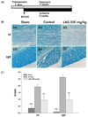
To examine whether LAG supplementation can reduce white matter injury, LAG (100 mg/kg/day) was supplemented for 5 days before the rats underwent BCCAO surgery, and supplementation continued for another 4 weeks after surgery (Fig. 2A). Of the regions in white matter, we focused on myelin injury incurred in the cc and opt because they are more vulnerable to hypoperfusion generated through BCCAO than other regions, including the internal capsule, because of the levels of blood supply to the regions [11].
To assess the white matter injury that manifests disarrangement and disappearance of nerve fibers consisting of axons and the myelin sheath, Luxol fast blue staining was used to observe changes in myelin structure. In the control group, white matter injury that manifests white matter rarefaction accompanying vacuolization was observed in both the cc and opt (Fig. 2B). In contrast, in the LAG-treated group, this white matter injury was reduced in both the cc and opt, compared with the control group (Fig. 2B). The severity of white matter injury was then quantitatively assessed using a grading system (0–3) (Fig. 2C). The grading scores were significantly reduced in the LAG-treated group by 51% (0.94 ± 0.29 vs. 1.92 ± 0.14, P < 0.01) in the cc, and by 63.5% (0.96 ± 0.35 vs. 2.63 ± 0.21, P < 0.01) in the opt, respectively, compared with the control group (Fig. 2C). These findings indicate that LAG supplementation protects against white matter injury attributed to hypoperfusion in the rat BCCAO model.
LAG supplementation improves PLR
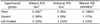
Because degeneration of the optic nerve that is connected to the opt causes PLR loss in a BCCAO model [811] (Fig. 1), we investigated whether LAG supplementation that reduced the opt injury could improve PLR. In the sham group, all six (100%) rats exhibited bilateral PLR, showing that all rats maintained normal PLR function in the sham group, as expected (Table 2). However, in the control group, five (83%) rats suffered bilateral PLR loss, while only one (17%) exhibited bilateral PLR exhibition. These findings indicate that five rats lost PLR function in both eyes, while one rat maintained normal PLR function in the control group. Conversely, two (33%) and two (33%) rats suffered bilateral and unilateral PLR loss, respectively, in the LAG-treated group, while two (33%) exhibited bilateral PLR. These findings indicate that two rats lost PLR function in both eyes, two lost PLR function in one eye, and two maintained normal PLR function in the LAG-treated group. Therefore, LAG supplementation improved PLR function by shifting two rats from bilateral PLR loss to unilateral PLR loss, and by shifting one rat from bilateral PLR loss to bilateral PLR exhibition. These findings suggest that LAG intake can improve the PLR.
LAG supplementation inhibits astrocytic activation in the cc
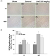
As chronic hypo-perfusion generated in a rat BCCAO model triggers astrocytic activation accompanying hypertrophy and proliferation of astrocytes [1617], we investigated whether LAG supplementation can modulate astrocytic activation through immunohistochemical staining of GFAP, a biomarker specific for astrocytes in the brain [18]. In the control group, increases in the number of GFAP-positive astrocytes were observed in both the cc and opt, compared with the sham group. In contrast, in the LAG-treated group, decreases in the number of GFAP-positive astrocytes were observed in the cc compared with the sham group (Fig. 3A). The degree of astrocytic activation was then quantitatively assessed through measurement of the area covered by GFAP-positive astrocytes (Fig. 3B). The relative total area in the LAG-treated group was significantly attenuated in the cc compared with the control group (106.3 ± 5.8 vs. 148.8 ± 13.1, P < 0.05), whereas the relative total area tended to be reduced in the opt compared with the control group (171.1 ± 25.3 vs. 209.9 ± 31.2, P > 0.05) (Fig. 3B). These findings showed that LAG intake can inhibit astrocytic activation.
LAG supplementation inhibits microglial activation in the opt
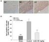
Because chronic hypo-perfusion generated in a rat BCCAO model also triggers microglial activation accompanying morphological transformation and proliferation of microglia [1617], we also investigated whether LAG supplementation can modulate microglial activation through immunohistochemical staining of Iba1, a biomarker specific for microglia in the brain [19]. In the control group, increases in the number of Iba1-positive microglia were observed in the opt compared with the sham group. In contrast, in the LAG-treated group, decreases in the number of Iba1-positive microglia were observed in the opt compared with the sham group (Fig. 4A). The degree of microglial activation was then quantitatively assessed through measurement of the area covered by Iba1-positive microglia (Fig. 4B). The relative total area in the LAG-treated group was significantly attenuated in the opt compared with the control group (186.5 ± 31.1 vs. 468.0 ± 70.7, P < 0.01) (Fig. 4B). These findings showed that LAG intake can inhibit microglial activation.
DISCUSSION
In this study, we showed that LAG supplementation reduced white matter injury in the cc and opt, bundles of myelinated axons, in a rat BCCAO model. This protection was associated with inhibition of astrocytic and microglial activation in the cc and opt, respectively. Additionally, LAG intake improved PLR. Similar observations were also made for WBE [10].
Previously, damage to the cc and opt were shown to be accompanied by activation of astrocytes and microglia in rat BCCAO models [20] and human VaD patients [2122]. Activation of nuclear factor kappa-B (NF-κB), an inflammatory cytokine, in astrocytes contributes to neuronal degeneration [23]. Inhibition of expression of NF-κB by astrocytes under conditions of astrocytic activation in a mouse model of bilateral common carotid artery stenosis ameliorated myelin injury and astrocytic activation, while preserving memory function [24]. These results suggest that astrocytic activation exerts deleterious effects on myelin injury. In addition, hot water extract of ground wheat [10] and WBE [11] also reduced myelin injury and astrocytic and microglial activation, and improved visuospatial memory [10] in a rat BCCAO model. These findings indicate that reduction of glial activation contributes to maintenance of white matter structure in VaD. Therefore, reduction of glial activation through LAG intake may also contribute to the reduced damage in the cc and opt observed in the present study.
PLR is mediated from the retina through the optic nerve, opt, pretectal nucleus, and oculomotor nerve to the pupillae muscle [25] (Fig. 1). Because LAG intake improved PLR in the model, it should protect axons involved in the PLR pathways. Therefore, injury reduction in the opt by LAG intake should help improve the PLR. In addition, it is highly likely that LAG intake also protected the retinal and optic nerves. This conclusion is supported by the findings that PLR loss was accompanied by retinal and optic nerve degeneration in rat BCCAO models [826]. Similar to those observed in rat BCCAO models, injury to the retinal nerve [27] and optic nerve [7] was also observed in human carotid artery occlusion, which is associated with dementia [28]. Moreover, patients with cerebral autosomal dominant arteriopathy with subcortical infarcts and leukoencephalopathy (CADASIL), a model of pure small-vessels disease that progresses to subcortical VaD, display damage to the retinal [2930] and optic nerves [2931]. Therefore, LAG intake may contribute to improved cognition in VaD patients through protection of the opt.
The cc is the principal interhemispheric commissure, and its integrity is crucial for cognition [32], including visual memory [33]. As expected, the cc in subcortical VaD patients shows axonal injury and astrocytic activation assessed through Luxol fast blue staining and GFAP immunohistochemical staining, respectively [34]. In addition, the cc in the subcortical VaD also shows reduced integrity when assessed through magnetic resonance imaging [3536]. Therefore, LAG intake may contribute to improved cognition in VaD patients through protection of the cc. Taken together, the findings from this study indicate that prophylactic LAG intake can prevent VaD by protecting myelinated axons. Consistent with these findings, intake of a polysaccharides blend, including arabinogalactan as a major constituent, improved memory for healthy middle-aged adults [37].
In our previous study, we showed that supplementation of hot water extract of ground wheat, arabinoxylan, and arabinose reduced white matter injury in a rat BCCAO model [10]. In this study, we showed that supplementation of the rat diet with LAG as a model polysaccharide for arabinogalactan-peptide in wheat also reduced white matter injury in the same BCCAO model. These results suggest that arabinogalactan-peptide in wheat is also an active component. Moreover, the results support our hypothesis that any polysaccharides consisting of arabinose can also act as active components. A proposed pathway in which arabinose reduces white matter injury is shown in Fig. 5 [38]. When LAG is ingested, arabinose attached to LAG can be released from LAG through acid hydrolysis in the stomach [39]. However, not much arabinose would be released in the small intestine because LAG is not digested in the small intestine owing to a lack of endogenously secreted digestive enzymes [40]. Conversely, arabinose can also be released from LAG by microorganisms inhabiting the large intestine such as Bacteroides thetaiotamicron, which can cleave bonds linking the arabinose and galactan backbone by α-arabinofuranosidase expressed by the microorganisms [3841]. The assumption that arabinose can be released from LAG is supported by observations it was detected in the feces of mice fed a diet containing arabinogalactan [42].
LAG was approved by the United States Food and Drug Administration (FDA) as Generally Recognized As Safe (GRAS), and is currently used as a food additive to improve bowel health [43] and as an immune enhancer [4344]. In this study, we provided further evidence of the feasibility for development of LAG as a functional food for preventing VaD, for which there are currently no treatments approved by the FDA [12]. Furthermore, the dose used in this study (33 days at 100 mg/kg/day for rats) was confirmed to be located in a safe range for use as a functional food based on the following findings of previous studies. Repetitive intravenous injection of LAG to rats at 500 mg/kg/day for 90 days showed no evidence of toxicity [45]. In addition, healthy human subjects ingested up to 30 g LAG for 3 weeks, which is equivalent to approximately 3 g/kg/day LAG for rats according to FDA guidelines for dose conversion between animals and human [47], and it was well tolerated [46].
 XML Download