

 PDF
PDF ePub
ePub Citation
 Print
Print


INTRODUCTION
Macrophages play an important role in inflammatory responses through the release of various inflammatory factors such as nitric oxide (NO), prostaglandins, inducible nitric oxide synthase (iNOS), cyclooxygenase (COX)-2 and cytokines such as tumor necrosis factor (TNF)-α and interleukin (IL)-6 [12]. Especially, macrophages can be stimulated by pro-inflammatory cytokines and endotoxin such as lipopolysaccharide (LPS) and it causes inflammatory responses [1]. In addition, it has been demonstrated that during the inflammatory process, the abnormal release of NO and prostaglandin E2 (PGE2) is caused by the activation of iNOS and COX-2 known as two pleiotropic inflammatory mediators which regulates platelet aggregation, vascular permeability, and thrombus formation [2]. Also, LPS stimulation can regulate the expression of COX-2 and iNOS and the secretions of TNF-α, IL-1β, and IL-6 known as the major pro-inflammatory cytokines by activating the nuclear factor-κB (NF-κB) signaling pathway as well as mitogen-activated protein kinase (MAPK) known as its upstream molecule [23]. Therefore, inhibition of these inflammatory mediators and cytokines might be an useful approach for the treatment of inflammatory diseases.
Marine environments are considered an invaluable treasure trove of bioactive compounds since they have large chemical and biological variations [4]. Among the studied marine organisms, brown seaweeds have numerous compounds with a broad spectrum of bioactivities, including anti-inflammatory, antioxidant, anti-microbial, and neuroprotective effects [5]. Recent in vitro and in vivo studies have shown that polysaccharides isolated from brown seaweeds have excellent therapeutic potential against inflammatory reactions [6]. The isolation of bioactive compounds from seaweed depends heavily on the extraction conditions. However, a large amount of cell wall polysaccharides in these seaweeds acts as a physical barrier that limits the use of conventional chemical and mechanical extraction methods. Therefore, enzyme-assisted extraction methods (EEMs) that can digest cell walls are often used to isolate bioactive compounds from seaweed [7]. These enzymatic extracts show increased food safety and water-solubility compared to organic solvent extracts, which is an added advantage of using EEMs over solvent extraction [8].
Sargassum horneri (S. horneri) is an edible brown alga that grows in the subtidal zone as an annual species along the coasts of China, Japan, and South Korea. The thallus of S. horneri can reach a length of more than 7 m and a fresh weight of 3 kg [9]. Previous reports have indicated that S. horneri consisted of vitamins, amino acids, and polysaccharides has utilized as a food source as well as an ingredient in traditional medicine for thousands of years in Asian countries including South Korea, Japan, and China [1011]. According to previous reports, the sulfated polysaccharide and some extracts from S. horneri China strain exerted potent bioactive properties in in vitro conditions such as antioxidant and anticancer properties [12]. However, there is a little information on anti-inflammatory potentials of polysaccharides isolated from S. horneri China strain and its biological mechanism. Moreover, within last few years, a huge amount of S. horneri moved into the coasts of Jeju Island from the east coast of China and caused considerable damages in the fishing industry and the beauty of the shores around Jeju Island. At this point, utilization of S. horneri is important to resolve the problems in South Korea have caused from this seaweed. Therefore, in this study, we prepared four crude polysaccharides (CPs) from S. horneri China strain by using four food grade enzymes including amyloglucosidase (AMG), Celluclast, Viscozyme, and Alcalase and evaluated their anti-inflammatory activities and its biological mechanism.
MATERIALS AND METHODS
Sample collection
S. horneri China strain was collected during May to June 2015 along the coast of Jeju Island in South Korea. S. horneri China strain was identified by Jeju Biodiversity Research Institute (Jeju, Korea).
Chemicals
The murine macrophage cell line RAW 264.7 was purchased from the Korean Cell Line Bank (KCLB; Seoul, Korea). Dulbecco's modified Eagle's medium (DMEM), penicillin-streptomycin, and fetal bovine serum (FBS) were purchased from Gibco BRL (Burlington, ON, Canada). 3-(4, 5-Dimethylthiazol-2-yl)-2, 5-diphenyltetrazolium bromide (MTT), dimethyl sulfoxide (DMSO), fucoidan (Product No. F5631-1G), and FT-IR-grade KBR powder were purchased from Sigma-Aldrich (St. Louis, MO, USA). Three carbohydrate-degrading enzymes (AMG, Celluclast, and Viscozyme) and one protease (Alcalase) were donated by Novo Nordisk (Bagsvaerd, Denmark). Enzyme-linked immunosorbent assay (ELISA) kits for TNF-α, IL-1β and PGE2 were purchased from R&D Systems Inc. (Minneapolis, MN, USA). All other chemicals and reagents used in these experiments were of analytical grade.
Preparation of enzymatic digests from S. horneri
The freeze-dried S. horneri samples were homogenized with a grinder to obtain a fine powder. The enzymatic hydrolytic reactions were performed by using the methods of Heo et al. [8]. Briefly, 2 g of S. horneri powder was incubated in 100 mL of distilled water (DW) with 20 µl or 20 mg of AMG, Celluclast, Viscozyme, or Alcalase at the optimum temperature and pH as described by Heo et al. [8]. After 24 h, the samples were centrifuged at 2,774 × g for 10 min and the supernatants were filtered with Watman No.4 (GE Healthcare, Buckinghamshire, UK). Then, the supernatants were freeze-dried and used as enzymatic digests.
Separation of crude polysaccharides from the enzymatic digests
Crude polysaccharides were prepared from the four enzymatic extracts according to the method described by Shao et al. [13] with slight modifications. Briefly, the enzymatic digests were mixed with 95% ethanol to a final concentration of 66%. The mixture was stored at 4℃ for 24 h, and then the precipitates were collected by centrifugation at 10,000 × g for 20 min at 4℃. The precipitates were freeze-dried and used as crude polysaccharides (CPs). The freeze-dried CPs were dissolved in PBS for use in the experiments. The CPs separated from the four enzymatic digests with AMG, Celluclast, Viscozyme, and Alcalase were nominated as follow. AMGCP, crude polysaccharides from AMG enzymatic digestion; CCP, crude polysaccharides from Celluclast enzymatic digestion; VCP, crude polysaccharides from Viscozyme enzymatic digestion; AlCP, crude polysaccharides from Alcalase enzymatic digestion.
Analysis of chemical composition
Total polysaccharide content was measured by the phenolsulfuric acid method as described by method described by DuBois et al. [14] using glucose as the standard. Total phenol content was quantified by using a protocol described by Chandler and Dodds [15] using gallic acid as the standard. The protein content of all samples was quantified using the Pierce™ BCA Protein Assay Kit (Thermo Scientific, Rockford, IL, USA) using bovine serum albumin as the standard. Finally, the sulfate content of the CPs was checked by the BaCl2 gelatin method using Na2SO4 as the standard as described by Saito et al. [16] with slight modifications.
Fourier transform infrared (FT-IR) spectroscopy
The IR spectrums of the CPs were recorded using a FT-IR spectrometer (Nicolet™ 6700 FT-IR spectrometer; Madison, WI, USA). The CPs were homogenized with KBr powder and then pressed into pellets for FT-IR measurement in the frequency range of 500-4,000 cm−1.
Cell culture
RAW 264.7 cells were grown in DMEM supplemented with 10% heat-inactivated FBS, 1% streptomycin (100 µg/mL), and penicillin (100 unit/mL). RAW 264.7 cells were incubated under 5% CO2 at 37℃ (Sanyo MCO-18AIC CO2 Incubator; Moriguchi, Japan). Cultured cells from passage 4–6 were used for the experiments.
Cell viability assay
The cytotoxicity of the CPs to RAW 264.7 cells was evaluated via the colorimetric MTT assay as described by Mosmann [17], with slight modifications. Briefly, the cells (1 × 10⁵ cells/mL) were seeded in a 24-well plate and incubated for 24 h. Then, the cells were treated with CPs (50 µg/mL and 100 µg/mL) for 24 h. MTT reagent (200 µg/mL) was added to each well. After 3 h of incubation, the formasan crystals were dissolved in DMSO, and the amount of blue-black formazan was determined by measuring the absorbance at 540 nm. The optical density of the formazan generated in non-treated control cells was considered to represent 100% viability. The data are expressed as mean percentages of the viable cells versus the respective control.
Determination of NO production
To determine the effect of CPs on NO production in LPS-stimulated RAW 264.7 cells, we performed griess assay described by Leiro et al. [18] with slight modifications. Briefly, the cells (1 × 10⁵ cells/mL) were seeded in 24-well plates and incubated for 24 h. Then, the cells were treated with CPs (50 µg/mL and 100 µg/mL) for 1 h and stimulated with LPS (1 µg/mL) for 24h. Finally, equal amounts of the culture medium and Griess reagent were reacted in a 96-well plate for 10 min, and the absorbance was measured at 540 nm using an ELISA plate reader machine (BioTek Instruments, Inc., Winooski, USA). The optical density of NO produced in only LPS-treated cells was considered to represent 100%. The data are expressed as mean percentages of the NO production versus the NO production of only LPS-treated cells.
Determination of PGE2 production
Prostaglandins play an important role in regulating inflammatory responses [19]. Cultured RAW-264.7 cells (1 × 10⁵ cells/mL) were treated with measured concentrations of polysaccharides, and after a 1 h incubation, the cells were stimulated with LPS (1 µg/mL). After 24 h of incubation, the PGE2 concentration in the supernatant was quantified by using a competitive enzyme immunoassay kit, according to the manufacturer's instruction.
Measurement of TNF-α and IL-1β production
RAW 264.7 cells (1 × 10⁵ cells/well) were pretreated with CCP (25 µg/mL, 50 µg/mL, and 100 µg/mL) for 1 h and then incubated with LPS (1 µg/mL) for 24 h. The production levels of TNF-α and IL-1β in macrophage cytoplasm were quantified using ELISA kits, according to the manufacturer's instructions.
Western blot analysis
To determine the effect of CCP on the protein expression levels of iNOS, COX-2, NF-κB, and MAPK in LPS-stimulated RAW 264.7 cells, western blot analysis was performed. Briefly, RAW 264.7 cells (1 × 10⁵ cells/mL) were seeded in 6-well plates and incubated for 24 h. The cells were treated with CCP (25 µg/mL, 50 µg/mL and 100 µg/mL) for 1 h. And then, the cells were stimulated with LPS (1 µg/mL) for 10 min or 20 min and 24 h. Nucleic and cytosolic proteins were extracted from the cells with the NE-PER® Nuclear and Cytoplasmic extraction kit (Thermo scientific, Rockford, USA).
After separation on a 10% SDS-polyacrylamide gel under denaturing conditions, the cytoplasmic proteins (40 µg) were electrotransferred onto a nitrocellulose membrane. After blocking with 5% nonfat milk for 1-h, the blots were separately incubated with the following primary antibodies: rabbit polyclonal antibodies (iNOS, p44/42 (ERK, extracellular signal-regulated kinase), phosphorylated p44/42 (ERK), p38, phosphorylated p38, NF-κB p65, NF-κB p50, and nucleolin), and mouse monoclonal antibodies (COX-2, and β-actin) (Cell Signaling Technology, Beverly, MA, USA) for 1-h. The blots were washed twice with Tween 20/Tris-buffered saline (TTBS) and then incubated with HRP-conjugated anti-mouse or anti-rabbit IgG for 45-min. Antibody binding was visualized by using enhanced chemiluminescence (ECL) reagents (Amersham, Arlington Heights, IL, USA). The basal levels of the each proteins were normalized by analyzing the level of β-actin or nucleolin protein by using ImageJ program.
Statistical analysis
All the data were expressed as the mean ± standard of three determinations. The collected data were analyzed by analysis of variance using the SPSS v20 statistical analysis package. The mean values of each experiment were compared using one-way analysis of variance. Duncan's multiple range test (DMRT) was used to determine mean separation. A P-value < 0.05 was considered to be statistically significant.
RESULTS
Extraction yields and general components.
First of all, we prepared the four kinds of enzymatic extracts from S. honeri and checked their extraction yields. The results showed the extraction yields of enzymatic digests (AMG digest; 16.00 ± 0.50%, Celluclast digest; 20.17 ± 0.76%, Viscozyme digest; 21.0 ± 1.00%, and Alcalase digest; 22.17 ± 0.76%) were significantly increased, compared to that of the DW extract (9.50 ± 0.87%).
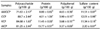
Next, we isolated crude polysaccharides from the enzymatic digests (CPs) and analyzed their isolation yields and proximate composition. Among the CPs, CCP led to the highest isolation yield (88.7%), compared to the others (AMGCP, VCP, and AlCP). As indicated in Table 1, the four kinds of CPs contained the major polysaccharide contents and minor protein and polyphenolic contents. Especially, CCP showed the highest polysaccharide and sulfate content (88.7 ± 2.44% and 12.01 ± 0.98%, respectively). From these results, we indicate that the isolated CCPs might be sulfated polysaccharides due to its plentiful polysaccharide and sulfate group contents.
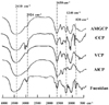
FT-IR spectrums of CPs
FT-IR spectroscopy is a useful analytical approach to identify the vibrations between the different atoms in molecules. The spectra obtained between 400-4,000 cm−1 can be used to analyze the structural features of polysaccharides, including glucosidic bonds and functional groups [2021]. In the present study, the FT-IR spectra of the four CPs were compared to that of a fucoidan purchased from Sigma-Aldrich. Interestingly, CCP had an IR spectrum similar to that of the commercial fucoidan (Fig. 1). In contrast, AlCP showed a completely different pattern than that of the other CPs and the commercial fucoidan. It has been reported that strong absorption at 840 cm−1 (a sulfate group at axial C-4) and shoulder absorption at 820 cm−1 (a sulfate group at C-2) are unique to native fucoidans. In addition, absorption at 1,240 cm−1 represents the sulfate ester groups found in commercial fucoidan [222324]. Moreover, the bands developed at 1,646-1,652 cm−1 were due to the bending vibrations of HOH and the broad bands centered at 2920-2940 cm−1 were assigned to C-H stretching vibrations. In addition, the bands in the region between 3,400-3,410 cm−1 are developed due to the stretching vibration of O-H which indicated hydroxyl groups existed in the fucoidan [2526].
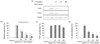
CCP showed the highest inhibitory effect on the NO production in LPS-stimulated RAW 264.7 cells without cytotoxicity
Cell viability experiments showed no cytotoxicity in RAW 264.7 cells for any of the CPs, with more than 90% viability (Fig. 2A). In addition, 100 µg/mL of AMGCP, CCP, VCP, and AlCP inhibited the NO production of RAW 264.7 cells induced by LPS stimulation (Fig. 2B). In particular, among the CPs, CCP had the highest inhibitory effect on the NO production in LPS-stimulated RAW 264.7 cells, with an IC50 value of 95.7 µg/mL and it was dose-dependent. Our results indicated that CCP has the inhibitory effect on the NO production in LPS-stimulated RAW 264.7 cells, without cytotoxicity. Therefore, CCP was used for all subsequent experiments.
CCP dose-dependently reduced the secretion of PGE2 from LPS-stimulated RAW 264.7 cells
Next, we evaluated effect of CCP on PGE2 production in LPS-stimulated RAW 264.7 cells. As shown in Fig. 2C, the LPS stimulation induced the PGE2 production in RAW 264.7 cells, whereas it was dose-dependently inhibited by the treatment of CCP.
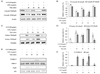
CCP reduced the expression levels of iNOS and COX-2 proteins in LPS-stimulated RAW 264.7 cells
The suppressive effect of CCP on the protein expression of iNOS and COX-2 was measured by western blot analysis. As shown in Fig. 3A and 3B, CCP dose-dependently down-regulated the expression levels of iNOS and COX-2 in LPS-stimulated RAW 264.7 cells, compared to the levels in the only LPS-treated cells.
CCP reduced the secretion of pro-inflammatory cytokines from LPS-stimulated RAW 264.7 cells
It is a well-established fact that the overproduction of pro-inflammatory cytokines, including TNF-α and IL-1β, has an important role in the pathogenesis of various inflammatory diseases [272829]. Therefore, compounds with inhibitory properties against TNF-α and IL-1β are considered anti-inflammatory agents. To examine the potential anti-inflammatory effects of CCP in LPS-stimulated RAW 264.7 cells, the levels of inflammatory cytokines, such as TNF-α and IL-1β, in culture supernatants were measured by ELISA. The results clearly demonstrated that the secretion of TNF-α and IL-1β was significantly increased by stimulation with LPS (Fig. 3C, 3D). Interestingly, the secretion of pro-inflammatory cytokines was inhibited by treatment with CCP in a dose-dependent manner.
CCP inhibited the NF-κB and MAPK signaling in LPS-stimulated RAW 264.7 cells
Using western blot analysis, we investigated the effect of CCP on NF-κB and MAPK activities in LPS-stimulated RAW 264.7 cells. As shown in Fig. 4A and 4B, the results demonstrated that the cytosolic NF-κB p50 and p65 protein levels were reduced by the stimulation of LPS in RAW 264.7 cells, compared to the non-treated control cells. However, the treatment of CCP (100 µg/mL) inhibited the reduction of cytosolic p50 and p65 protein levels in LPS-stimulated RAW 264.7 cells. CCP also inhibited the translocation of p50 and p65 to the nucleus compared to the only LPS-stimulated RAW 264.7 cells (Fig. 4C and 4D). In further study, we examined the effect of CCP on MAPK signal pathway known as an upstream molecule. Interestingly, CCP down-regulated the phosphorylation of p38 and ERK increased by LPS stimulation in RAW 264.7 cells (Fig. 4E and 4F). From these results, we suggest that CCP has anti-inflammatory effects via inhibiting the activation of MAPK and NF-κB signaling by down-regulating the phosphorylation of p38 and ERK and the translocation of NF-κB p50 and p65 into nucleus.
DISCUSSION
EEM is a popular green extraction method due to the increased extraction efficiency compared to water extraction and nontoxic nature compared to the organic solvent extraction methods. Thus, compounds from EEM are cheaper and safer to use as material in functional foods, cosmeceuticals or nutraceuticals [7]. According to the extraction data, we also observed that EEM techniques significantly increased the extraction efficiency without alerting cell viability and showed better NO inhibition in LPS-stimulated RAW 264.7 cells in tested concentrations compared to water extraction (data not shown).
Numerous anti-inflammatory polysaccharides have been isolated from marine brown algae and their role as anti-inflammatory agents has been well documented [30]. Fucoidan is a natural sulfated polysaccharide found in brown algae. Recently, a number of studies demonstrated that the anti-inflammatory activity of fucoidan occurred by suppressing the activation of NF-κB and MAPK pathways [3132]. However, the anti-inflammatory potential of sulfate-rich polysaccharides separated from S. horneri via enzymatic digestion has not yet been reported. Therefore, in the present study, we evaluated the anti-inflammatory activities of sulfate-rich CPs separated from the enzymatic digests of S. horneri on LPS-stimulated RAW 264.7 cells. Interestingly, the crude sulfated polysaccharides separated from the enzymatic digests exhibited the higher NO inhibitory activities than the CP isolated from DW extract. Based on these results, our results indicate that the enzyme extraction technique improved the extraction yields and the polysaccharide contents as well as the inhibition of NO production in LPS-stimulated macrophages. Our results were also similar to the suggestions of Wijesinghe et al. [7] that CP obtained from enzymatic digestions had better performance and high polysaccharide yield compared to the CPs obtained from DW extract.
During inflammatory responses, the expression of iNOS and COX-2 in macrophages leads to the production of NO and PGE2. Additionally, NO and PGE2 produced during inflammation can be cytotoxic to the host cells, and prolonged production of NO and PGE2 in inflammatory cells can cause a range of inflammatory diseases and cancers [3334]. Moreover, recent studies have shown the exposure of macrophages into inflammatory stimulant can cause significant up-regulation of pro-inflammatory cytokines such as IL-1β and TNF-α [35]. Finally, the excessive production of pro-inflammatory cytokines by activated macrophages play an important role in pathogenesis inflammatory diseases such as rheumatoid arthritis, Alzheimer's disease, and Parkinson's disease as well as inflammatory mediators [36]. Therefore, reducing levels of these mediators might be an effective strategy for treat inflammatory diseases [37]. Indeed, previous studies have demonstrated that sulfated polysaccharides including fucoidan derived from E. cava and Fucus vesiculosus inhibited the expression of inflammatory mediators such as iNOS and COX-2 in activated RAW 264.7 cells and BV2 microglia [31]. In this study, we revealed that CCP decreased the NO and PGE2 productions via reducing the expression levels of inflammatory mediators such as iNOS and COX2 as well as the production of inflammatory cytokines such as TNF-α and IL-1β. These results suggest that the inhibitory effect of CCP on the inflammatory mediators and cytokines is one of the mechanisms responsible for its anti-inflammatory activities and CCP might have potential to develop as a functional food ingredient or as a therapeutic agent to treat inflammatory diseases.
Normally, LPS stimulation leads to inflammation via activation of the NF-κB and MAPK signaling pathway [3138]. The NF-κB transcription factor family comprises five members, namely c-Rel, RelB, p50, p52, and p65. NF-κB is an important transcription factor involved in inflammatory responses. NF-κB is normally sequestered in the cytosol and is activated by inflammatory stimulants, such as LPS [31]. After stimulation of macrophages, the cytosolic NF-κB proteins are transported into the nucleus where they induce the transcription of pro-inflammatory genes, including iNOS and COX-2, as well as the genes encoding pro-inflammatory cytokines [31]. Furthermore, altered NF-κB activation is caused by deregulated, and often constitutive, NF-κB phosphorylation, which is major contributor of the pathogenesis of chronic inflammatory diseases and cancer. Therefore, inhibition of NF-κB signaling cascade related proteins found to be a promising target for treatment of inflammation associated diseases [39]. Interestingly, our results indicated that CCP suppressed the activation of NF-κB signaling via the inhibiting the phosphorylation of NF-κB as well as its translocation to the nucleus in the LPS-stimulated RAW 264.7 cells.
Exposure of RAW 264.7 cells into the LPS can activate the MAPK signaling cascade associated proteins such as ERK, p38, and JNK [40]. Moreover, a number of studies have reported that the MAPK signaling pathway regulates the activation of NF-κB [4142]. In addition, studies have demonstrated that the phosphorylation of p38 kinase and ERK1/2 are involved in the transcriptional regulation of pro-inflammatory cytokines such as IL-1β and TNF-α [4344]. Thus, inhibition of MAPK related protein expression is also considered a useful approach to treating inflammatory diseases as MAPK signaling cascade capable of regulating NF-κB and pro-inflammatory cytokine production [414245]. Our results also demonstrated that CCP inhibited the phosphorylation of ERK1/2 and p38. Recently, many researchers have reported that various polysaccharides, including fucoidan derived from seaweeds, exert anti-inflammatory effects by inhibiting activation of the NF-κB and MAPK signaling pathway in LPS-stimulated mouse macrophages [304647]. With these results, we demonstrated that the anti-inflammatory capacity of CCP, a sulfated polysaccharide via the inhibition of NF-κB signaling may be due to its inhibitory effect on the MAPK signaling.
In conclusion, this study suggests that CCP led to the anti-inflammatory effects by blocking the activation of the NF-κB and MAPK signaling pathway. Based on these findings, we conclude that CCP is a potential candidate for the formulation of a functional food ingredient or/and drug to treat inflammatory diseases.

 XML Download