Journal List > Nutr Res Pract > v.11(4) > 1050889

Park and Lee: Monocyte chemoattractant protein-1 polymorphism interaction with spirulina immunomodulatory effects in healthy Korean elderly: A 16 week, double-blind randomized clinical trial

Abstract

BACKGROUND/OBJECTIVES

Spirulina is a known a functional food related to lipid profiles, immune functions, and antioxidant capacity. Circulating monocyte chemoattractant protein-1 (MCP-1) level is associated with inflammation markers. Single nucleotide polymorphism in the MCP-1 promoter region -2518 have been identified and shown to affect gene transcription. Gene variation may also impact functional food supplementary effects. The current study investigated the interaction of MCP-1 -2518 polymorphism with spirulina supplements on anti-inflammatory capacity in Korean elderly.

SUBJECTS/METHODS

After genotyping, healthy elderly subjects (n = 78) were included in a randomized, double blind, and placebo controlled study. Baseline characteristic, body composition, and dietary intake were measured twice (baseline vs. week 16). For 16 weeks, subjects consumed 8 g either spirulina or placebo daily. Plasma MCP-1, interleukin (IL) -2, IL-6, tumor necrosis factor (TNF)-α, complement (C) 3, immunoglobulin (Ig) G, and Ig A concentrations and lymphocyte proliferation rate (LPR) were analyzed as inflammatory markers.

RESULTS

In the placebo group with A/A genotype, MCP-1 level was significantly increased, but the spirulina group with A/A genotype was unchanged. IL-2 was significantly increased only in subjects with spirulina supplementation. TNF-α was significantly reduced in subjects with the G carrier. C3 was significantly increased in the placebo group, particularly when A/A increased more than G, but not when spirulina was ingested. LPR was significantly different only in subjects with A/A genotype; there was a significant increase in phytohemagglutinin and lipopolysaccharide induced LPR in the spirulina group.

CONCLUSION

In healthy Korean elderly, spirulina supplementation may influence different inflammatory markers by the MCP-1 genotype. These results may be useful for customized dietary guidelines to improve immune function in Koreans.

INTRODUCTION

Chemokines (CCs) play a variety of roles in intercellular signaling, particularly in the recruitment of leukocytes to inflammation sites [1]. Monocyte chemotactic protein-1 (MCP-1), also referred to as chemokine ligand 2 (CCL2), promotes transmigration and emigration of circulating monocytes into tissues, and exerts various effect on monocytes, including the induction of superoxide anions and expression of various proinflammatory genes [2345]. MCP-1 levels are elevated in the plasma of older persons in the apparent absence of cardiovascular disease [6]. Single nucleotide polymorphism (SNP) in the MCP-1 promoter region has been shown to affect gene transcription [7]. The -2518 G/A polymorphism in the MCP-1 is associated with inflammatory related diseases, including rheumatoid arthritis, and diabetic retinopathy [89]. It has also been reported that the G/G genotype of -2518 MCP-1 was associated with susceptibility to cardiovascular disease (CVD) and was identified as a genetic risk factor for severe coronary artery disease [10].
Dietary factors may influence gene expression [111213]. One case control study [11] reported a nutrigenetic interaction between dietary vitamin E and thioredoxin and catechol-Omethyltransferase gene polymorphisms on waist circumference. Dietary factors may also alter the risk of inflammatory disease. Many studies have reported that dietary factors such as fish oil, broccoli, and probiotics were associated with anti-inflammatory effects [141516]. However, clinical trial outcomes are mixed, which may be due to an interaction between genetic background and dietary intake. Gene variation may also influence dietary requirements, e.g. the new field of nutritional genomics, and raises the possibility of individual nutritional management for optimal health and disease prevention on the basis of an individual genome. Multiple studies have already shown significant gene × diet interactions. Ortega-Azorín et al. [17] reported that the association of fat mass and obesity associated (FTO) gene polymorphism with type 2 diabetes depends on diet, and that adherence to the Mediterranean diet counteracts the genetic predisposition. Serrano et al. [18] showed that metabolic response to 25-hydroxyvitamin D and soybean supplementation was dependent on vitamin D receptor GG genotype. Giacconi et al. [19] showed the effect of zinc transporter protein 2 Gln/Arg/Leu polymorphism on proinflammatory mediators and zinc homeostasis in the elderly population. Park et al. [20] showed gene-nutrient interactions between transient receptor potential vanilloid-1 variants re161364 and preference for oily taste, intake of oily foods, and fat intake. Thus supplements may have different effects depending on genetic predisposition. However, little is known about the association of MCP-1 polymorphism and dietary factors as nutrients or supplements in the Korean population.
Spirulina is a cyanobacterial blue-green algae, commonly used as a dietary supplement [21]. Several studies have demonstrated spirulina's beneficial effects and therapeutic functions [2223]. Other studies have shown that spirulina has beneficial effects on the treatment of malnutrition and other pathologies such as obesity, hypercholesterolemia, hypertension, and diabetes mellitus [242526]. Studies have reported spirulina was associated with modulation of the host immune system. Hayashi et al. [27] first published detailed studies on the immunomodulatory properties of dietary spirulina in mice. Dietary spirulina enhanced responses of phagocytic activity responses, interleukin (IL) -1b expression, and tumor necrosis factor (TNF)-α genes in carp [28]. Spirulina treated chicken macrophages showed increased spreading and vacuolization with minimal cytotoxicity [29]. In humans, spirulina increases natural killer cell cytotoxicity [30] and ameliorates immunosenescence in older subjects [31]. Spirulina supplementation produced a significant rise in plasma IL-2 level, and a significant reduction in IL-6 level in health elderly [32]. These results suggest that spirulina somehow stimulates the immune system.
From the perspective of personalized nutrition, the effect of spirulina on immune function is different depending on the individual's obesity [33]. No studies have investigated the effect of MCP-1 polymorphism on the effect of spirulina supplementation on immune function. Therefore, the aim of the current study was to explore interactions between spirulina supplementation and MCP-1 -2518 G/A polymorphism on immunity in Korean elderly.

SUBJECTS AND METHODS

Subjects and experimental design

Subjects were recruited through an advertisement in local newspapers (Dec 2005 to June 2006). The subjects were first interviewed by telephone for screening. Exclusion criteria were: current user of vitamin supplements, current drug-user for inflammatory disease (e.g. Crohn disease, rheumatoid arthritis), dyslipidemia, hypertension, concurrent or recent participant in any other intervention study.
The study protocol was approved by the Institutional Review Board of Ewha Women's University (ECT 109-02-01), and written informed consent was obtained from all study subjects. A randomized, controlled, double blinded, parallel group design study was performed for 78 healthy older adult subject (43 male, 35 female). Subjects consumed spirulina or placebo, 8 g per day, for 16 consecutive weeks. At the first visit of the study, blood was drawn after a minimum of 12 h fasting, defined as the baseline. Blood samples were taken again at the end of the study period. During the intervention period, subjects were asked to keep their usual diet, and prohibited to take any functional food or supplements. Both spirulina and placebo (100% starch) were provided by Earth Spirulina Biotech (Seoul, Korea). Spirulina and placebo were suppled every 2 weeks and compliance was confirmed by telephone twice a week.

Food consumption and anthropometric measures

The elderly subjects were interviewed individually to obtain data on food consumption and general characteristics. Food consumption was assessed using the 24 hour recall method. Food intake data was analyzed using CAN-pro 4.0 software [34].
Standing height was measured using an anthropometer (Seca 213, Seca Inc. Birmingham, UK). Body weight and composition [body fat (kg), body fat (%), and lean body mass (kg)] were measured using INBODY 2.0 (Biospace co, Seoul, Korea) with subjects wearing light clothing without shoes or socks.

Blood collection and analysis

Blood samples collected before and after supplementation from overnight were taken on the beginning day (pretest) and 16th week (posttest) of the study period. Fasting blood samples were obtained and sera were kept at 80℃ until measurement. Total cholesterol and triglyceride level were measured using an autoanalyzer (Ekachem DTSC module, Johnson & Johnson, New Brunswick, NJ, USA). HDL cholesterol level was determined by autoanalyzer after treating UC infranatant with phosphotungstic acid Mg. LDL cholesterol was calculated as described by Friedewald [35].
Plasma MCP-1, IL-2, IL-6, and TNF-α concentrations were determined by enzyme linked immunosorbent assay (ELISA) (Quantikine, R&D Systems Inc., Minneapolis, MN, USA) using an ELISA reader (Spectra Max 340, Molecular Devices, CA, USA). Radial immunodiffusion plates (Norpartigen, Behring Co., Marburg, Germany) were used to measure Ig and C3 level. Assays were performed as recommended by the manufactures.
The proliferation capacity of lymphocytes was analyzed by the Mosmann method [36]. Fresh blood samples treated with heparin were diluted in phosphate buffered saline (PBS) 1:1, and then cells were separated in 3 mL Ficoll-paque gradient (lymphocyte isolation solution, Histopaque 1077, Sigma, MO, USA) by centrifuge at 1,000 g for 30 min. The isolated peripheral blood mononuclear cells (PBMCs) were seeded at 2 × 10⁵ cell per well in a 96-well plate. PBMCs were treated with concanavalin A (ConA, Sigma, MO, USA, 0.5 µg/10 µl), phytohemagglutinin (PHA, ThermoFisher Scientific co., MA, USA, 0.5 µg/10 µl), and lipopolysaccharide (LPS, Sigma, MO, USA, 0.5 µg/10 µl), and then incubated for 68 h in a CO2 incubator. Viable cell count was estimated by 3-[4, 5-dimethylthizol-2-yl]-2,5-diphenyl tetrazoliumbromide (MTT, Sigma, MO, USA) assay. Peripheral blood lymphocytes proliferation rate (LPR) was estimated by the stimulation index, SI = absorbance of wells with mitogens / absorbance of wells without mitogen. All tests were run in triplicate.

DNA extraction and analysis of the MCP-1 -2518 G/A polymorphism

All subjects were genotyped for MCP-1 polymorphism (-2518 G/A) (dbSNP: rs1024611). DNA was extracted from whole blood (QIAamp DNA Blood Kit, Qiagen, Hilden, Germany), and a 649 bp segment of MCP-1 gene including polymorphism sites was amplified by polymerase chain reaction (forward primer: 5′-AGT CCA ACC AAG GTT TGT GC-3′, and reverse primer 5′-TCA TGC TTC GGG TTT TCT CT-3′). The presence of MCP-1 polymorphism was determined by restriction fragment length polymorphism (RFLP) assay using PvuII enzyme.

Statistical analysis

Statistical analysis was conducted using SAS Statistics version 9.4 (SAS Institute, NC, USA). Data are presented as mean ± standard error (SE) for placebo and spirulina groups by genotype. Paired t-test was used to analyze mean differences for all measured parameters between baseline and week 16. Repeated measure ANOVA was conducted to examine treatment effects.

RESULTS

Subjects' baseline characteristics

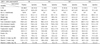
Seventy-eight healthy subjects were divided into placebo (p, n = 37) and spirulina groups (s, n = 41). The proportions of A/A, G/A, and G/G genotypes were 17% (n = 14, p/s = 5/9), 56% (n = 44, p/s = 19/25), and 25% (n = 20, p/s = 13/7), respectively. Selected baseline characteristics are summarized in Table 1. Average subject age was 66.0 years. There were no significant differences in age, BMI, lipid profile, and dietary intake between 2 groups within the same genotype. Three day dietary records showed no significant intergroup and intragroup differences in mean daily intakes of energy, protein, carbohydrates, total fat, vitamin A, vitamin C, and vitamin E.

Spirulina effects on plasma cytokine based on genotype

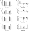
Plasma MCP-1 level did not differ significantly among the intervention group at baseline (placebo 80.9 pg/dL, spirulina 77.8 pg/dL) and after supplementation (placebo 77.2 pg/dL, spirulina 74.3 pg/dL) (Fig. 1A). When the plasma MCP-1 level of subjects was analyzed by genotype, MCP-1 level was significantly increased in the placebo group (P < 0.05) with AA genotype, whereas there was no change in the spirulina group (Fig. 1B). No significant difference in the IL-6 level was observed for the intervention group or genotype groups (Figs. 1C, 1D). Plasma TNF-α level was significantly decreased after spirulina supplementation for all subjects (Fig. 1E, P < 0.05), but this significance was not observed for genotype subgroups (Fig. 1F). IL-2 level was significantly increased in the spirulina group after supplementation (Fig. 1G), and the significant was observed in all genotypes (Fig. 1H).
Table 2 shows the cytokine changes after spirulina intervention between the A/A genotype (n = 14) and G/A + G/G MCP-1 genotypes (n = 64). There were significant differences in plasma MCP-1 levels for MCP-1 genotype and MCP-1 genotype × spirulina treatment (P < 0.01, P < 0.05), whereas there were no significant differences in IL-6 levels. The level of IL-2 increased in the spirulina group (P < 0.05) but not in the placebo group, irrespectively of MCP-1 genotype. Although TNF-α level was significantly decreased (P < 0.05) in both groups with G carriers, there was no significant difference by genotype after supplementation.

Spirulina effects on complement and Ig based on genotype

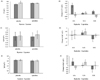
C3 level significantly increased after placebo supplementation for 16 weeks (Fig. 2A, P < 0.05). However, C3 level was not changed in all groups after spirulina supplementation, but only for subjects with A genotype (P < 0.05) (Fig. 2B). There were no significant differences of IgA and IgG levels in all genotypes after spirulina supplementation (Figs. 2D and 2F).
Table 3 shows C3 and Ig changes after placebo or spirulina supplementation for the A and G genotype subgroups. There was significant spirulina supplementation effect on C3 level (P < 0.01), and also MCP-1 genotype × treatment for C3 (P < 0.05).

Spirulina effects on lymphocyte proliferation rate based on genotype

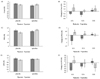
Lymphocyte proliferation rate showed no significant difference for all subjects after spirulina or placebo supplementation (Figs. 3A, 3C, 3E). Changes in stimulation index of mitogen induced LPR from PBMC are shown in Figs. 3B, 3D, and 3F by genotype. PHA induced SI was significantly changed for A/A and G/A genotypes (Fig. 3D, P < 0.05). LPS induced SI was significantly changed only for the A/A genotype (Fig. 3F, P < 0.05).
A significant difference of intervention between the A and G genotype subgroups appeared in PHA and LPS induced proliferation rate with A carrier (Table 4). Mitogen induced proliferation of lymphocyte was not significantly changed for both G carrier groups. A significant supplementary effect was observed for the case of PHA induced SI, regardless of genotype (P < 0.01).

DISCUSSION

MCP-1 is a small cytokine that belongs to the CC chemokine family [37]. The concentration gradient of MCP-1 is responsible for the movement of mononuclear cells to inflammation sites [38]. MCP-1 is produced by a variety of cell types, including immune and non-immune cells [3473940], in response to stimulation by a broad range of mediators, including cytokines, growth factors and mitogens [4]. MCP-1 recruits monocytes, memory T cells, and dendritic cells to inflammation sites produced by either tissue injury or infection [3841]. MCP-1 is also implicated in pathogeneses of several diseases characterized by monocytic infiltrates, such as psoriasis, rheumatoid arthritis, and atherosclerosis [40]. Epidemiological evidence suggests that MCP-1 plasma levels are associated with traditional risk factors for CVD, such as hyperlipidemia, hypertension, and diabetes [4243]. MCP-1 may also contribute to thrombotic aspects of advanced atherosclerotic lesions, a late complication of atherosclerosis responsible for myocardial infarctions and strokes [37]. MCP-1 expression is up-regulated in human atherosclerotic plaques [37], suggesting that genetic factors leading to MCP-1 production deregulation could affect development of atherosclerosis and associated diseases.
The human MCP-1 gene is located on chromosome 17q11.2-q21.1 [37]. Several MCP-1 SNPs, such as -1811 A/G, -2136 A/T, -2518 A/G, -2835 C/A, -927 G/, 764 C/G, and 3726 T/C have been identified [44]. Among these, MCP-1 -2518 A/G SNP, located in the MCP-1 regulatory region, has been most frequently studied for its relevance to diet and disease. Szalai et al. [44] found that MCP-1 -2518*G allele homozygotes frequency was significantly higher in coronary artery disease (CAD) patients than in healthy controls. The higher MCP-1 -2518*G homozygotes prevalence was observed in patients with history of stroke [45] and myocardial infarction [46]. Angeles-Martinez showed that MCP-1 polymorphisms were associated with premature coronary artery disease risk [47]. Other studies [4849] have reported MCP-1 polymorphism association with disease related inflammation susceptibility.
Several studies have investigated the relationship between MCP-1 -2518A/G polymorphism and CVD risk in various populations worldwide [50515253]. However, the results are inconsistent, and MCP-1 -2518 A/G polymorphism appears to be ethnically different. Caucasian or African subjects are less likely to express MCP-1 -2518 G/G polymorphism than Asian or Mexican [7]. On the other hand, Caucasian or African subjects with MCP-1 -2518 G/G polymorphism show higher MCP-1 plasma levels and higher CVD risk, such as hypertension, than those with other genotypes [54]. In Chinese subjects, MCP-1 -2518 A/G polymorphism was not associated with CAD risk [50]. In a previous study, we reported that subjects with A/A genotype exhibited higher MCP-1 levels than other genotypes in Korean elderly, and had higher CVD risk [55]. These results suggest that MCP-1 concentration and CVD risk vary with MCP-1 polymorphism.
In the current study, the effects of spirulina supplementation were different depending on MCP-1 genotypes. Anti-inflammatory spirulina supplementation effects in all subjects were not significant, but the effect of intervention was significantly different when analyzed by genotype. For A/A genotype subjects, spirulina supplementation showed significant changes for plasma IL-2 level and PHA or LPS induced LPR. Placebo subjects showed significant MCP-1 increase, but those who took spirulina did not. Circulating MCP-1 levels are associated with CVD risk factors, since MCP-1 is expressed at high levels in atherosclerotic plaque [52535457]. IL-2 is a representative anti-inflammatory cytokine that decreases with aging [58].
C3 is a protein with a central role in the innate immune system, and C3 levels have an aging dependent increase in general. Several studies have reported that C3 was positively related to CVD [5960]. In the current study, placebo subjects showed significant C3 level increase after 16 weeks, but there was no significant difference in spirulina subjects. Subjects with A/A genotypes also had significantly more C3 compared with G/A and G/G genotypes when analyzed by genotype and placebo supplementation: A/A increased 32.8%, and G/A and G/G increased 7.5%. These results indicate that the degree of C3 increase depends on the MCP-1 genotype and that C3 increase was suppressed when spirulina was ingested.
Mitogens, such as PHA, ConA, and LPS induced stimulation index, are generally reduced by aging [61]. The current study shows that lymphocyte proliferation was improved after spirulina supplementation. Subjects who consumed spirulina for 16 weeks showed significantly increased PHA and LPS induced stimulation index. Significantly, the PHA and LPS induced stimulation index also increased in A/A genotype subjects.
Thus, spirulina supplementation shows anti-inflammatory effects, such as decreasing MCP-1 level and increasing IL-2 level in healthy subjects, and improved LPS and PHA induced mitogenesis. Theses anti-inflammatory effects were larger for A/A genotype subjects. Therefore, spirulina may be considered beneficial for anti-inflammatory capacity, particularly for A/A genotype subjects.
In conclusion, functional food supplementation has different effects by genotype. Spirulina supplementation affects anti-inflammatory capacity and may help immune function. Although the population size of the current study was small, the study confirmed that supplement response and anti-inflammatory profiles were different by MCP-1 genotype. These results suggest that personalized management could be advantageous for different genotypes. While the current study is insufficient to make specific personalized nutritional recommendations based on genetic information, the results provide a valuable basis for further study to investigate the relationship between MCP-1 polymorphism and diet. Careful attention should be paid for subsequent studies to include sufficient subjects for each phenotype and genotype with appropriate statistical power.

Figures and Tables

Fig. 1

Plasma cytokine levels for (A) MCP-1, (C) IL-6, (E) TNF-α, and (G) IL-2; and changes (B, D, F, H, respectively) after placebo or spirulina supplementation in subject grouped by MCP-1 polymorphism.

MCP-1, monocyte chemoattractant protein -1; IL, interleukin; TNF, tumor necrosis factor; A/A, MCP-1 -2518 AA genotype; G/A, MCP-1 -2518 GA genotype; G/G, MCP-1 -2518 GG genotype. Data are expressed as mean ± SE. *P < 0.05, **P < 0.01.
nrp-11-290-g001
Fig. 2

Plasma level of (A) C3, (C) IgA, and (E) IgG; and changes (B, D, and F, respectively) after placebo or spirulina supplementation in subjects grouped by MCP-1 polymorphism.

Blood samples were collected before and after supplementation (pretest: beginning day, post-test: 16 weeks). C3, Ig A, and IgG plasma levels were analyzed by radical immunodiffusion plate. MCP-1, monocyte chemoattractant protein 1; C3, complement 3; Ig A, immunoglobulin A; Ig G, immunoglobulin G; A/A, MCP-1 -2518 AA genotype; G/A, MCP-1 -2518 GA genotype; G/G, MCP-1 -2518 GG genotype. Data are expressed as mean ± SE. *P < 0.05.
nrp-11-290-g002
Fig. 3

Lymphocyte proliferation rate: (A) Con A, (B) PHS, and (C) LPS induced; and changes (B, D, and F, respectively) after placebo or spirulina supplementation in subjects grouped by MCP-1 polymorphism.

Isolated lymphocytes were seeded at 2 × 10⁵ cells per well in a 96-well plate and treated with ConA, PHA, or LPS. After incubating for 68 hours in CO2 incubator, mitogen induced LPR was determined by ELISA. MCP-1, monocyte chemoattractant protein 1; ConA, concanavalin A; PHA, phtyhemagglutinin; LPS, lipopolysaccharide; A/A, MCP-1 -2518 AA genotype; G/A, MCP-1 -2518 GA genotype; G/G, MCP-1 -2518 GG genotype. Data are expressed as mean ± SE. *P < 0.05
nrp-11-290-g003
Table 1

Selected characteristics of study subjects

nrp-11-290-i001

1)Mean ± SE

A/A, MCP-1 -2518 AA genotype; G/A, MCP-1 -2518 GA genotype; G/G, MCP-1 -2518 GG genotype.

Table 2

Cytokine changes after spirulina supplementation for the A and G genotype subgroups

nrp-11-290-i002

1)Mean ± SE

2)Significantly different by paired t-test between pre-test and post-test within the same genotype (*P < 0.05).

3)Determined by two-way ANOVA for main effects of MCP-1 genotype and spirulina supplementation, and interaction effects of MCP-1 genotype and spirulina supplementation.

A/A, MCP-1 -2518 AA genotype; G/A, MCP-1 -2518 GA genotype; G/G, MCP-1 -2518 GG genotype; NS, Not significant.

Table 3

Changes in C3 and Ig after supplementation for A and G genotype subgroups

nrp-11-290-i003

1)Mean ± SE.

2)Significantly different by paired t-test between pre-test and post-test within the same genotype (*P < 0.05).

3)Determined by two-way ANOVA for main effects MCP-1 genotype and spirulina supplementation, and interaction effects of MCP-1 genotype and spirulina supplementation.

A/A, MCP-1 -2518 AA genotype; G/A, MCP-1 -2518 GA genotype; G/G, MCP-1 -2518 GG genotype; NS, Not significant.

Table 4

Changes in lymphocyte proliferation rate from peripheral blood mononuclear cell based on MCP-1 genotype

nrp-11-290-i004

1)Mean ± SE.

2)Significantly different by paired t-test between pre-test and post-test within the same genotype (*P < 0.05).

3)Determined by two-way ANOVA for main effects MCP-1 genotype and spirulina supplementation, and interaction effects of MCP-1 genotype and spirulina supplementation.

A/A, MCP-1 -2518 AA genotype; G/A, MCP-1 -2518 GA genotype; G/G, MCP-1 -2518 GG genotype; NS, Not significant.

ACKNOWLEDGEMENTS

Financial aid from the Korean Research Foundation and ES Biotech is gratefully acknowledged. The authors would like to thank the elderly who participated in this study, as well as the staff of Ewha Women's University for their invaluable efforts to make the study possible.

Notes

This work was supported by the Korean Research Foundation Fund grant No. R04-2003-000-10119-0.

CONFLICT OF INTEREST The authors declare no potential conflicts of interests.

References

1. Muller WA. Getting leukocytes to the site of inflammation. Vet Pathol. 2013; 50:7–22.
crossref
2. Xu L, Rocnik E, Rahlpour R, Hunter N, Pickering G, Kelvin DJ. MCP-1 induces proliferation and migration of vascular smooth muscle cells. FASEB J. 1996; 10:A1932.
3. Luster AD. Chemokines--chemotactic cytokines that mediate inflammation. N Engl J Med. 1998; 338:436–445.
crossref
4. Rossi D, Zlotnik A. The biology of chemokines and their receptors. Annu Rev Immunol. 2000; 18:217–242.
crossref
5. Salcedo R, Ponce ML, Young HA, Wasserman K, Ward JM, Kleinman HK, Oppenheim JJ, Murphy WJ. Human endothelial cells express CCR2 and respond to MCP-1: direct role of MCP-1 in angiogenesis and tumor progression. Blood. 2000; 96:34–40.
crossref
6. Inadera H, Egashira K, Takemoto M, Ouchi Y, Matsushima K. Increase in circulating levels of monocyte chemoattractant protein-1 with aging. J Interferon Cytokine Res. 1999; 19:1179–1182.
crossref
7. Rovin BH, Lu L, Saxena R. A novel polymorphism in the MCP-1 gene regulatory region that influences MCP-1 expression. Biochem Biophys Res Commun. 1999; 259:344–348.
crossref
8. Martin ES, Schneeberger EE, Aranda FM, Peres SW, Del Carmen Valerio M, de Los Angeles Correa M, Pra FD, Martinez L, Remondino G, de Larrañaga G, Citera G. The -2518 A/G polymorphism in the monocyte chemoattractant protein 1 gene (MCP-1) is associated with an increased risk of rheumatoid arthritis in Argentine patients. Clin Rheumatol. 2016; 35:3057–3061.
crossref
9. Wang W, He M, Huang W. Association of monocyte chemoattractant protein-1 gene 2518A/G polymorphism with diabetic retinopathy in type 2 diabetes mellitus: a meta-analysis. Diabetes Res Clin Pract. 2016; 120:40–46.
crossref
10. Niu J, Kolattukudy PE. Role of MCP-1 in cardiovascular disease: molecular mechanisms and clinical implications. Clin Sci (Lond). 2009; 117:95–109.
crossref
11. Mansego ML, De Marco G, Ivorra C, Lopez-Izquierdo R, Morcillo S, Rojo-Martínez G, González-Albert V, Martinez F, Soriguer F, Martín-Escudero JC, Redon J, Chaves FJ. The nutrigenetic influence of the interaction between dietary vitamin E and TXN and COMT gene polymorphisms on waist circumference: a case control study. J Transl Med. 2015; 13:286.
crossref
12. Pase CS, Teixeira AM, Roversi K, Dias VT, Calabrese F, Molteni R, Franchi S, Panerai AE, Riva MA, Burger ME. Olive oil-enriched diet reduces brain oxidative damages and ameliorates neurotrophic factor gene expression in different life stages of rats. J Nutr Biochem. 2015; 26:1200–1207.
crossref
13. Pietropaolo S, Goubran MG, Joffre C, Aubert A, Lemaire-Mayo V, Crusio WE, Layé S. Dietary supplementation of omega-3 fatty acids rescues fragile X phenotypes in Fmr1-Ko mice. Psychoneuroendocrinology. 2014; 49:119–129.
crossref
14. Nigam A, Talajic M, Roy D, Nattel S, Lambert J, Nozza A, Jones P, Ramprasath VR, O'Hara G, Kopecky S, Brophy JM, Tardif JC. AFFORD Investigators. Fish oil for the reduction of atrial fibrillation recurrence, inflammation, and oxidative stress. J Am Coll Cardiol. 2014; 64:1441–1448.
crossref
15. Yanaka A, Fahey JW, Fukumoto A, Nakayama M, Inoue S, Zhang S, Tauchi M, Suzuki H, Hyodo I, Yamamoto M. Dietary sulforaphane-rich broccoli sprouts reduce colonization and attenuate gastritis in Helicobacter pylori-infected mice and humans. Cancer Prev Res (Phila). 2009; 2:353–360.
crossref
16. Vicari E, La Vignera S, Castiglione R, Condorelli RA, Vicari LO, Calogero AE. Chronic bacterial prostatitis and irritable bowel syndrome: effectiveness of treatment with rifaximin followed by the probiotic VSL#3. Asian J Androl. 2014; 16:735–739.
crossref
17. Ortega-Azorín C, Sorlí JV, Asensio EM, Coltell O, Martínez-González MA, Salas-Salvadó J, Covas MI, Arós F, Lapetra J, Serra-Majem L, Gómez-Gracia E, Fiol M, Sáez-Tormo G, Pintó X, Muñoz MA, Ros E, Ordovás JM, Estruch R, Corella D. Associations of the FTO rs9939609 and the MC4R rs17782313 polymorphisms with type 2 diabetes are modulated by diet, being higher when adherence to the Mediterranean diet pattern is low. Cardiovasc Diabetol. 2012; 11:137.
crossref
18. Serrano JC, De Lorenzo D, Cassanye A, Martín-Gari M, Espinel A, Delgado MA, Pamplona R, Portero-Otin M. Vitamin D receptor BsmI polymorphism modulates soy intake and 25-hydroxyvitamin D supplementation benefits in cardiovascular disease risk factors profile. Genes Nutr. 2013; 8:561–569.
crossref
19. Giacconi R, Costarelli L, Malavolta M, Cardelli M, Galeazzi R, Piacenza F, Gasparini N, Basso A, Mariani E, Fulop T, Rink L, Dedoussis G, Herbein G, Jajte J, Provinciali M, Busco F, Mocchegiani E. Effect of ZIP2 Gln/Arg/Leu (rs2234632) polymorphism on zinc homeostasis and inflammatory response following zinc supplementation. Biofactors. 2015; 41:414–423.
crossref
20. Park S, Zhang X, Lee NR, Jin HS. TRPV1 gene polymorphisms are associated with type 2 diabetes by their interaction with fat consumption in the Korean genome epidemiology study. J Nutrigenet Nutrigenomics. 2016; 9:47–61.
crossref
21. Thengodkar RR, Sivakami S. Degradation of Chlorpyrifos by an alkaline phosphatase from the cyanobacterium Spirulina platensis. Biodegradation. 2010; 21:637–644.
crossref
22. Lee JB, Srisomporn P, Hayashi K, Tanaka T, Sankawa U, Hayashi T. Effects of structural modification of calcium spirulan, a sulfated polysaccharide from Spirulina platensis, on antiviral activity. Chem Pharm Bull (Tokyo). 2001; 49:108–110.
crossref
23. Marcel AK, Ekali LG, Eugene S, Arnold OE, Sandrine ED, von der Weid D, Gbaguidi E, Ngogang J, Mbanya JC. The effect of Spirulina platensis versus soybean on insulin resistance in HIV-infected patients: a randomized pilot study. Nutrients. 2011; 3:712–724.
crossref
24. Nkengfack G, Torimiro J, Ngogang J, Heike E. Effects of nutritional education and lifestyle modification on the nutritional status of HIV positive patients: results of a cluster randomized intervention over a period of six months. Glob Epidemi Obes. 2013; 1:5.
crossref
25. Moura LP, Puga GM, Beck WR, Teixeira IP, Ghezzi AC, Silva GA, Mello MA. Exercise and Spirulina control non-alcoholic hepatic steatosis and lipid profile in diabetic Wistar rats. Lipids Health Dis. 2011; 10:77.
crossref
26. Layam A, Reddy CL. Antidiabetic property of Spirulina. Diabetol Croat. 2007; 35:29–33.
27. Hayashi O, Katoh T, Okuwaki Y. Enhancement of antibody production in mice by dietary Spirulina platensis. J Nutr Sci Vitaminol (Tokyo). 1994; 40:431–441.
crossref
28. Watanuki H, Ota K, Tassakka AC, Kato T, Sakai M. Immunostimulant effects of dietary Spirulina platensis on carp, Cyprinus carpio. Aquaculture. 2006; 258:157–163.
crossref
29. Qureshi MA, Kidd MT, Ali RA. Spirulina platensis extract enhances chicken macrophages functions after in vitro exposure. J Nutr Immunol. 1996; 3:35–45.
30. Hirahashi T, Matsumoto M, Hazeki K, Saeki Y, Ui M, Seya T. Activation of the human innate immune system by Spirulina: augmentation of interferon production and NK cytotoxicity by oral administration of hot water extract of Spirulina platensis. Int Immunopharmacol. 2002; 2:423–434.
crossref
31. Selmi C, Leung PS, Fischer L, German B, Yang CY, Kenny TP, Cysewski GR, Gershwin ME. The effects of Spirulina on anemia and immune function in senior citizens. Cell Mol Immunol. 2011; 8:248–254.
crossref
32. Park HJ, Lee YJ, Ryu HK, Kim MH, Chung HW, Kim WY. A randomized double-blind, placebo-controlled study to establish the effects of Spirulina in elderly Koreans. Ann Nutr Metab. 2008; 52:322–328.
crossref
33. Park HJ, Lee HS. The influence of obesity on the effects of Spirulina supplementation in the human metabolic response of Korean elderly. Nutr Res Pract. 2016; 10:418–423.
crossref
34. The Korean Nutrition Society. CAN-Pro 4.0: nutritional assessment program. Seoul: The Korean Nutrition Society;2010.
35. Friedewald WT, Levy RI, Fredrickson DS. Estimation of the concentration of low-density lipoprotein cholesterol in plasma, without use of the preparative ultracentrifuge. Clin Chem. 1972; 18:499–502.
crossref
36. Mosmann T. Rapid colorimetric assay for cellular growth and survival: application to proliferation and cytotoxicity assays. J Immunol Methods. 1983; 65:55–63.
crossref
37. Bielinski SJ, Pankow JS, Miller MB, Hopkins PN, Eckfeldt JH, Hixson J, Liu Y, Register T, Myers RH, Arnett DK. Circulating MCP-1 levels shows linkage to chemokine receptor gene cluster on chromosome 3: the NHLBI family heart study follow-up examination. Genes Immun. 2007; 8:684–690.
crossref
38. Deshmane SL, Kremlev S, Amini S, Sawaya BE. Monocyte chemoattractant protein-1 (MCP-1): an overview. J Interferon Cytokine Res. 2009; 29:313–326.
crossref
39. Yamamoto T, Eckes B, Mauch C, Hartmann K, Krieg T. Monocyte chemoattractant protein-1 enhances gene expression and synthesis of matrix metalloproteinase-1 in human fibroblasts by an autocrine IL-1 alpha loop. J Immunol. 2000; 164:6174–6179.
crossref
40. Xia M, Sui Z. Recent developments in CCR2 antagonists. Expert Opin Ther Pat. 2009; 19:295–303.
crossref
41. Rollins BJ. Chemokines. Blood. 1997; 90:909–928.
crossref
42. de Lemos JA, Morrow DA, Sabatine MS, Murphy SA, Gibson CM, Antman EM, McCabe CH, Cannon CP, Braunwald E. Association between plasma levels of monocyte chemoattractant protein-1 and long-term clinical outcomes in patients with acute coronary syndromes. Circulation. 2003; 107:690–695.
crossref
43. Deo R, Khera A, McGuire DK, Murphy SA, Meo Neto, Morrow DA, de Lemos JA. Association among plasma levels of monocyte chemoattractant protein-1, traditional cardiovascular risk factors, and subclinical atherosclerosis. J Am Coll Cardiol. 2004; 44:1812–1818.
crossref
44. Navratilova Z. Polymorphisms in CCL2&CCL5 chemokines/chemokine receptors genes and their association with diseases. Biomed Pap Med Fac Univ Palacky Olomouc Czech Repub. 2006; 150:191–204.
crossref
45. Szalai C, Kozma GT, Nagy A, Bojszkó A, Krikovszky D, Szabó T, Falus A. Polymorphism in the gene regulatory region of MCP-1 is associated with asthma susceptibility and severity. J Allergy Clin Immunol. 2001; 108:375–381.
crossref
46. Arakelyan A, Zakharyan R, Hambardzumyan M, Petrkova J, Olsson MC, Petrek M, Boyajyan A. Functional genetic polymorphisms of monocyte chemoattractant protein 1 and C-C chemokine receptor type 2 in ischemic stroke. J Interferon Cytokine Res. 2014; 34:100–105.
crossref
47. Bjarnadottir K, Eiriksdottir G, Aspelund T, Gudnason V. Examination of genetic effects of polymorphisms in the MCP-1 and CCR2 genes on MI in the Icelandic population. Atherosclerosis. 2006; 188:341–346.
crossref
48. Angeles-Martínez J, Posadas-Sánchez R, Álvarez-León E, Villarreal-Molina T, Cardoso-Saldaña G, Fragoso JM, Juárez-Rojas JG, Medina-Urrutia A, Posadas-Romero C, Vargas-Alarcón G. Monocyte chemoattractant protein-1 gene (MCP-1) polymorphisms are associated with risk of premature coronary artery disease in Mexican patients from the Genetics of Atherosclerotic Disease (GEA) study. Immunol Lett. 2015; 167:125–130.
crossref
49. Xu J, Liao YF, Zhou WP, Ming HL, Wang QH. The MCP-1 gene A-2518G polymorphism confers an increased risk of vascular complications in type 2 diabetes mellitus patients. Genet Test Mol Biomarkers. 2015; 19:411–417.
crossref
50. Hou S, Yang P, Du L, Jiang Z, Mao L, Shu Q, Zhou H, Kijlstra A. Monocyte chemoattractant protein-1 -2518 A/G single nucleotide polymorphism in Chinese Han patients with ocular Behçet's disease. Hum Immunol. 2010; 71:79–82.
crossref
51. Zuo S, Wang H, Wang B. Association of monocyte chemoattractant protein-1-2518A/G polymorphism and risk of coronary artery disease among the Chinese population: a meta-analysis. Int J Clin Exp Med. 2015; 8:15182–15189.
52. Jemaa R, Rojbani H, Kallel A, Ben Ali S, Feki M, Chabrak S, Elasmi M, Taieb SH, Sanhaji H, Souheil O, Mechmeche R, Kaabachi N. Association between the -2518G/A polymorphism in the monocyte chemoattractant protein-1 (MCP-1) gene and myocardial infarction in Tunisian patients. Clin Chim Acta. 2008; 390:122–125.
crossref
53. Kaur R, Matharoo K, Arora P, Bhanwer AJ. Association of -2518A>G promoter polymorphism in the monocyte chemoattractant protein-1 (MCP-1) gene with type 2 diabetes and coronary artery disease. Genet Test Mol Biomarkers. 2013; 17:750–755.
crossref
54. Okopień B, Haberka M, Cwalina L, Kowalski J, Belowski D, Madej A, Zieliński M, Krysiak R, Labuzek K, Kalina Z, Herman ZS. Plasma cytokines as predictors of coronary heart disease. Res Commun Mol Pathol Pharmacol. 2002; 112:5–15.
55. Gao HH, Gao LB, Wen JM. Correlations of MCP-1 -2518A>G polymorphism and serum levels with cerebral infarction risk: a meta-analysis. DNA Cell Biol. 2014; 33:522–530.
crossref
56. Park HJ. Association of MCP-1 polymorphism with cardiovascular disease risk factors in Korean elderly. J Nutr Health. 2013; 46:511–520.
crossref
57. Nelken NA, Coughlin SR, Gordon D, Wilcox JN. Monocyte chemoattractant protein-1 in human atheromatous plaques. J Clin Invest. 1991; 88:1121–1127.
crossref
58. Nikolich-Žugich J. T cell aging: naive but not young. J Exp Med. 2005; 201:837–840.
59. Halkes CJ, van Dijk H, de Jaegere PP, Plokker HW, van Der Helm Y, Erkelens DW, Castro Cabezas M. Postprandial increase of complement component 3 in normolipidemic patients with coronary artery disease: effects of expanded-dose simvastatin. Arterioscler Thromb Vasc Biol. 2001; 21:1526–1530.
crossref
60. Muscari A, Bozzoli C, Puddu GM, Sangiorgi Z, Dormi A, Rovinetti C, Descovich GC, Puddu P. Association of serum C3 levels with the risk of myocardial infarction. Am J Med. 1995; 98:357–364.
crossref
61. Hoskinson CD, Chew BP, Wong TS. Age-related changes in mitogen-induced lymphocyte proliferation and polymorphonuclear neutrophil function in the piglet. J Anim Sci. 1990; 68:2471–2478.
crossref
TOOLS
Similar articles