Abstract
BACKGROUND/OBJECTIVES
Chronic ultraviolet (UV) exposure-induced reactive oxygen species (ROS) are commonly involved in the pathogenesis of skin damage by activating the metalloproteinases (MMP) that break down type I collagen. Adenophora remotiflora (AR) is a perennial wild plant that inhabits Korea, China, and Japan. The present study investigated the protective effects of AR against UVB-induced photo-damage in keratinocytes.
MATERIALS/METHODS
An in vitro cell-free system was used to examine the scavenging activity of 2,2-diphenyl-1-picrylhydrazyl (DPPH) free radical and nitric oxide (NO). The effect of AR on ROS formation, antioxidant enzymes, elastase, MMP-1 level, and mRNA expression of MMP-1 were determined in UVB-irradiated human keratinocyte HaCaT cells.
RESULTS
AR demonstrated strong DPPH free radical and NO scavenging activity in a cell-free system exhibiting IC50 values of 1.88 mg/mL and 6.77 mg/mL, respectively. AR pretreatment dose-dependently attenuated the production of UVB-induced intracellular ROS, and antioxidant enzymes (catalase and superoxide dismutase) were enhanced in HaCaT cells. Furthermore, pretreatment of AR prevented UVB-induced elastase and collagen degradation by inhibiting the MMP-1 protein level and mRNA expression. Accordingly, AR treatment elevated collagen content in UVB-irradiated HaCaT cells.
CONCLUSION
The present study provides the first evidence of AR inhibiting UVB-induced ROS production and induction of MMP-1 as a result of augmentation of antioxidative activity in HaCaT human keratinocytes. These results suggest that AR might act as an effective inhibitor of UVB-modulated signaling pathways and might serve as a photo-protective agent.
UV irradiation is the most common environmental factor involved in human skin damage, leading to conditions such as skin carcinogenesis, inflammation, solar erythema and premature senescence [1]. The UV spectrum is classified by wavelength as UVA (315-400 nm), UVB (280-315 nm), and UVC (100-280 nm). UVA and UVB reach the earth's surface but UVC is filtered out by the ozone layer [2]. UVA accounts for 90-99% of the UV energy that reaches the earth's surface and UVB contributes the other 1-10% [3]. However, UVB has been reported to be 1,000-10,000 fold more carcinogenic than UVA [3]. Furthermore, ozone depletion due to the emission of halogen-containing compounds caused by human activities has increased the level of UVB absorbed by human skin.
Excessive exposure to UVB irradiation generates reactive oxygen species (ROS) in skin [4], and increased ROS generation can overwhelm the antioxidant-defense mechanism resulting in oxidative stress and oxidative photo-damage in the skin [5]. An enzymatic antioxidant defense system composed of catalase and superoxide dismutase (SOD) is therefore crucial for the protection of skin from UVB-induced oxidative stress [6]. ROS also leads to the activation of transcription factors that induce the expression of pro-inflammatory cytokines and metalloproteinase (MMPs) [1]. Collagen-degrading MMP-1, also known as collagenase, is up-regulated and serves as the primary MMP in UV-exposed skin. Therefore, excessive degradation of collagen and matrix by UV-induced MMPs is a characteristic feature of photo-damaged skin, and MMP is used as a major marker of UVB-induced photoaging as well as skin inflammation. Elastin, another fibrous protein of the skin, has an influence on skin elasticity although the distribution is lower than that of collagen [7]. Because this elastic fiber is easily decomposed by elastase secretion and activation due to UV or ROS, an approach that inhibits elastase activity could also be used as a useful method in protecting against photoaging [8].
Natural agents, with potential antioxidant, anti-inflammatory, anti-mutagenic, anti-carcinogenic and immunomodulatory properties are gaining considerable attention in relation to the prevention of UV-induced skin damage [9]. Adenophora remotiflora (AR) is a perennial wild plant of the Campanulaceae family that inhabits Korea, China, and Japan. It has traditionally been used in Korea as alternative medicine for the treatment of certain diseases including expectorations, chills, fever, poisoning, and phlegm discharge [10]. Recently, the antioxidant and chemopreventive activity of AR has been reported in several in vitro studies [1112]. However, the preventive effect of photoaging has never been examined. The present study therefore investigated the protective effects of AR against UVB-induced photo-damage in human epidermal keratinocytes.
Two-month-old vacuum freeze-dried AR leaves were donated by Eco-Sprout Co. Ltd. (Gyeonggido, Korea), an environmentally friendly agricultural company. The samples were extracted with 80% ethanol at 65℃ for 5 h, filtered through a 0.45 µm filter (Osmonics, Minnetonka, MN), and lyophilized.
Antioxidant activities in a cell-free system were evaluated by free radical and NO scavenging activities. The free radical scavenging activity of AR extracts on DPPH radical was determined using the method described by Huang et al. [13], with slight modification. Briefly, DPPH ethanol solution was added to various concentrations of AR extract (0.4-50 mg/mL) in 96-well plates. After 30 min incubation at room temperature in the dark, the absorbance at 515 nm was measured by a plate reader (BioTek Inc., Winooski, VT). The free radical scavenging activity of the sample was calculated by the following formula:
Where As is the absorbance of the sample and Ab is the absorbance of the blank.
NO production was assessed by measuring the nitrite content. Briefly, Griess reagent (0.1% N-1-naphthylenediamine dihydrochloride and 5% H3PO4 solution) was added to AR extracts in a 1:1 (v/v) manner. After gentle mixing and 15 min incubation in the dark, NO levels were subsequently measured and compared with a nitrate standard curve. Absorbance values at 560 nm were measured using a microplate reader. The NO scavenging activity of the sample was calculated using the same formula as used for the DPPH scavenging activity. The IC50 values were obtained using GraphPad Prizm (Ver. 6, La Jolla, CA, USA).
An immortalized human keratinocyte cell line, HaCaT (ATCC, Rockville, MD), was cultured in Dulbecco's Modification of Eagle's Medium (DMEM) containing 10% heat-inactivated Fetal bovine serum (FBS), 100 U/mL penicillin and 100 µg/mL streptomycin in a humidified atmosphere with 5% CO2 at 37℃. Cells were exposed to UVB (30 mJ/cm2) with a thin layer of PBS using a UVB lamp (312 nm, Spectroline Model EB-160C, New York, NY). After UVB irradiation for 5 min, the cells were washed with warm PBS, and incubated with serum-free DMEM for 24 h. Mock-irradiated controls followed the same schedule of medium changes without UVB irradiation.
UVB irradiation-induced cytotoxicity was determined using a colorimetric lactate dehydrogenase (LDH) leakage assay kit (LK100, Oxford Biomedical Research, Rochester Hills, MI) in accordance with the manufacturer's instruction. Cells in 96-well plates (2 × 10⁵ cells/well) were pretreated with AR extract at a concentration of 10, 30 or 100 µg/mL prior to UVB irradiation. After incubation for 24 h, reaction buffer was added. The intensity of color obtained from the reaction is proportional to LDH activity. The cytotoxicity was expressed as a percentage of the absorbance at 490 nm of a non-UV-irradiated control.
2',7'-dichlorodihydrofluorescein diacetate (DCFH-DA) was
used to detect ROS production in cells [14]. DCFH-DA, which had entered the cells, was cleaved to form 2',7'-dichlorodihydrofluorescein (DCFH). Trapped DCFH was oxidized by oxygen free radicals to produce fluorescent',7'-dichlorofluorescein (DCF). Cells which had been treated with AR extract prior to UV irradiation were incubated with 20 µM DCFH-DA for 30 min and harvested after 24 h. ROS formation was analyzed with a fluorometer (TECAN,SER-NR 94572, Salzburg, Austria) using 485 nm of excitation and 530 nm of emission filters. ROS production was expressed as a percentage of the fluorescence of a non-UV-irradiated control.
To estimate the effects of AR on catalase and SOD activity after UVB irradiation, cells were pretreated with AR extract for 24 h prior to UVB irradiation. Catalase activity was measured using Amplex Red Catalase assay kit (Molecular Probes, Invitrogen, Eugene, OR). Catalase first reacts with H2O2 to produce water and oxygen, and residual H2O2 then reacts with Amplex Red reagent in the presence of horseradish peroxidase to produce the highly fluorescent oxidation product, resorufin. One unit of catalase was defined as the amount of enzyme required to decompose 1 µM of H2O2 per minute. The rate of decomposition of H2O2 was measured spectrophotometrically at 560 nm for 1 min.
SOD activity was measured using a commercially available SOD assay kit (Dojindo Molecular Technologies, Rockville, MD). Xanthine and xanthine oxidase were used to generate superoxide radicals reacting with 2-(4-iodophenyl)3-(4-nitrophenol)-5-phenyl tetrazolium chloride to form a red formazan dye. SOD activity was then measured at 505 nm.
The cells were planted in 48-well plates (5 × 10⁴ cells/well), pretreated with AR extracts (0-100 µg/mL) for 24 h, and exposed to UVB irradiation. The activity of porcine pancreatic elastase type IV (Sigma Chem. Co., USA) was determined using a spectrophotometric method [15] with N-Succ-(Ala)3-p-nitroanilide as a substrate. The reaction mixture was pre-incubated for 15 min before addition of the substrate, and the release of p-nitroaniline was monitored by measuring the absorbance at 410 nm.
HaCaT cells were cultured in 24-well plates (1 × 10⁶ cells/well), pretreated with AR extract (10-100 µg/mL) for 24 h, and exposed to UVB. The production of MMP-1 was determined using a commercial ELISA kit (Human total MMP-1 kit; R&D systems, Minneapolis, MN).
The collagen content in HaCaT cells after UVB irradiation was quantified using a SirCol collagen assay kit (Biocolor Ltd., Belfast, Northern Ireland). Anionic Sirus red dye, which reacts specifically with basic side chain groups of collagen, was added to cell lysates and then incubated under gentle rotation for 30 min at room temperature. After centrifugation at 12,000 × g for 10 min, collagen-bound dye was dissolved with 0.5 mM NaOH, and the absorbance was measured at 540 nm. Collagen content was expressed as a percentage of the non-UV-irradiated control.
Total RNA, isolated using RNeasy® Protect Mini kit (Qiagen, Valencia, CA, USA), was reverse transcribed to cDNA with the SuperScript First-Strand Synthesis System (Invitrogen). The primer sequences for MMP-1 were: forward, 5'-ATT CTA CTG ATA TCG GGG CTT TGA-3'; and reverse, 5'-ATG TCC TTG GGG TAT CCG TGT AG-3'. The primer sequences for GAPDH were: forward, 5'-TCA TCA ATG GAA ATC CCA TCA CC-3'; and reverse, 5'-TGG ACT CCA CGA CGT ACT CAG C-3'. PCR amplification was carried out using a QuantiTectTM SYBR Green PCR kit (Qiagen, Valencia, CA, USA). The PCR cycle was 94℃ for 10 min, followed by 40 cycles of reaction at 94℃ for 10 s, 58℃ for 15 s, and 72℃ for 20 s. The level of MMP-1 mRNA was normalized to the level of GAPDH, and compared with a control (untreated sample) using the ΔΔCT method [16].
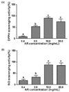
The effect of AR on free radical and NO scavenging capacities were determined in a cell-free system. DPPH radical and NO scavenging activities were both dose-dependently increased with AR treatment, reaching a saturation point at 10 mg/mL concentration and exhibiting scavenging activities of 90.4 ± 5.0% and 87.4 ± 9.0%, respectively (Fig. 1). The level of scavenging activity for DPPH radical was greater than that for NO. The IC50 values for the DPPH radical and NO scavenging activities were 1.88 mg/mL and 6.77 mg/mL, respectively.
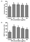
The protective effect of AR against UVB-induced cytotoxicity was tested by incubating human keratinocyte HaCaT cells with AR extract (10-100 µg/mL) for 24 h before UVB treatment. Cytotoxicity was evaluated by LDH measurement. The LDH leakage assay is based on the measurement of LDH activity in the extracellular medium. The loss of intracellular LDH and its release into the culture medium is an indicator of irreversible cell death due to cell membrane damage [17]. As shown in Fig. 2, UVB irradiation caused cytotoxicity as measured by LDH release from keratinocytes, and AR treatment had no effect on cytotoxicity.
The UVB-induced intracellular oxidative stress level was determined using the redox sensitive dye DCFH-DA. UVB irradiation caused a significant 2-fold increase in ROS generation as compared with non-irradiated control cells, indicating massive oxidant generation. The increase in ROS was significantly reduced (P < 0.05) in the presence of AR in a concentrationdependent manner (Fig. 2).
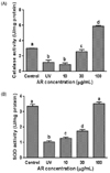
To investigate whether the ROS scavenging activity of AR was mediated by the activity of antioxidant enzymes, catalase and SOD activities were measured in UVB-exposed HaCaT cells. As shown in Fig. 3, UVB irradiation markedly reduced catalase and SOD activities by 62.02 ± 8.4% (P < 0.05) and 68.94 ± 3.7% (P < 0.05), respectively, compared to non-irradiated control cells. However, addition of AR extracts prior to UVB exposure was able to dose-dependently reverse inactivation of both catalase and SOD activities. The effect of AR on catalase activity was greater than the effect on SOD. The antioxidant enzyme activities after 30 and 100 µg/mL AR treatment were increased by 2.2- and 5.0-fold for catalase, and 1.7- and 3.4-fold for SOD, respectively. Thus, AR reduced photo-oxidative stress by scavenging ROS through enhanced catalase and SOD activities. The antioxidative enzyme-enhancing actions of AR may also contribute to its beneficial effects against cell damage caused by UVB exposure. The numbers of viable activated keratinocytes were not altered by AR as determined by LDH assays (Fig. 2), indicating that the effect on catalase and SOD activities were not simply due to cytotoxic effects.
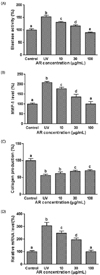
The effects of AR on elastase activity, MMP-1 level, collagen content, and mRNA expression of MMP-1 were determined using a spectrophotometric method, ELISA, a dye binding method, and quantitative real time RT-PCR, respectively. Exposure of HaCaT cells to UVB significantly increased elastase and MMP-1 levels by 1.5-fold and 2.0-fold, respectively, both of which were reversed by AR treatment in a dose-dependent manner (Fig. 4). Elastase activity and the MMP-1 level were reduced to 89.4 ± 2.7% and 100.6 ± 11.6%, respectively, of non-irradiated control cells with 100 µg/mL AR treatment. Accordingly, collagen content, assessed by SirCol collagen staining assay, was reduced to 56% of the non-irradiated control cells by UVB irradiation, and 100 µg/mL AR pretreatment dose-dependently restored it to 70.5% of the control value. Furthermore, mRNA expression of MMP-1, determined by quantitative real time RT-PCR, was dramatically increased by 3-fold after UVB exposure, but was normalized by AR treatment in a dose-dependent manner. The effects of AR seemed not to originate from its cytotoxicity as AR showed no significant cytotoxicity (Fig. 2).
ROS and reactive nitrogen species (RNS) production by UVB irradiation contributes to photo-damage, inflammation, immune suppression, gene mutation and carcinogenesis. Therefore, substances able to inhibit these reactive species could be used in the prevention of photoaging and skin cancer [18]. The results of the present study demonstrate the hydrogen donating capability as well as NO scavenging activity of AR, suggesting that AR is a photo-protective agent.
DPPH is a stable free radical donor that is widely used to test the free radical scavenging effects of natural antioxidants. NO is a free radical that reacts with oxygen to form oxides of nitrogen. NO and RNS have been shown to be associated with common forms of skin diseases. NO, liberated following UV irradiation, plays a significant role in initiating erythema and inflammation [19]. NO can combine with UV-induced superoxide to form peroxynitrite, which exists in equilibrium with peroxynitrous acid. These reactive nitrogen species are very toxic, and can cause DNA damage.
UV irradiation is also associated with formation of ROS leading to skin aging and photo-carcinogenesis [14]. UVB rays interact with cellular chromophores and photosensitizers, resulting in the generation of singlet oxygen, superoxide anions, hydroxyl radicals and hydrogen peroxide [1]. The results demonstrate the protective potential of AR from antioxidant activity through the inhibition of ROS formation induced by UVB in human keratinocytes HaCaT cells.
Several reports have suggested that botanical antioxidants, including polyphenols, have been shown to be associated with a reduced incidence of ROS-mediated photocarcinogenesis and photoaging [2920]. Recently, Kim et al. reported on AR containing a high total phenolic content, a well-known antioxidant compound [11]. Therefore, the high polyphenol content in AR could be partly responsible for its antioxidant effect. The antioxidant properties of polyphenols are due to the presence of their many phenolic hydroxyl groups, which have a high potential for scavenging free radicals [21]. Phenolic compounds donate hydrogen to reactive radicals and break the chain reaction of lipid oxidation at the initiation step [22].
Under normal conditions, skin produces enzymes such as elastase and collagenase at a similar rate as the aging process occurs and age increases. However, these enzymes are produced at a faster rate with overexposure to UV and excessive ROS, resulting in faster degradation of elastin and collagen, which form the main foundation of the extracellular matrix (ECM) of the dermis and epidermis [2]. ROS accumulation in photoaged skin has been suggested to associate with increased MMP-1 expression, which could be reversed by promoting the capacity of antioxidant defenses including catalase [23], SOD, and glutathione [24]. Therefore, the redox balance accountable for the protection against photo-oxidative stress in keratinocytes [24] and redox regulation of MMP-1 might represent strategies for photoaging prevention.
The mechanisms by which AR suppressed activation of MMP-1 in HaCaT cells exposed to UVB are probably attributed to the attenuation of UVB-mediated ROS accumulation as a result of the augmentation of endogenous antioxidant capacity as well as high polyphenol content in AR. The functional components in AR other than total phenolic content [11] have not been thoroughly investigated. Polyphenols have been shown to inhibit photoaging by preventing the expression of MMPs. Polyphenol-rich pomegranate fruit extract has been shown to inhibit UVB-induced up-regulation of MMP-1 [25], polyphenol-rich fraction of Quinoa significantly inhibited MMP-1 mRNA expression in human dermal fibroblast cells [26], and extracts of Coffea arabica [27] prevented skin cells from UVB-induced photoaging by inhibiting the expression of MMP-1, 3, and 9. Furthermore, polyphenols such as epigallocatechin-3-gallate (EGCG) from green tea, equol from soy, and Salvianolic acid B, have been reported to inhibit UVB-induced expression of MMP-1 [282930].
UV irradiation damages the antioxidant defense system, impairs signal transduction pathways in skin cells, and degrades ECM proteins including collagen, elastin, proteoglycans, and fibronectin [3132]. Therefore, collagenase as well as elastase inhibitors have been identified as potential therapeutic agents that protect against photoaging and wrinkle formation. The results of the present study clearly demonstrate that AR treatment significantly attenuates ROS production and elastase activity, MMP-1 at the mRNA and protein levels, and collagen content in a dose-dependent manner. These data suggest that AR is a potential candidate for the prevention and treatment of skin photoaging.
In conclusion, the present study provides the first evidence that AR inhibited UVB-induced ROS production and induction of MMP-1 as a result of augmentation of antioxidant enzymes in HaCaT human keratinocytes. These results suggest that AT might act as an effective inhibitor of UVB-modulated signaling pathways and might serve as a photo-protective agent.
Figures and Tables
 | Fig. 1Antioxidant effect of AR on DPPH radical and nitric oxide scavenging in a cell-free system.(A) DPPH radical scavenging activity of AR. (B) Nitric oxide scavenging activity of AR. The level of DPPH radical was measured spectrophotometrically at 515 nm. The NO scavenging capacity was assessed by Griess assay. The IC50 values for DPPH radical and NO scavenging activities were 1.88 mg/mL and 6.77 mg/mL, respectively. Each bar represents the mean ± SD (n = 6). The bars with a different letter are significantly different from each other at the level of P < 0.05.
|
 | Fig. 2Effect of AR on UVB-induced cell cytotoxicity and ROS formation of human keratinocytes.(A) Cells were pretreated with AR prior to UVB irradiation (30 mJ/cm2) and harvested 24 h later. Cytotoxicity was determined by LDH leakage assay. (B) Intracellular ROS levels induced by UVB were determined by the DCF-DA method. Cells, treated with AR prior to UV irradiation, were incubated with 20 µM of DCF-DA for 30 min, and harvested after 24 h. ROS formation was analyzed with a fluorometer (excitation; 486 nm, emission; 530 nm). Each bar represents the mean ± SD (n = 3). The bars with a different letter are significantly different from each other at the level of P < 0.05.
|
 | Fig. 3Effect of AR on antioxidative enzyme activity.Cells were pretreated with AR for 24 h prior to UVB irradiation (30 mJ/cm2) and harvested 24 h later. (A) Catalase activity was measured using a colorimetric assay kit. One unit of catalase was defined as the amount of enzyme required to decompose 1 µM of H2O2 per minute. (B) SOD activity was measured using a colorimetric assay kit. Enzyme activity is expressed as average enzyme activity unit per mg protein ± SD (n = 3). The bars with a different letter are significantly different from each other at the level of P < 0.05.
|
 | Fig. 4Effect of AR on UVB-induced degradation of elastin and collagen, and collagen synthesis in human keratinocytes.Cells were pretreated with AR for 24 h prior to UVB irradiation (30 mJ/cm2) and harvested 24 h later. (A) Elastase activity was measured spectrophotometrically with N-Succ-(Ala)3-p-nitroanilide as a substrate, and the release of p-nitroaniline was monitored. (B) MMP-1 level was determined using an ELISA kit. (C) Collagen production was determined using a SirCol collagen assay kit. (D) Expression of MMP-1 mRNA was determined by quantitative real time RT-PCR. GAPDH was used as an internal control. Each bar represents the mean ± SD (n = 3). The bars with a different letter are significantly different from each other at the level of P < 0.05.
|
References
1. Pillai S, Oresajo C, Hayward J. Ultraviolet radiation and skin aging: roles of reactive oxygen species, inflammation and protease activation, and strategies for prevention of inflammation-induced matrix degradation - a review. Int J Cosmet Sci. 2005; 27:17–34.

2. Svobodová A, Psotová J, Walterová D. Natural phenolics in the prevention of UV-induced skin damage. A review. Biomed Pap Med Fac Univ Palacky Olomouc Czech Repub. 2003; 147:137–145.

3. de Gruijl FR. Photocarcinogenesis: UVA vs UVB. Methods Enzymol. 2000; 319:359–366.
4. Podhaisky HP, Riemschneider S, Wohlrab W. UV light and oxidative damage of the skin. Pharmazie. 2002; 57:30–33.
5. Sander CS, Chang H, Salzmann S, Müller CS, Ekanayake-Mudiyanselage S, Elsner P, Thiele JJ. Photoaging is associated with protein oxidation in human skin in vivo. J Invest Dermatol. 2002; 118:618–625.

6. Jang J, Ye BR, Heo SJ, Oh C, Kang DH, Kim JH, Affan A, Yoon KT, Choi YU, Park SC, Han S, Qian ZJ, Jung WK, Choi IW. Photo-oxidative stress by ultraviolet-B radiation and antioxidative defense of eckstolonol in human keratinocytes. Environ Toxicol Pharmacol. 2012; 34:926–934.

7. Meyer W, Neurand K, Radke B. Elastic fibre arrangement in the skin of the pig. Arch Dermatol Res. 1981; 270:391–401.

8. Wiedow O, Schröder JM, Gregory H, Young JA, Christophers E. Elafin: an elastase-specific inhibitor of human skin. Purification, characterization, and complete amino acid sequence. J Biol Chem. 1990; 265:14791–14795.

9. Nichols JA, Katiyar SK. Skin photoprotection by natural polyphenols: anti-inflammatory, antioxidant and DNA repair mechanisms. Arch Dermatol Res. 2010; 302:71–83.

10. Kim TC. Korean Resources Plants, IV. Seoul: Seoul National University Press;1998. p. 183–189.
11. Kim AJ, Han MR, Kim MH, Lee M, Yoon TJ, Ha SD. The antioxidant and chemopreventive potentialities of Mosidae (Adenophora remotiflora) leaves. Nutr Res Pract. 2010; 4:30–35.

12. Kim SH, Choi HS, Lee MS, Chung MS. Volatile compounds and antioxidant activities of Adenophora remotiflora. Korean J Food Sci Technol. 2007; 39:109–113.
13. Huang D, Ou B, Prior RL. The chemistry behind antioxidant capacity assays. J Agric Food Chem. 2005; 53:1841–1856.

14. Chan WH, Wu CC, Yu JS. Curcumin inhibits UV irradiation-induced oxidative stress and apoptotic biochemical changes in human epidermoid carcinoma A431 cells. J Cell Biochem. 2003; 90:327–338.

15. Kraunsoe JA, Claridge TD, Lowe G. Inhibition of human leukocyte and porcine pancreatic elastase by homologues of bovine pancreatic trypsin inhibitor. Biochemistry. 1996; 35:9090–9096.

16. Pfaffl MW. A new mathematical model for relative quantification in real-time RT-PCR. Nucleic Acids Res. 2001; 29:e45.

17. Wawszczyk J, Kapral M, Hollek A, Węglarz L. In vitro evaluation of antiproliferative and cytotoxic properties of pterostilbene against human colon cancer cells. Acta Pol Pharm. 2014; 71:1051–1055.
18. Russo PA, Halliday GM. Inhibition of nitric oxide and reactive oxygen species production improves the ability of a sunscreen to protect from sunburn, immunosuppression and photocarcinogenesis. Br J Dermatol. 2006; 155:408–415.

19. Deliconstantinos G, Villiotou V, Stavrides JC. Nitric oxide and peroxynitrite released by ultraviolet B-irradiated human endothelial cells are possibly involved in skin erythema and inflammation. Exp Physiol. 1996; 81:1021–1033.

20. Afaq F, Mukhtar H. Botanical antioxidants in the prevention of photocarcinogenesis and photoaging. Exp Dermatol. 2006; 15:678–684.

21. Sawa T, Nakao M, Akaike T, Ono K, Maeda H. Alkylperoxyl radical-scavenging activity of various flavonoids and other phenolic compounds: implications for the anti-tumor-promoter effect of vegetables. J Agric Food Chem. 1999; 47:397–402.

22. Gülçin I, Beydemir S, Alici HA, Elmastaş M, Büyükokuroğlu ME. In vitro antioxidant properties of morphine. Pharmacol Res. 2004; 49:59–66.

23. Shin MH, Rhie GE, Kim YK, Park CH, Cho KH, Kim KH, Eun HC, Chung JH. H2O2 accumulation by catalase reduction changes MAP kinase signaling in aged human skin in vivo. J Invest Dermatol. 2005; 125:221–229.

24. Pluemsamran T, Onkoksoong T, Panich U. Caffeic acid and ferulic acid inhibit UVA-induced matrix metalloproteinase-1 through regulation of antioxidant defense system in keratinocyte HaCaT cells. Photochem Photobiol. 2012; 88:961–968.

25. Zaid MA, Afaq F, Syed DN, Dreher M, Mukhtar H. Inhibition of UVB-mediated oxidative stress and markers of photoaging in immortalized HaCaT keratinocytes by pomegranate polyphenol extract POMx. Photochem Photobiol. 2007; 83:882–888.

26. Graf BL, Cheng DM, Esposito D, Shertel T, Poulev A, Plundrich N, Itenberg D, Dayan N, Lila MA, Raskin I. Compounds leached from quinoa seeds inhibit matrix metalloproteinase activity and intracellular reactive oxygen species. Int J Cosmet Sci. 2015; 37:212–221.

27. Chiang HM, Lin TJ, Chiu CY, Chang CW, Hsu KC, Fan PC, Wen KC. Coffea arabica extract and its constituents prevent photoaging by suppressing MMPs expression and MAP kinase pathway. Food Chem Toxicol. 2011; 49:309–318.

28. Bae JY, Choi JS, Choi YJ, Shin SY, Kang SW, Han SJ, Kang YH. (-)Epigallocatechin gallate hampers collagen destruction and collagenase activation in ultraviolet-B-irradiated human dermal fibroblasts: involvement of mitogen-activated protein kinase. Food Chem Toxicol. 2008; 46:1298–1307.

29. Reeve VE, Widyarini S, Domanski D, Chew E, Barnes K. Protection against photoaging in the hairless mouse by the isoflavone equol. Photochem Photobiol. 2005; 81:1548–1553.

30. Sun Z, Park SY, Hwang E, Zhang M, Jin F, Zhang B, Yi TH. Salvianolic acid B protects normal human dermal fibroblasts against ultraviolet B irradiation-induced photoaging through mitogen-activated protein kinase and activator protein-1 pathways. Photochem Photobiol. 2015; 91:879–886.

31. Rittié L, Fisher GJ. UV-light-induced signal cascades and skin aging. Ageing Res Rev. 2002; 1:705–720.

32. Uitto J. The role of elastin and collagen in cutaneous aging: intrinsic aging versus photoexposure. J Drugs Dermatol. 2008; 7:s12–s16.


 PDF
PDF ePub
ePub Citation
 Print
Print


 XML Download