Abstract
The objective of this study was to evaluate the inhibitory activity of natural products, against growth of Escherichia coli (ATCC 25922) and Salmonella typhimurium (KCCM 11862). Chitosan, epigallocatechin gallate (EGCG), and garlic were used as natural bioactives for antibacterial activity. The testing method was carried out according to the disk diffusion method. All of chitosan, EGCG, and garlic showed inhibitory effect against the growth of E. coli and Salmonella typhi. To evaluate the antibacterial activity of natural products during storage, chicken skins were inoculated with 106 of E. coli or Salmonella typhi. The inoculated chicken skins, treated with 0.5, 1, or 2% natural bioactives, were stored during 8 day at 4℃. The numbers of microorganisms were measured at 8 day. Both chitosan and EGCG showed significant decrease in the number of E. coli and Salmonella typhi in dose dependent manner (P < 0.05). These results suggest that natural bioactives such as chitosan, EGCG may be possible to be used as antimicrobial agents for the improvement of food safety.
When people ingested food that was spoiled or contaminated with bacterial microorganisms, food poisoning frequently occurred. Industrialized civilization has led to dramatically increased standards of hygiene today, but food poisoning remains a major cause of death worldwide (Sibel, 2003). In recent years, there has been a dramatic increase in the number of reported cases of food-borne illnesses. A variety of microorganisms, including Escherichia coli and Salmonella typhimurium may lead to food spoilage, one of the most important concerns of the food industry. Many attempts, such as use of synthetic chemicals, have been made to control microbial growth and to reduce the incidence of food poisoning and spoilage with antimicrobial chemicals.
Much has been reported about the potential for natural antimicrobial compounds to replace or reduce reliance on synthetic food preservatives. In the last 20 years, hundreds of studies demonstrated antimicrobial activity of natural compounds against pathogenic or spoilage organisms. However, few of these have been translated into real food applications. There has been a tendency to infer from a zone of inhibition on an agar plate that a putative antimicrobial agent has potential for use in real foods. Rarely has the investigator bothered to demonstrate antagonism or biocidal activity in foods (Sibel, 2003). Recently, consumers have concerned about the side effects and want safer materials for preventing and controlling pathogenic microorganisms in foods. Some natural substances of plant origin have good antimicrobial properties and have been used as seasonings for centuries. Spices and aromatic vegetable materials have long been used in food not only for their flavor and fragrance qualities and appetizing effects but also for their preservative and medicinal properties. Since ancient times throughout the world, these have been used for preventing food spoilage and deterioration and also for extending shelf life of foods, while attempts to characterize these properties in the laboratory date back to the early 1900s. It has been extensively reported that the natural substances have shown antimicrobial functions against food-borne pathogens (Bin et al., 2007). The ultimate goal of our study is to develop by using natural compounds to reduce pathogens in foods and to extend the shelf life of foodstuffs.
Chitosan is a polycationic polymer with specific structure and properties. It contains more than 5000 glucosamine units and is obtained commercially from shrimp and crabshell chitin (a N-acetylglucosamine polymer) by alkaline deacetylaion. It has been shown to be useful in many different areas as an antimicrobial compound in agriculture, as potential elictor of plant defense responses, as an additive in the food industry, as a hydrating agent in cosmetics, and more recently as a pharmaceutical agent in biomedicine. The antimicrobial activity of chitosan against different groups of microorganisms, such as bacteria has received considerable attention (Entsar et al., 2003).
Green tea (Camellia sinensis L.) and its extract in particular have shown many health benefits to humans and animals, including chemo-preventive, anticarcinogenic, antioxidant and antimicrobial activities (Cooper; 2005). In terms of the antimicrobial activity, major food-borne pathogens such as Escherichia coli, Salmonella typhimurium, Listeria monocytogenes, Staphylococcus aureus, and Camphlobacter jejuni have been reported to be inhibited by tea components (Hamilton-Miller; 1995) which may be from different types of tea or tea extract, including oolong, jasmine and black tea. Most studies have used crude extracts of tea for the evaluation of antimicrobial activity against pathogens including food-borne pathogens, and many measured the activities of pure standards of tea polyphenols to confirm the activities found in tea extract. (Weidou, 2006).
Garlic is used world-wide as a spice, food, and folk medicine (Yoshida et al., 1999). In vitro evidence of the antimicrobial activity of fresh and freeze-dried garlic extracts against many bacterial, fungi, and viruses supports these applications. Early steps involved in identifying the active constituents of garlic were the discovery of the compound allicin formed when garlic cloves are crushed and that its formation depends upon the action of the enzyme alliinase of the bundle sheath cells upon the alliin of mesophyll cells. The classic studies attributed the antibacterial properties of garlic clove homogenates for allicin. These properties were confirmed against Escherichia coli and Staphylococcus aureus for garlic clove homogenates plus related garlic compounds (Ross et al., 2001). However, antimicrobial activities of chitosan, EGCG against E. coli and Salmonella typhi have not yet been fully studied. Also, E. coli and Salmonella typhi is indicator organism of food borne pathogenic bacteria. Therefore, the aim of the present study was to investigate the antibacterial effects of bioactives against two common pathogenic bacteria (E. coli and Salmonella typhi).
E. coli (ATCC 25922) and Salmonella typhi (KCCM 11862) were purchased from KCCM. The medium for growth and preservation of bacteria was nutrient agar (NA, Kisan Biotech) and Luria-Bertani medium (LB, Kisan Biotech).
Acetic acid was purchased from Sigma-Aldrich (Saint Louis, MO, USA), chitosan (70% water-soluble) from Iljin Pharmaceuticals, and EGCG (98%, TEAVIGO™) from DSM Nutritional Products (Basel, Switzerland). Garlic was purchased in bulb from a supermarket in Seoul. The skin was removed and it was washed with water, mashed, freeze dried, sealed airtight, and stored it in a freezer. Chitosan, EGCG, and garlic powder were all dissolved in 100 ml of distilled water and set at concentrations of 0.5%, 1%, and 2%. Each solution was filtered with Whatman paper No. 3 (Whatman Ltd, Maidstone, England), and sterilized with 0.22 µm PVDF membrane (Millipore corporation, Bedford, USA), and stored at 4℃.
Screening of chitosan, EGCG and garlic powder for antibacterial activity against E. coli or Salmonella typhi was done using the paper disc method (Davidsom & Parish, 1989). For the treatment, 50 µl each of 0.5%, 1%, and 2% chitosan, EGCG, and garlic powder solutions were slowly absorbed into the sterilized paper disc (diameter: 8 mm, Watman, England) and adhered to the surface of the plate on which 106 CFU/ml E. coli or Salmonella typhi had been inoculated. Sterilized distilled water was used as a control. After culturing for 24 hours in an incubator at 37℃, the clear zone around the disk was measured and the antibacterial activity compared and analyzed.
Chicken was purchased from a chicken processing vendor near Seoul, transported on ice and used for experimentation. The chicken skin was cut uniformly to 20 cm2 pieces. In order to maintain those in a sterile state, the pieces were then immersed in 100 ppm sodium hypochlorite (NaClO) for 30 min and rinsed with distilled water. They were placed on a sterilized stainless steel net to remove efflux and used as samples. 106 CFU/ml of E. coli or Salmonella typhi was inoculated into each 20 cm2/piece chicken skin in a 60 mm dish. Chitosan, EGCG or garlic solutions were then added at the 0.5%, 1%, and 2% concentrations in a 60 mm dish, and stored in a refrigerator at 4℃. The amount of bacteria on each piece of chicken was measured at 8 days.
The results for the day 0 sample were analyzed immediately after the natural compounds treatment. Each chicken piece was moved and 10 ml sterilized 0.1% (w/v) peptone water was added at a ratio of 1:1. It was mixed thoroughly for 60 seconds, after which a 0.1 ml of solution was taken for analysis. The 0.1 ml of sample solution was diluted in steps from 10-1 to 10-5 and inoculated to the NA medium, which was then cultured at 37℃ for 24-48 hours. To measure number of bacteria, colonies of 30-300 formed on the medium were counted using a colony counter. The bacteria count on the medium was then multiplied by the multiple of dilution and converted to Log10 CFU/ml.
We used the statistics program SPSS 12.0 and ANOVA (analysis of variance) to analyze the antibacterial effects and the mean values of the samples for E. coli count, pH and generation time for the chicken pieces to which natural additives were added. The analysis of differences by various factors used Turkey's test, every experiment was repeated three times, and the results were expressed as mean ± standard error.
To study its antimicrobial efficiency, various concentrations of the natural bioactivies were added to cultures of E. coli and compared with that of the commonly used preservative acetic acid. Table 1 shows the mean ± standard error for inhibitory diameters of bioactives against E. coli. The clear zone sizes of the 0.5% and 1% acetic acid treatments were 16.3 mm and 20.0 mm, respectively, showing significant increase of 2 to 2.5 times in comparison with that of control (P < 0.05) (Table 1, Fig 1A). The clear zone size of the 0.5% chitosan treatment was 10.6 mm, 1.3 times greater than that of the control. The clear zone size of 1% chitosan treatment was 11.3 mm, 1.4 times greater than that of the control (Table 1, Fig 1 B). The clear zone size of the 0.5% EGCG treatment was 17.0 mm, 2.1 times greater than that the control, and 1% EGCG treatment also was 2.4 times greater in comparison with that of control (P < 0.05) (Table 1, Fig 1 C). The clear zone size of the 1% garlic treatment was 9.7 mm, 1.2 times greater than that of the control, and 2% was 11.7 mm, 1.5 times greater in comparison with that of control (Table 1, Fig 1 D).
To study its antimicrobial efficiency, various concentrations of the natural bioactivies were added to cultures of Salmonella typhi and compared with that of the commonly used preservative acetic acid. Table 2 shows the mean ± standard error for inhibitory diameters of bioactives against Salmonella typhi.
The clear zone sizes of the 0.5% and 1% ascetic acid treatments were 16.7 mm and 21.3 mm, respectively, showing significant increase of 2.1 to 2.7 times in comparison with that of the control (P < 0.05) (Table 2, Fig 2 A). The clear zone size of the 0.5% chitosan treatment was 10.7 mm, 1.3 times greater than that of the control. The clear zone size of 1% chitosan treatment was 11.3 mm, 1.4 times greater that of the control (Table 2, Fig 2 B). The clear zone size of the 0.5% EGCG treatment was 22.7 mm, 2.8 times greater than that of the control, and 1% EGCG treatment also was 3.4 times greater in comparison with that of control (P < 0.05) (Table 2, Fig 2 C). The clear zone size of the 0.5% garlic treatment was 9.7 mm, 1.2 times greater than that of the control, and 1% was 14.0 mm, 1.8 times greater (Table 2, Fig 2 D).
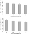
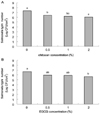
During storage at 4℃, results are presented in order to determine the effects on microbiological decontamination of chicken skins treated with different concentration of bioactives. For 0.5% chitosan treatment, the E. coli count decreased by 85% vs. control, showing dose-dependent effects (Fig 3A), with similar results obtained in the number of E. coli count by EGCG treatment after 8 days storage (P < 0.05) (Fig 3B). The numbers of Salmonella typhi on chicken skins treated with 0.5% chitosan or EGCG were also significantly lower than those of controls after 8 days storage (P < 0.05) (Fig 4A and 4B).
Synthetic chemicals are often used as antimicrobials in food processing and storage to eliminate food-borne pathogens, many of which have built resistance to the antibiotics. However consumer's awareness and concerns over the potential risks of synthetic food bioactivies to human health have renewed the interests in using naturally occurring alternatives. Therefore, much attention in recent years has been focused on natural bioactives. Numerous studies have shown the effect of chitosan on microbial inhibition. Higher antibacterial activity of chitosan was reported by several workers (No et al., 2002). For example, No et al. (2002) observed that chitosan and its enzymatic hydrolyzates inhibited growth of Gram-negative bacteria. And chitosan acted mainly on the outer surface of the bacteria. At a lower concentration (< 0.2 mg/mL), the polycationic chitosan does probably bind to the negatively charged bacterial surface to cause agglutination, while at higher concentrations, the larger number of positive charges may have imparted a net positive charge to the bacterial surfaces to keep them in suspension (Entsar et al., 2003).
Our present study revealed that chitosan showed significantly highly antimicrobial activities against E. coli and Salmonella typhi. Also, E. coli and Salmonella typhi numbers of chicken skins treated with chitosan were significantly lower (P < 0.05) than controls during storage.
Most studies have used crude extract of tea for the evaluation of antimicrobial activity against pathogens including food-borne pathogens (Weiduo et al., 2006), and many measured the activities of pure standards of tea polyphenols to confirm the activities found in tea extracts. Recently purified EGCG demonstrated an increased antimicrobial activity towards E. coli (Weiduo, et al., 2006).
In this study, EGCG showed significantly antimicrobial activities against E. coli and Salmonella typhi. Also, E. coli and Salmonella typhi numbers of chicken skins treated with EGCG were significantly lower than controls during storage.
Garlic is known to have antibacterial activity, and Bakri (2005) were the first to demonstrate that the antibacterial action of garlic is mainly due to allicin. The sensitivity of various bacterial and clinical isolates to pure preparations of allicin (Bakri et al., 2005) is significant. In the present study, garlic showed antimicrobial activities against E. coli and Salmonella typhi. The weak antimicrobial activity of 0.5% garlic may be due to the losses of sulfur compounds, and also due to the nature of garlic itself, which is volatile and hydrophobic. It appears that chitosan, EGCG, strongly inhibits the growth of E. coli and Salmonella typhi.
Even though bioactives have been considered as versatile biopolymers in agriculture applications, its potential uses as an animicrobial compound and its mode of action need to be further studied in depth. Furthermore, it is a current matter of discussion as to whether these bioactivies may have the potential to influence physiological functions or metabolism in the microorganisms. Therefore, a significant increase in the number of scientific studies to obtain evidence to support this can expected.
Figures and Tables
Fig. 1
Effects of natural bioactives on the growth of E. coli
Antimicrobial activity of various bioactives was carried out according to disk diffusion method by measuring the inhibitory zone size. a; 0%, b; 0.5%, c; 1.0%, d; 2.0% of selected bioactive compound solution

Fig. 2
Effects of natural bioactives on the growth of Salmonella typhi
Antimicrobial activity of various bioactives was carried out according to disk diffusion method by measuring the inhibitory zone size. a; 0%, b; 0.5%, c; 1.0%, d; 2.0% of selected bioactive compound solution

Fig. 3
Inhibitory effect of chitosan and EGCG on the growth of E. coli on the chicken skin during storage at 4℃
The chicken skin surfaces (20cm2/piece) were inoculated with 106 CFU/ml of Salmonella typhi. The natural bioactives used were 0.5, 1 or 2% water soluble chitosan (A) and EGCG (B). The chicken skins treated with chitosan or EGCG were stored at 4℃ and the numbers of Salmonella typhi were counted at 8 days. The number of Salmonella typhi was expressed as mean Log10CFU/ml for the triple treatments.

Fig. 4
Inhibitory effect of chitosan and EGCG on the growth of Salmonella typhi on chicken skin during storage at 4℃
The chicken skin surfaces (20cm2/piece) were inoculated with 106 CFU/ml of Salmonella typhi. The natural bioactives used were 0.5, 1 or 2% water soluble chitosan (A) and EGCG (B). The chicken skins treated with chitosan or EGCG were stored at 4℃ and the numbers of Salmonella typhi were counted at 8 days. The number of Salmonella typhi was expressed as mean Log10CFU/ml for the triple treatments.

References
1. Bakri IM, Douglas CWI. Inhibitory effect of garlic extract on oral bacteria. Arch Oral Biol. 2005. 50:645–651.

2. Bin S, Yi ZC, John DB, Harold C. Antibacterial properties and major bioactive components of Cinnamon Stick (Cinnamomum burmannii): activity against food-borne pathogenic bacteria. J Agric Food Chem. 2007. 55:5484–5490.
3. Cho MH, Bae EK, Ha SD, Park JY. Application of natural antimicrobials to food industry. Food Science and Industry. 2005. 38:36–45.
4. Chung KT, Lu Z, Chou MW. Mechanism of inhibition of tannic acid and related compounds on the growth of intestinal bacteria. Food Chem Toxicol. 1998. 36:1053–1060.

5. Cooper R, Morre DJ, Morre J. Medicinal Benefits of Green Tea: Part I. Review of Noncancer Health Benefits. J Altern Complement Med. 2005. 11:521.

6. Daljit SA, Jasleen Kaur. Antimicrobial activity of spices. Int J Antimicrob Agents. 1999. 2:257–262.
7. Davidsom PM, Parish ME. Method for testing the efficacy of food antimicrobials. Food Technology. 1989. 43:148–155.
8. Dimitrios G, Ioannis A, Panagiota K, Georgios B, Spyridon AG. Effect of rosemary extract, chitosan and a-tocopherol on microbiological parameters and lipid oxidation of fresh pork sausages stored at 4℃. Meat Sci. 2007. 76:172–181.
9. Elena DR, Rosa C, Miguel P, Carlos AC. Comparison of pathogenic and spoilage bacterial levels on refrigerated poultry parts following treatment with trisodium phosphate. Food Microbiol. 2006. 23:195–198.

10. Elena DR, Monica PM, Miguel P, Carlos AC, Rosa C. Effect of various chemical decontamination treatments on natural microflora and sensory characteristics of poultry. Int J Food Microbiol. 2007. 115:268–280.

11. Entsar IR, Mohamed ET, Christian VS, Guy S, Walter S. Chitosan as antimicrobial agent: Applications and mode of action. Biomacromolecules. 2003. 6:1457–1465.

12. Fernandez LJ, Zhi N, Aleson CL, Perez JA, Kuri V. Antioxidant and antibacterial activities of natural extracts: application in beef meatballs. Meat Sci. 2005. 69:371–380.

13. Hamilton-Miller JM. Antimicrobial properties of tea (Camellia sinensis L.). Antimicrob Agents Chemother. 1995. 39:2375–2377.

14. Kong YJ, Park BK, Oh DH. Antimicrobial activity of Quercus mongolica leaf ethanol extracts and organic acids against food-borne microorganisms. Korean Journal of Food Science and Technology. 2001. 33:178–183.
15. Mau JL, Chen CP, Hsieh PC. Antimicrobial effect of extracts from Chinese Chive, Cinnamon, and Corni Fructus. J Agric Food Chem. 2001. 49:183–188.

16. No HK, Park NY, Lee SH, Hwang HJ, Meyers SP. Antibacterial activities of chitosans and chitosan oligemers with different molecular weights on Spoilage Bacteria isolated from Tofu. J Food Sci. 2002. 67:1511–1514.

17. Olasupo NA, Fitzgerald DJ, Gasson MJ, Narbad A. Activity of natural antimicrobial compounds against Escherichia coli and Salmonella enterica serovar Tuphimurium. Lett Appl Microbiol. 36:448–451.
18. Patsias A, Chouliara I, Badeka A, Savvaidis IN, Kontominas MG. Shelf-life of a chilled precooked chicken product stored in air and under modified atmospheres: microbiological, chemical, sensory attributes. Food Microbiol. 2006. 23:423–429.

19. Ross ZM, O'gara EA, Hill DJ, Sleightholme H.V, Maslin DJ. Antimicrobial properties of Garlic oil against human enteric bacteria: Evaluation of methodologies and comparisons with Garlic oil sulfides and Garlic powder. Appl Environ Microbiol. 2001. 67:475–480.

20. Shin JH, Lee SY, Dougherty RH, Rasco B, Kang DH. Combined effect of mild heat and acetic acid treatment for inactivating Escherichia coli 0157:H7, Listeria monocytogenes and Salmonella typhimurium in an asparagus puree. J Appl Microbiol. 2006. 101:1140–1151.

21. Sibel R. Natural antimicribials for the minimal processing of foods. Camb Munic Code 1988 Camb Mass. 2003. 1–295.
22. Sallam KhI, Ishioroshi M, Samejima K. Antioxidant and antimicrobial effects of garlic in chicken sausage. Lebenson Wiss Technol. 2004. 37:849–855.

23. Sallam KI, Samejima K. Effects of Trisosdium phosphate and Sodium chloride dipping on the microbial quality and shelf life of refrigerated tray-packaged chicken breasts. Food Sci Biotechnol. 2004. 13:425–438.
24. Sallam KI, Samejima K. Microbiological and chemical quality of ground beef treated with sodium lactate and sodium chloride during refrigerated storage. Lebenson Wiss Technol. 2004. 37:865–892.

25. Sami F, Pierluigi C, Valentina C, Carlo IG, Tuberoso CI, Alberto A, Sandro D, Nejib M, Paolo C. Antimicrobial activity of Tunisian Quince (Cydonia oblonga miller) pulp and peel polyphenolic extracts. J Agric Food Chem. 2007. 55:963–969.

26. Serge A, David M. Antimicrobial properties of allicin from garlic. Microbes Infect. 1999. 125–129.
27. Weiduo SI, Joshua G, Rong T, Milosh K, Raymond Y, Yulong Y. Bioassay-guided purification and identification of antimicrobial components in Chinese green tea extract. J Chromatogr A. 2006. 1125:204–210.

28. Wang Y, Zhou P, Yu J, Pan X, Wang P, Lan W, Tao S. Antimicrobial effect of Chitooligosaccharides produced by Chitosanase from Pseudomonas CUY8. Asia Pac J Clin Nutr. 2007. 16:174–177.


 PDF
PDF ePub
ePub Citation
 Print
Print


 XML Download