Abstract
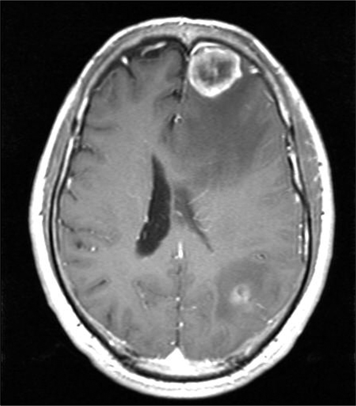
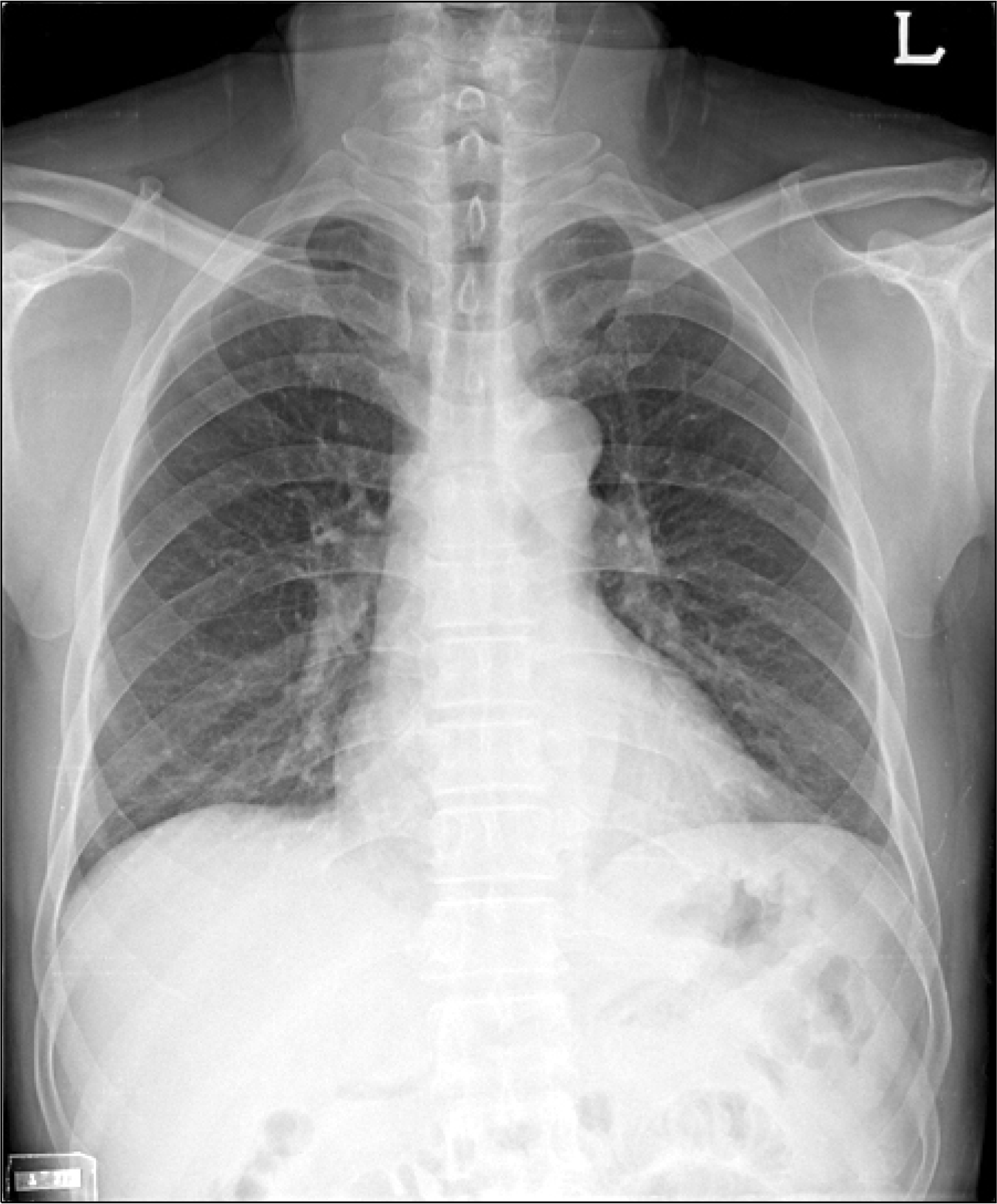
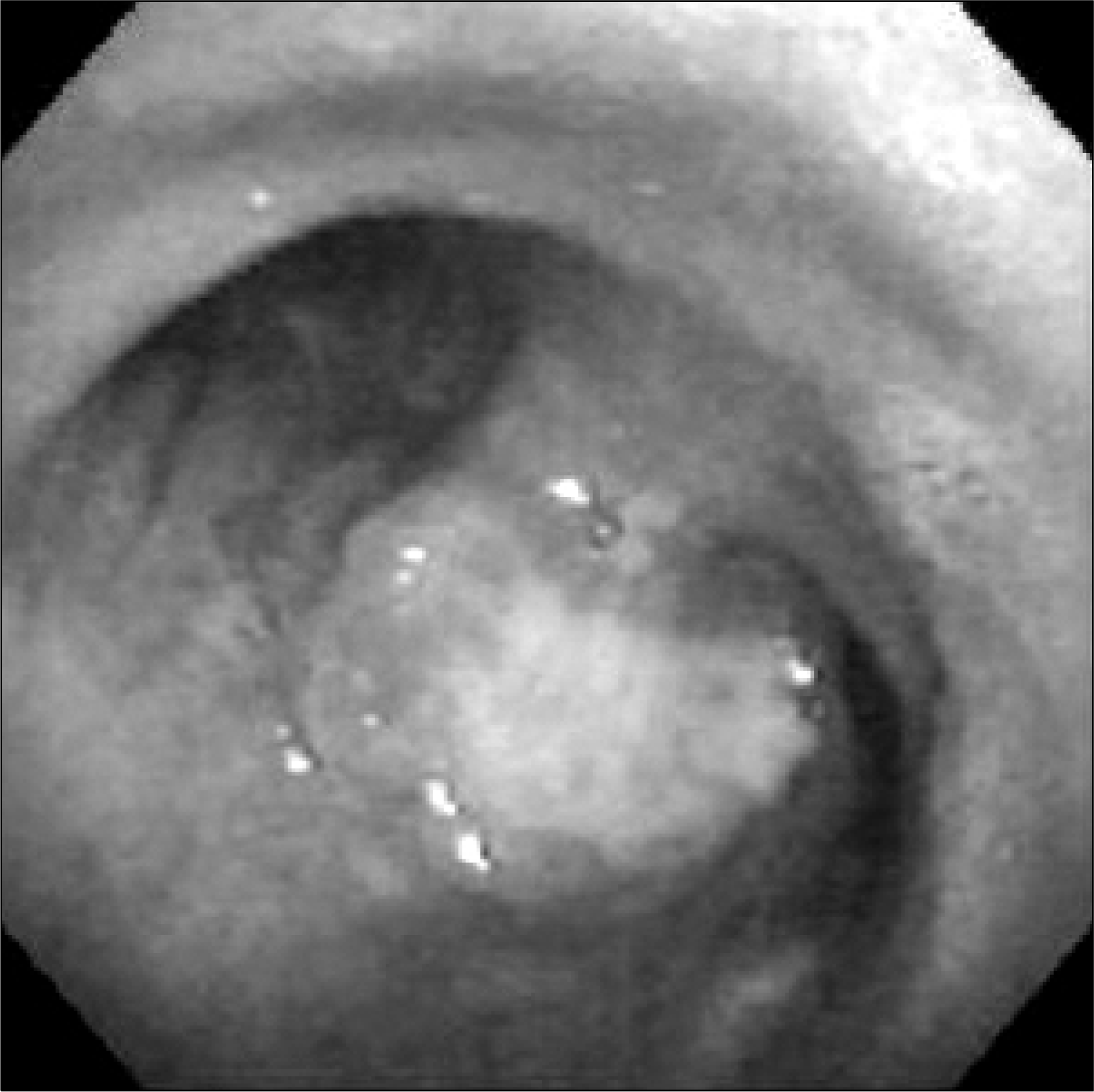
A 59-year-old man was rushed to the emergency room. The patient complained of headache with impaired memory function. Brain MRI showed a necrotic tumor in Lt cerebral hemisphere, with severe peritumoral edema (Fig. 1). Pathologic examination of the brain lesion confirmed that the tumor was a small cell lung cancer (SCLC). Chest computed tomography revealed a large soft tissue mass with central necrosis at subcarinal area in spite of an initial normal chest X-ray (Fig. 2). Bronchoscopic biopsy of the polypoid mass at subcarina revealed that the mass was a SCLC (Fig. 3). This is the case of SCLC only with an extrapulmonary symptoms despite of a normal chest X-ray. When metastatic brain tumor was found, appropriate chest evaluation should be performed even though chest X-ray was normal because brain is a common site of invasion of lung cancer.
Go to : 


 PDF
PDF ePub
ePub Citation
 Print
Print


 XML Download