Abstract
Immunoglobulin G4 (IgG4)-related sclerosing disease involving the lung is a rare condition, and this is characterized by an elevated serum IgG4 level, fibrotic inflammation with numerous IgG4-positive plasma cells and a response to steroid therapy. We present here a case of pulmonary IgG4-related disease in a 75-year-old man who presented with cough and yellowish sputum for the previous 3 months. The chest images showed a consolidative mass in the right lower lobe that suggested mucinous bronchioloalveolar carcinoma. The wedge resected specimen revealed an ill-defined graytan, firm lesion. Microscopically, the lesion showed a diffuse lymphoplasmacytic infiltration with irregular fibrosis in the alveolar interstitium and bronchovascular bundles. There were numerous IgG4-positve plasma cells and these cells were diffusely distributed. The serum IgG4 level was elevated on the postoperative check-up (249 mg/dL). After corticosteroid therapy for 7 months, the patient's symptoms and radiologic abnormalities were improved.
References
1. Hamed G, Tsushima K, Yasuo M, et al. Inflammatory lesions of the lung, submandibular gland, bile duct and prostate in a patient with IgG4-associated multifocal systemic fibrosclerosis. Respirology. 2007; 12:455–457.

2. Kobayashi H, Shimokawaji T, Kanoh S, et al. IgG4-positive pulmonary disease. J Thorac Imaging. 2007; 22:360–362.

3. Takato H, Yasui M, Ichikawa Y, et al. Nonspecific interstitial pneumonia with abundant IgG4-positive cells infiltration, which was thought as pulmonary involvement of IgG4-related autoimmune disease. Intern Med. 2008; 47:291–294.

4. Shigemitsu H, Koss MN. IgG4-related interstitial lung disease: a new and evolving concept. Curr Opin Pulm Med. 2009; 15:513–516.

5. Kamisawa T, Okamoto A. IgG4-related sclerosing disease. World J Gastroenterol. 2008; 14:3948–3955.

6. Yamashita K, Haga H, Kobashi Y, et al. Lung involvement in IgG4-related lymphoplasmacytic vasculitis and interstitial fibrosis: report of 3 cases and review of the literature. Am J Surg Pathol. 2008; 32:1620–1626.
Figures
Fig. 1.
(A) The chest X-ray shows parenchymal consolidation at the right costophrenic angle with pleural effusion. (B, C) The chest computed tomography scans reveal air space consolidation and ground-glass attenuations.

Fig. 2.
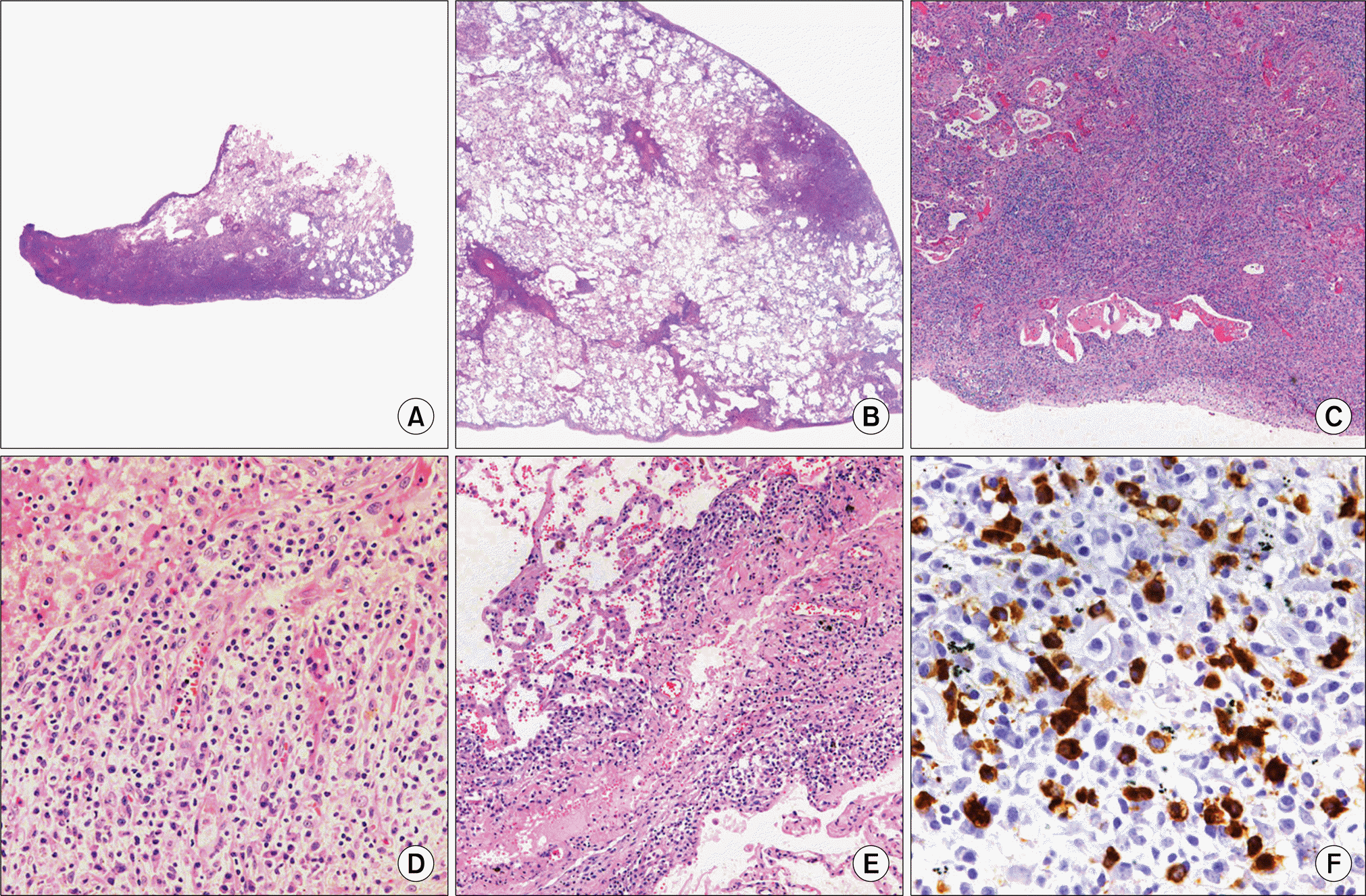
(A, B) Scanning views of the VATS wedge resected specimen show a subpleural patchy mononuclear cells infiltration. (C) The low power view shows a dense lymphoid cell infiltration with fibrosis in the alveolar interstitium and bronchovascular bundles (H&E satin, ×40). (D) The lesion is composed of mature plasma cells and some lymphocytes (H&E stain, ×200). (E) Phlebitis is also present in the interlobular septal vein (H&E stain, ×100). (F) Many plasma cells are positive for IgG4 (×400).


 PDF
PDF ePub
ePub Citation
 Print
Print


 XML Download