Abstract
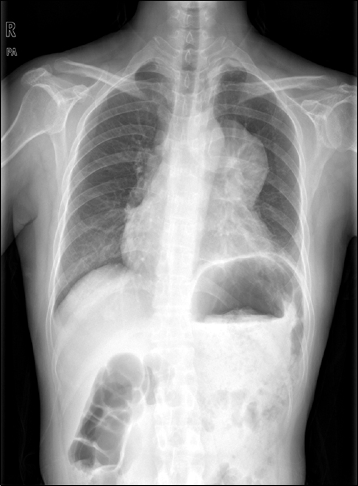
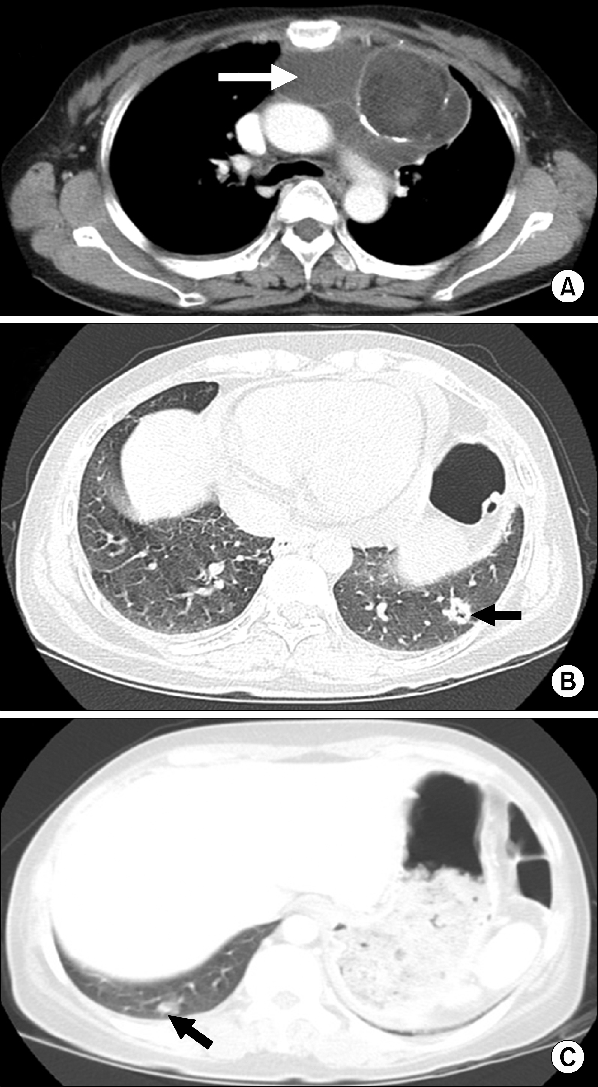
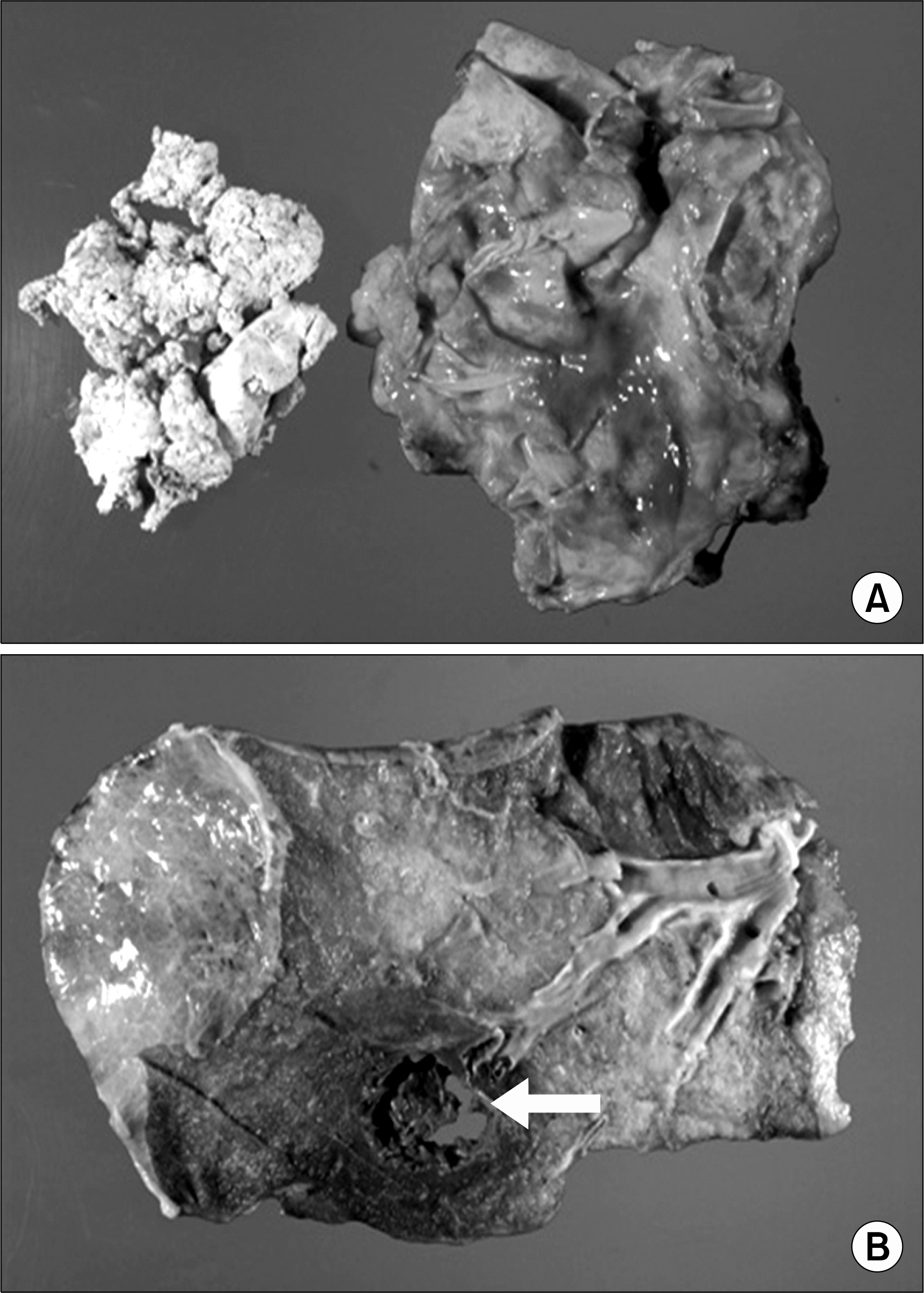
A mediastinal mass and 2 lung masses on the lower lobes were detected by chest CT in a 44-year old woman. The mass on the left lower lobe was diagnosed as an adeno type of cancer by preoperative lung biopsy. But the preoperative lung biopsy exam for the mass on the right lower lobe couldn’t perform because of patient's noncooperation. Removal of the mediastinal mass, left lower pulmonary lobectomy and wedge resection of the right lower pulmonary lobe were done. The histopathologic diagnosis of the resected mediastinal mass was mature cystic teratoma and both lung masses were the adeno type of cancer with the same histopathologic patterns. We experienced a rare case of lung adeno cancer together with mediastinal mature cystic teratoma.
References
1. Golash V. A giant anterior mediastinal teratoma presenting as orthopnea and dysphagia in an adult. J Thorac Cardiovasc Surg. 2005; 130:612–613.

2. Lewis BD, Hurt RD, Payne WS, Farrow GM, Knapp RH, Muhm JR. Benign teratomas of the mediastinum. J Thorac Cardiovasc Surg. 1983; 86:727–731.

3. Arai K, Ohta S, Suzuki M, Suzuki H. Primary immature mediastinal teratoma in adulthood. Eur J Surg Oncol. 1997; 23:64–67.

4. Nichols CR. Mediastinal germ cell tumors: clinical features and biologic correlates. Chest. 1991; 99:472–479.
5. Jun YB, Sohn ST, Jeon SH, et al. Medistinal teratoma with pleural and pericardial effusion teratoma with pleural and pericardial effusion. Korean J Thorac Cardiovasc Surg. 1998; 31:436–439.
6. Choe JW, Kim YL. Benign mediastinal cystic teratoma complicated by cardiac tamponade due to trauma. Korean J Thorac Cardiovasc Surg. 2006; 39:729–732.


 PDF
PDF ePub
ePub Citation
 Print
Print


 XML Download