

 PDF
PDF ePub
ePub Citation
 Print
Print


Abstract
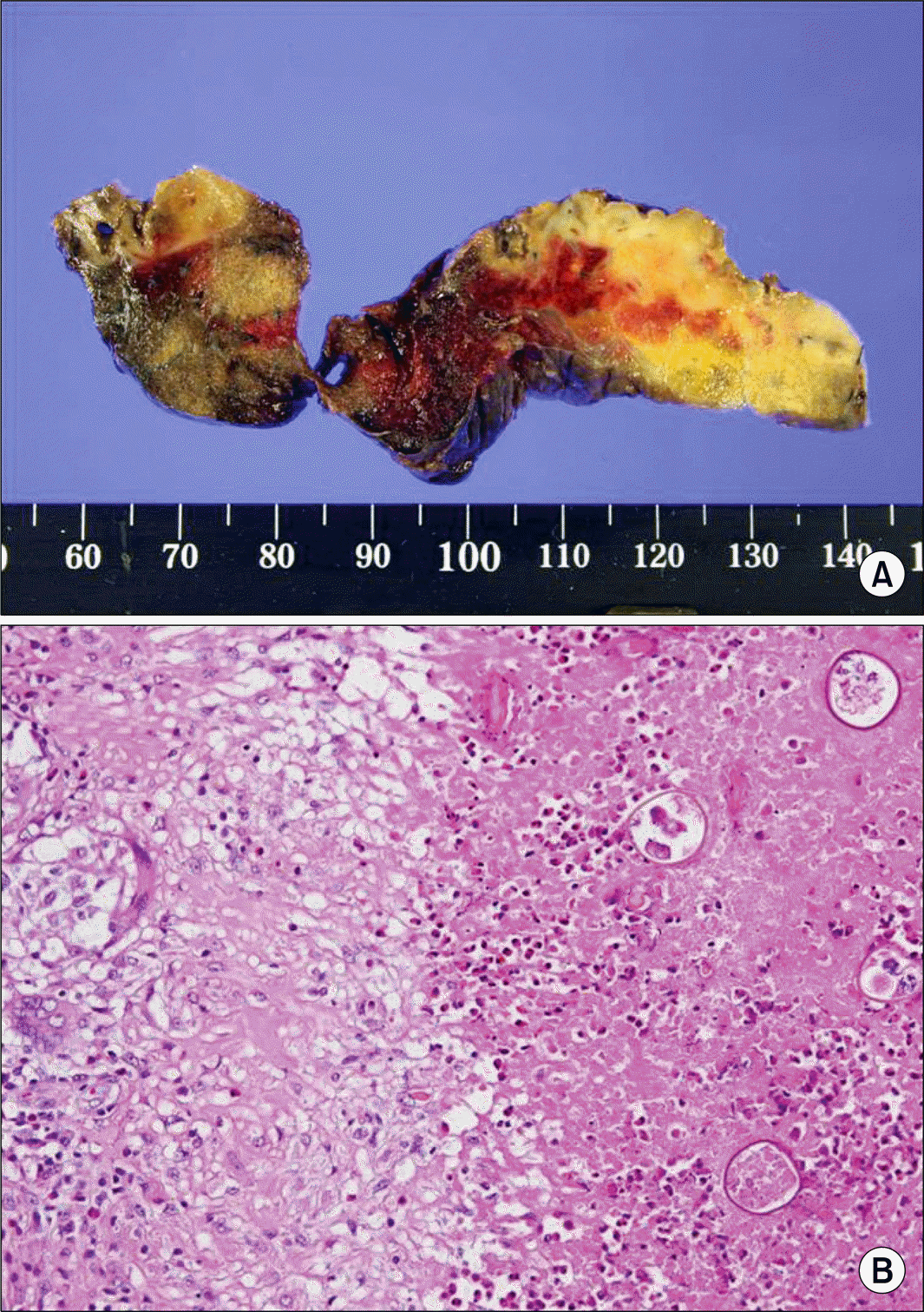
A 50-year-old man was admitted to our hospital with a complaint of blood-tinged sputum. Chest computed tomography (CT) showed a 37 mm sized heterogeneously lobulated enhancing mass in the central aspect of the left upper lobe and this mass was abutting the adjacent mediastinum (Fig. 1). There was either a focal fibrotic pleural thickening or fissural thickening adjacent to the pulmonary mass. Conglomerated enlarged lymph nodes were observed in the left paraaortic and left anterior cardiophrenic angle areas. 18-Fluorodeoxyglucose positron emission tomography demonstrated hyper-metabolic activity in the left upper lobe and the right thyroid gland (Fig. 2). The pathology of the fine needle aspiration from the thyroid gland revealed papillary carcinoma. Since primary or metastatic lung cancer could not be ruled out, video-assisted thoracic surgery was performed. The microscopic findings demonstrated numerous eosinophil infiltrations and many eggs of paragonimiasis westermani, which were observed in the necrotized granuloma area (Fig. 3). He was underwent subtotal thyroidectomy. Moreover, he subsequently received iodine ablation therapy due to papillary carcinoma with regional lymph node invasion.
Fig. 1.
(A) The chest CT shows a 37 mm sized heterogeneously lobulated enhancing mass in the central aspect of the LUL and the mass is abutting the adjacent mediastinum (arrow). (B) There is either a focal fibrotic pleural thickening or fissural thickening adjacent to a pulmonary nodule (arrow). (C, D) Conglomerated enlarged lymph nodes are observed in the left paraaortic area and an additional 34 mm sized heterogeneously enhancing mass is located in the left anterior cardiophrenic angle (arrowhead).
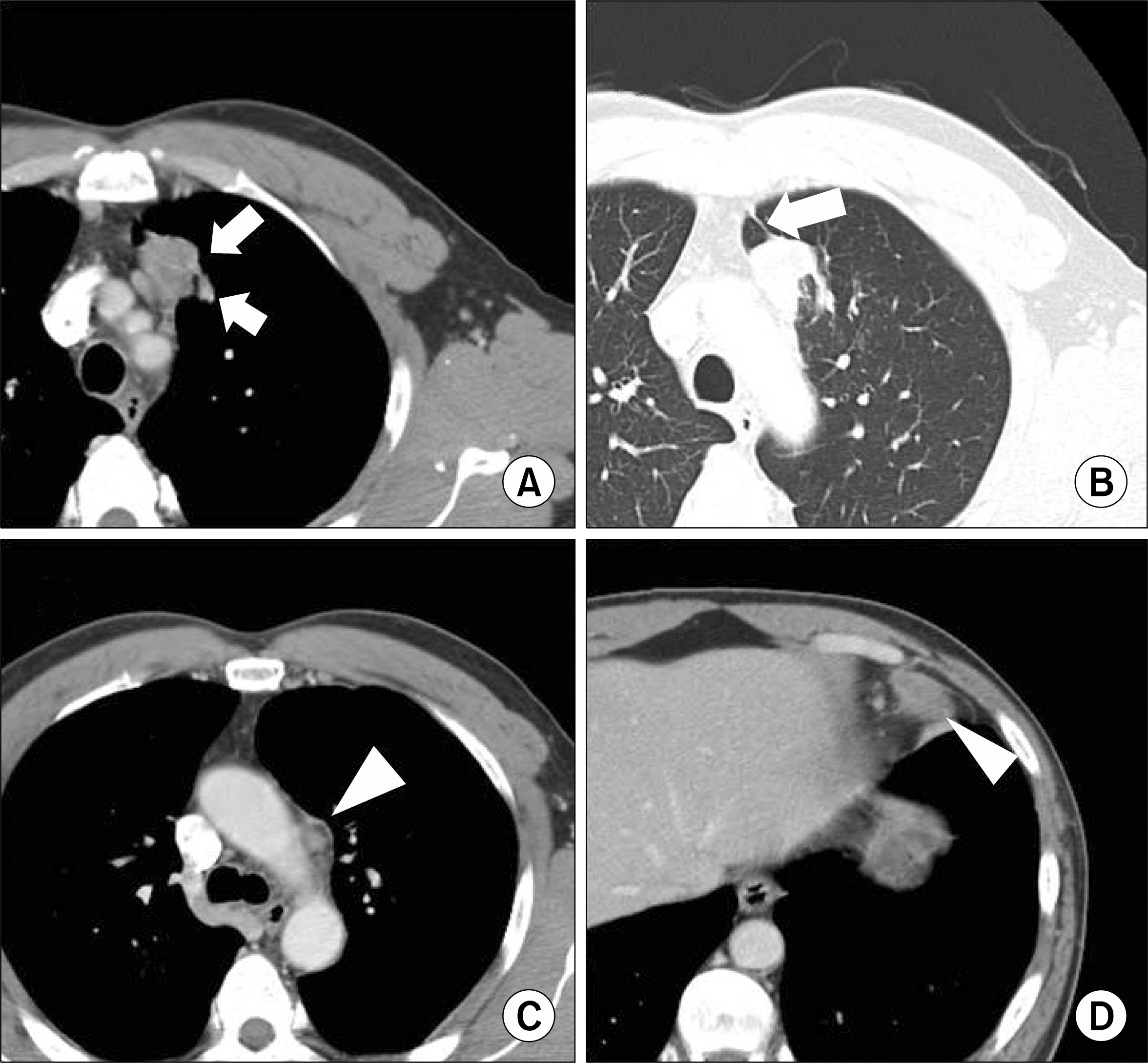
Fig. 2.
The findings of 18-Fluorodeoxyglucose PET-CT reveal hypermetabolic nodules on the left upper lobe (maxSUV: 6.8), the mediastinum and the cardiophrenic angle (maxSUV: 5.2) (arrow). There is another hyper-metabolic lesion (maxSUV: 4.0) with an irregular margin on the right thyroid gland (arrowhead). maxSUV: maximal standardized uptake value.
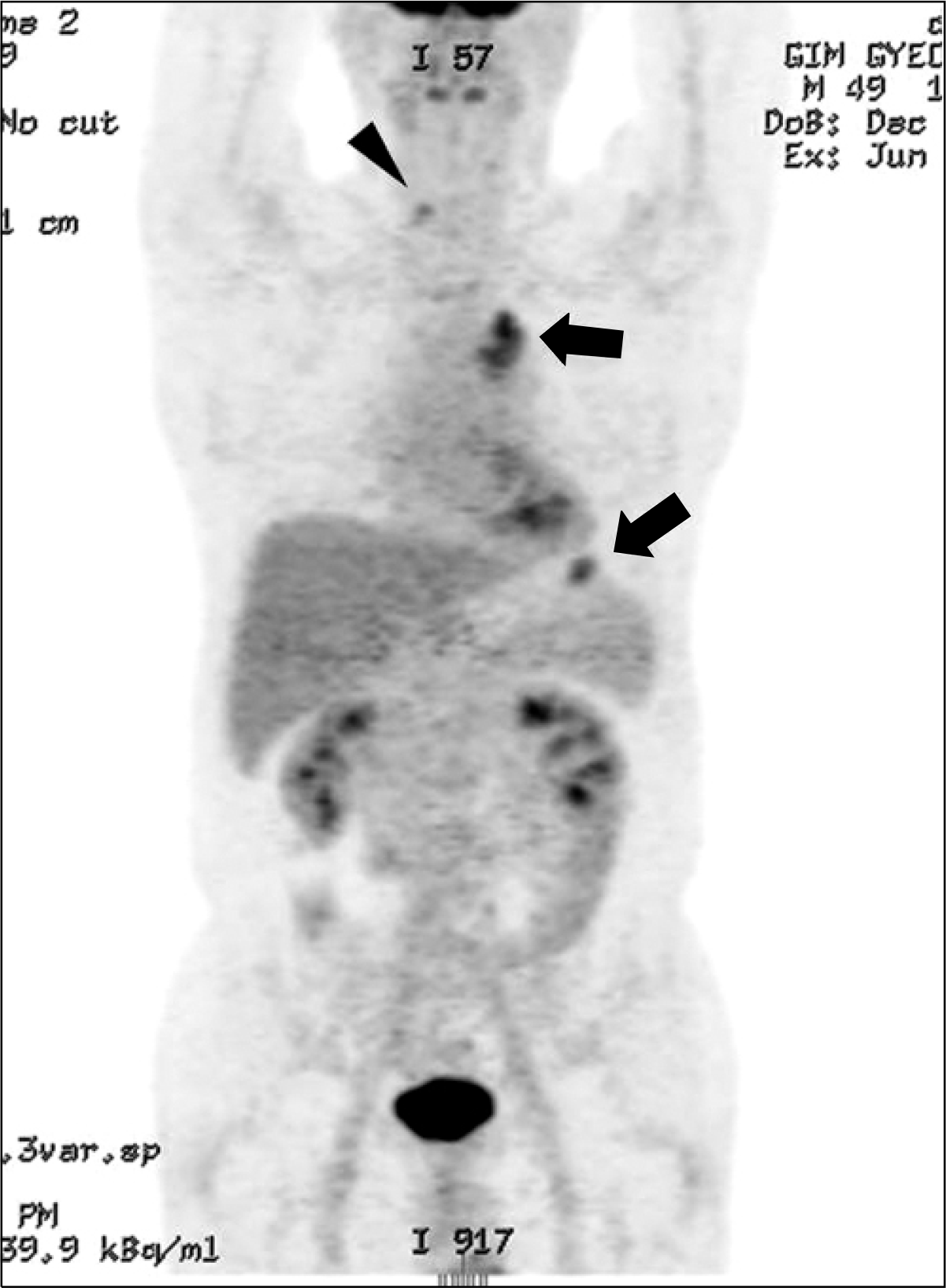
Fig. 3.
(A) The resected lung tissue displays various sized necrotic areas and focal hemorrhagic foci. (B) The microscopic findings demonstrate some small granulomas with central necrotic foci being identified. Numerous eosinophil infiltrations are detected, and many eggs of paragonimiasis westermani are observed in the necrotic area (H&E stain, ×200).
 XML Download