Abstract
Epithelioid trophoblastic tumor is a rare type of gestational trophoblastic disease that is distinct from placental site trophoblastic tumor and choriocarcinoma, and epithelioid trophoblastic tumor has features resembling a carcinoma. We report here on an epithelioid trophoblastic tumor that was discovered as a solitary pulmonary nodule in the lung of a 50-year-old woman. The patient had suffered from a hydatidiform mole 20 years previously. Wedge resection of the lung was done and this showed a 1.9×1.5 cm sized, relatively well defined mass composed of mononuclear tumor cells admixed with hyaline-like material and necrosis. The tumor cells were positive for EMA, Cam5.2, α-inhibin, PLAP and hCG. After consulting the gynecologic department, a 7.5×6.5 cm sized mass was discovered in the uterine fundus. Hysterectomy was then done. The tumor cells were same to those of the lung mass. The lung mass is considered to be metastasis from the epithelioid trophoblastic tumor of the uterus. She has been an uneventful clinical course for three years.
Go to : 
References
1. Mazur MT, Lurain JR, Brewer JI. Fatal gestational choriocarcinoma: clinicopathologic study of patients treated at a trophoblastic disease center. Cancer. 1982; 50:1833–1846.

2. Mazur MT, Kurman RJ. Gestational trophoblastic disease and related lesions. Blaustein A, Kurman RJ, editors. Blaustein's pathology of the female genital tract. 4th ed.New York: Springer-Verlag;1994. p. 1049.

3. Oh HS, Shin JH, Song SH, et al. Epithelioid trophoblastic tumor: a case report and review of the literature. Korean J Obstet Gynecol. 2001; 44:1330–1335.
4. Lee EJ, Lee HW, Lee JS, et al. A case of epithelioid trophoblastic tumor. Korean J Gynecol Oncol Colposc. 2001; 12:152–155.

5. Urabe S, Fujiwara H, Miyoshi H, et al. Epithelioid trophoblastic tumor of the lung. J Obstet Gynaecol Res. 2007; 33:397–401.

6. Shih IM, Kurman RJ. Epithelioid trophoblastic tumor: a neoplasm distinct from choriocarcinoma and placental site trophoblastic tumor simulating carcinoma. Am J Surg Pathol. 1998; 22:1393–1403.
7. Hamazaki S, Nakamoto S, Okino T, et al. Epithelioid trophoblastic tumor: morphological and immunohistochemical study of three lung lesions. Hum Pathol. 1999; 30:1321–1327.

8. Kuo KT, Chen MJ, Lin MC. Epithelioid trophoblastic tumor of broad ligament: a case report and review of the literature. Am J Surg Pathol. 2004; 28:405–409.
Go to : 
Figures
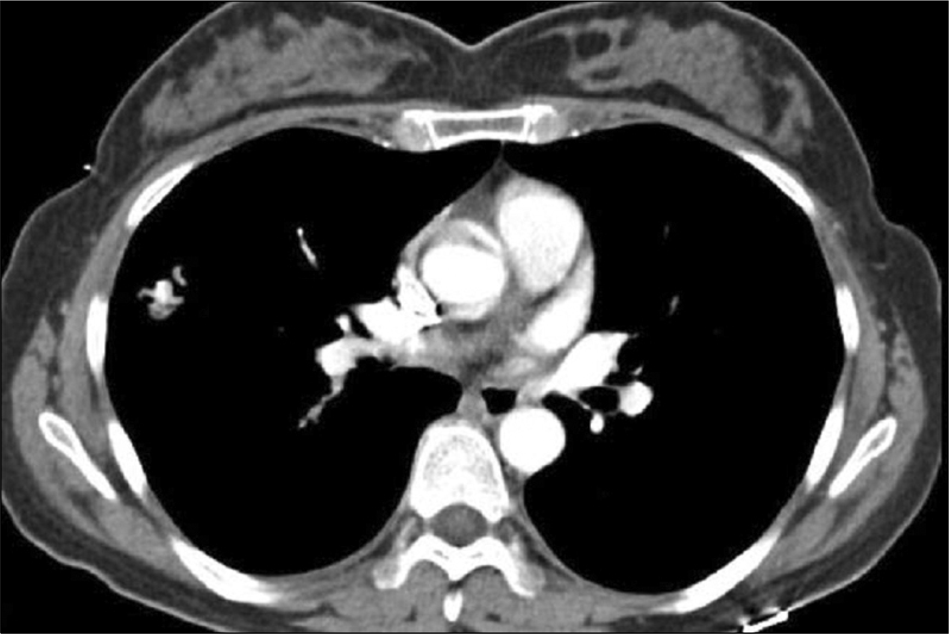
 | Fig. 1.Chest CT showed a focal consolidating lesion with central calcification in the right middle lobe. |
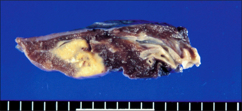
 | Fig. 2.Grossly, there was an ovoid yellowish white mass with necrosis and whitish calcification. It was not connected with the bronchus. |
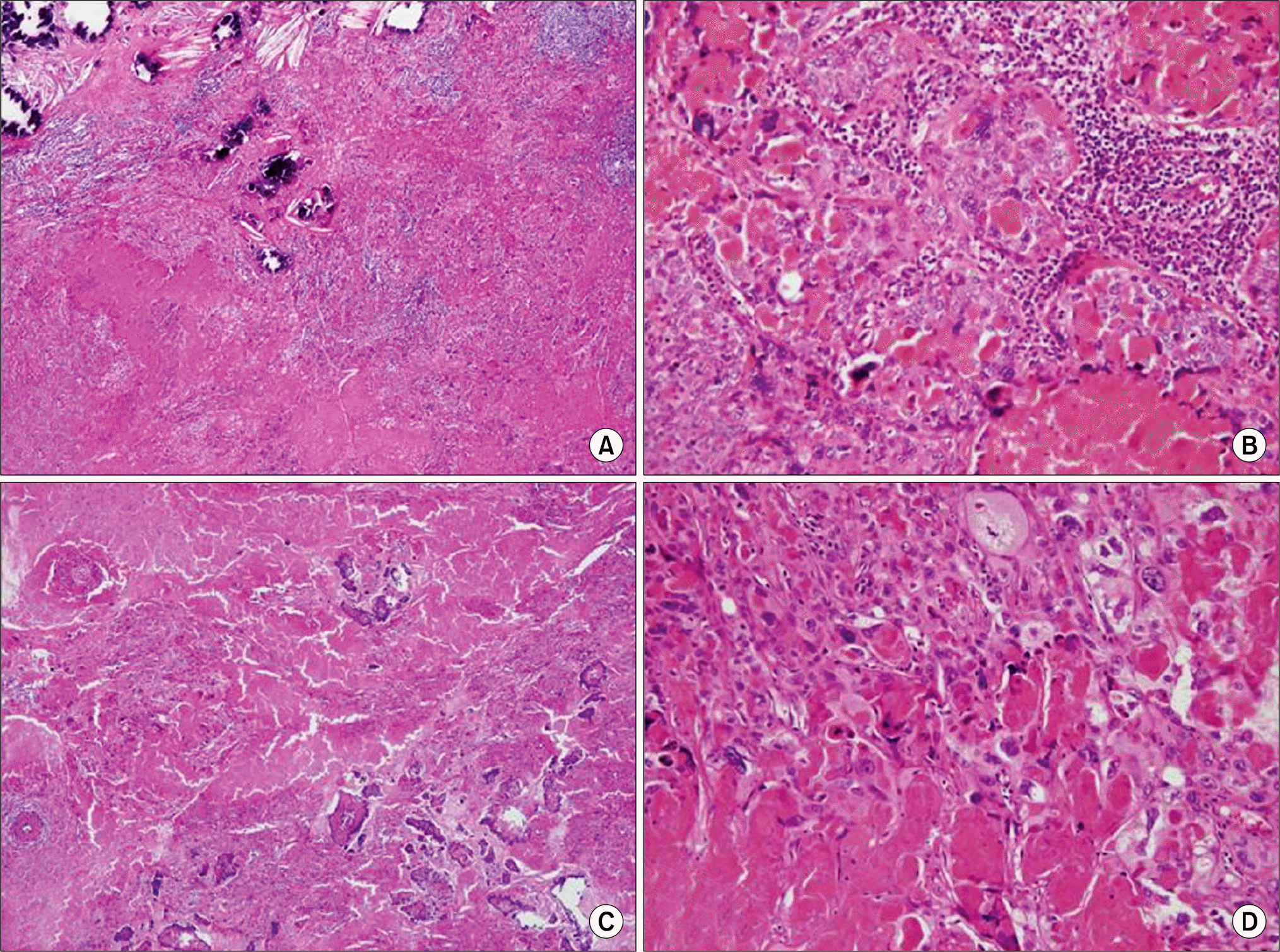
 | Fig. 3.Histological findings for the lung mass (A, B). The low power view showed nests of atypical cells with necrosis and multifocal calcification (A, H&E stain, ×40). The tumor cells were mononucleate trophoblasts admixed with dense eosinophilic material (B, H&E stain, ×200). The histological findings for the mass in the uterus (C, D). Microscopically, there was massive necrosis and a solid mass of atypical cells (C, H&E stain, ×40). The tumor showed infiltrating nests of cells surrounded by dense hyaline material (D, H&E stain, ×200). |


 PDF
PDF ePub
ePub Citation
 Print
Print


 XML Download