

 PDF
PDF ePub
ePub Citation
 Print
Print


Abstract
We report a rare case of a primary mediastinal synovial sarcoma. A 44-year-old man had a well-defined tumor in the left posterior mediastinum involving the left lower lobe of the lung, as detected on chest computed tomography, and underwent an en bloc excision and a left lower lobectomy. Grossly, the tumor measured 8.0 cm in the greatest diameter, with a solid and tan-white cut surface. Histologically, the tumor was mainly composed of spindle-shaped cells with a few foci of epithelial differentiation. Immunohistochemical studies were focally positive for cytokeratin, and diffusely positive for vimentin and bcl-2. Epithelial membrane antigen, S-100 protein, desmin, smooth muscle actin, calretinin, and CD34 were all negative. The SYT-SSX1 gene fusion transcript was detected by a reverse transcription-polymerase chain reaction, which was diagnostic of primary synovial sarcoma of the mediastinum. We also reviewed the literature with regard to the clinicopathologic, immunohistochemical, and molecular studies of primary intrathoracic synovial sarcoma.
References
1. Witkin GB, Miettinen M, Rosai J. A biphasic tumor of the mediastinum with feature of synovial sarcoma: a report of four cases. Am J Surg Pathol. 1989; 13:490–499.
2. Weiss SW, Goldblum JR. Enzinger and Weiss's Soft Tissue Tumors. 4th ed.St Louis, MO: Mosby;2001. p. 1487–1503.
3. Essary LR, Vargas SO, Fletcher CD. Primary pleuropulmonary synovial sarcoma: reappraisal of a recently described anatomic subset. Cancer. 2002; 94:459–469.
4. Keel SB, Bacha E, Mark EJ, Nielsen GP, Rosenberg AE. Primary pulmonary sarcoma: a clinicopathologic study of 26 cases. Mod Pathol. 1999; 12:1124–1131.
5. Okamoto S, Hisaoka M, Daa T, Hatakeyama K, Iwamasa T, Hashimoto H. Primary pulmonary synovial sarcoma: a clinicopathologic, immunohistochemical, and molecular study of 11 cases. Hum Pathol. 2004; 35:850–856.

6. Zeren H, Moran CA, Suster S, Fishback NF, Koss MN. Primary pulmonary sarcomas with features of monophasic synovial sarcoma: a clinicopathological, immunohistochemical, and ultrastructural study of 25 cases. Hum Pathol. 1995; 26:474–480.
7. Hisaoka M, Hashimoto H, Iwamasa T, Ishikawa K, Aoki T. Primary synovial sarcoma of the lung: report of two cases confirmed by molecular detection of SYT-SSX fusion gene transcripts. Histopathology. 1999; 34:205–210.
8. Guillou L, Coindre JM, Gallagher G, et al. Detection of the synovial sarcoma translocation t(X;18)(SYT;SXX) in paraffin-embedded tissues using reverse transcriptase-polymerase chain reaction: a reliable and powerful diagnostic tool for the pathologists. A molecular analysis of 221 mesenchymal tumors fixed in different fixatives. Hum Pathol. 2001; 32:105–112.
9. Begueret H, Galateau-Salle F, Guillou L, et al. Primary intrathoracic synovial sarcoma: a clinicopathologic study of 40 t(X;18)-positive cases from the French Sarcoma Group and the Mesopath group. Am J Surg Pathol. 2005; 29:339–346.
10. Guillou L, Benhattar J, Bonichon F, et al. Histologic grade, but not SYT-SSX fusion type, is an important prognostic factor in patients with synovial sarcoma: a multicenter, retrospective analysis. J Clin Oncol. 2004; 22:4040–4050.
11. Hirakawa N, Naka T, Yamamoto I, Fukuda T, Tsuneyoshi M. Overexpression of bcl-2 protein in synovial sarcoma: a comparative study of other soft tissue spindle cell sarcomas and an additional analysis by fluorescence in situ hybridization. Hum Pathol. 1996; 27:1060–1065.
12. Suster S, Fisher C, Moran CA. Expression of bcl-2 oncoprotein in benign and malignant spindle cell tumors of soft tissue, skin, serosal surface, and gastrointestinal tract. Am J Surg Pathol. 1998; 22:863–872.
13. Saito T, Oda Y, Sakamoto A, et al. Prognostic value of the preserved expression of the E-cadherin and catenin families of adhesion molecules and of beta-catenin mutations in synovial sarcoma. J Pathol. 2000; 192:342–350.
Figures
Fig. 1.
Computed tomography (CT) images showed a soft tissue mass in the left posterior mediastinum.
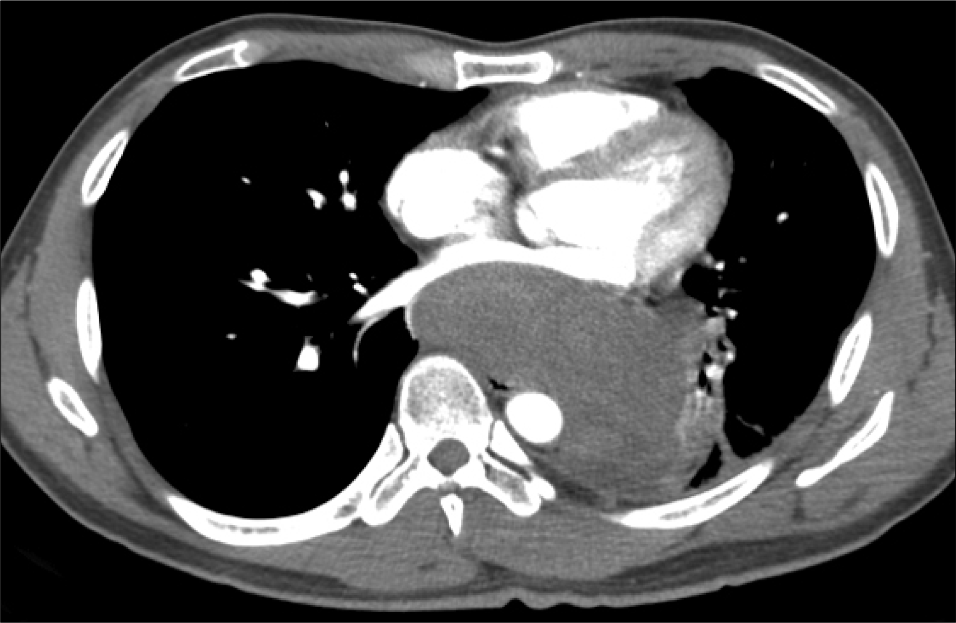
Fig. 2.
Gross photograph of the resected synovial sarcoma. The mass was relatively well-demarcated and showed expansile growth to the lung.
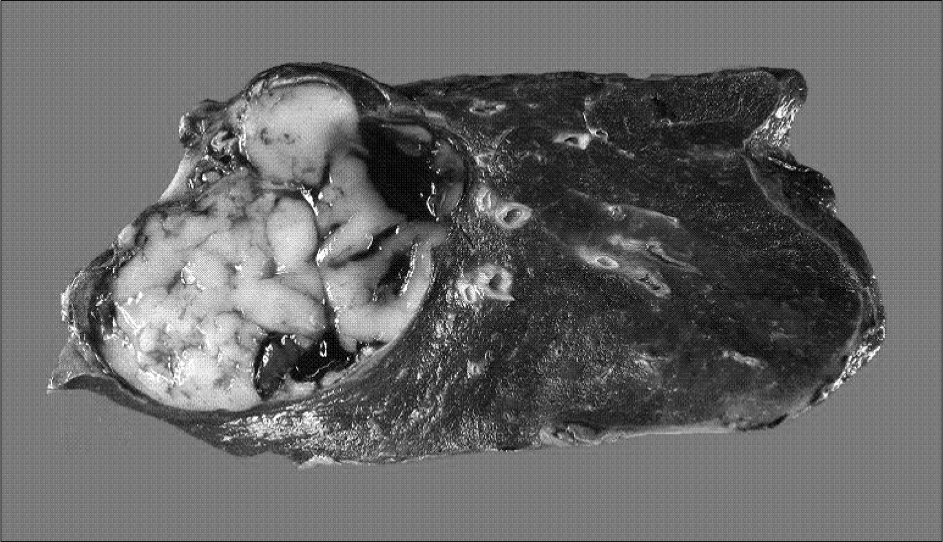
Fig. 3.
Histologic features of synovial sarcoma; most areas consisted of monophasic fibrous type (x200) (A), A transition between pale staining epithelial and dark staining spindle cells is identified (x100) (B).


 XML Download