

 PDF
PDF ePub
ePub Citation
 Print
Print


Abstract
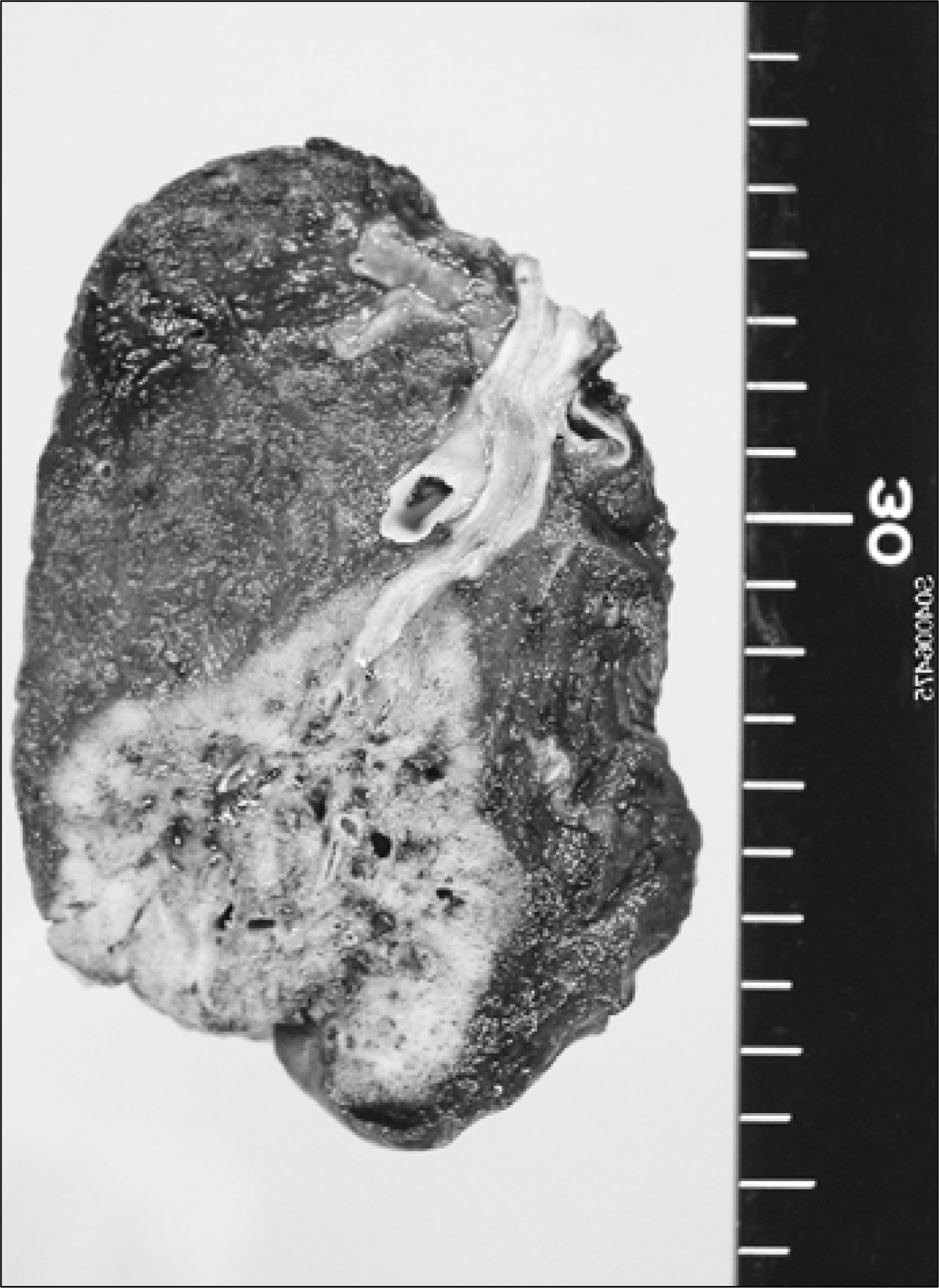
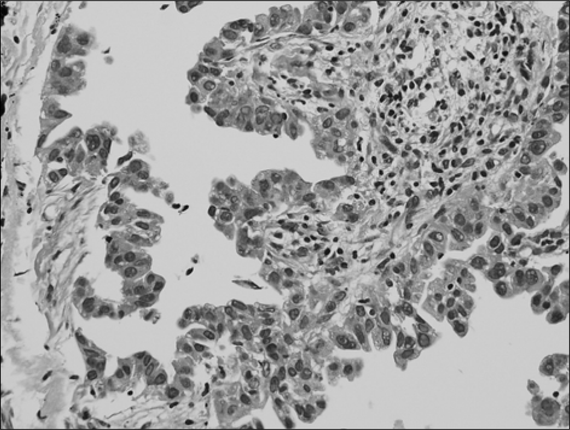
The prognosis of lung cancer is very poor. Patients with lung cancer have usually no symptom in early stage or some mild cough, sputum. When patient feel weight loss or dyspnea, majority of patients with lung cancer are advanced stage and inoperable. The growth rate of lung cancer is different according to cell type of tumor and related to prognosis. Generally, tumor doubling time (TDT) of lung cancer has been known that small cell lung cancer is about 65 days, squamous cell carcinoma is about 90 days, and adenocarcinoma is about 185 days. There has been rarely reported of lung cancer with very fast or very slow growth. The prognosis of a slow growing lung cancer is relatively good but rapidly growing cancer is not. We report a very rare case that s䴸rgically treated early stage non-small cell lung cancer (adenocarcinoma) with 4-year- TDT without invasion or distant metastasis, (J Lung Cancer 2006;5(1):51 -54)
Go to : 
REFERENCES
1.Freiberg S., Mattso S. On the growth rates of human malignant tumors: implications for medical decision making. J Surg Oncol. 1997. 65:784–797.
2.Yamaguchi M: Sugio K: Ondo K, et al. Slowly progressive adenocarcinoma of the lung: report case. Ann Thorac Cardiovasc Surg. 2002. 8:160–162.
3.Ching-Lih S., Yu-Chin L., Reury-Pcmg P. Fast-growing squamous cell lung cancer. Lung Cancer. 2002. 36:199–202.

4.Schwartz M. A biomathmatical approach to clinical tumor growth. Cancer. 1961. 14:1272–1294.
5.Mattem J., Volm M. Clinical estimation of the growth rate of lung cancer. Anticancer Res. 2001. 21:4067–4070.
6.Hasegawa M., Sone S: Takashima S, et al. Growth rate of small lung cancers detected on mass CT screening. Br J Radiol. 2000. 73:1252–1259.
7.Takatoshi A., Hajime N., Hideyuki W, et al. Evolution of peripheral lung adenocarcinomas: CT findings correlated with histology and tumor doubling time. AJR. 2000. 174:763–768.
8.Morihito 0., Wataru N., Toshihiko S, et al. Correlation between computed tomographic findings, bronchioloalveolar carcinoma component, and biologic behavior of small sized lung adenocarcinomas. J Thorac Cardiovasc Surg. 2004. 127:857–861.
9.Wang JC: Sone S., Feng L, et al. Rapidly growing small peripheral lung cancers detected by screening CT: correlation between radiological appearance and pathological features. Br J Radiol. 2000. 73:930–937.
10.Noguchi M., Morikawa A., Kawasaki M, et al. Small adenocarcinoma of the lung. Cancer. 1995. 75:2844–2852.
11.Aquino SL: Chiles C: Halford P. Distinction of consolidative bronchioloalveolar carcinoma from pneumonia: do CT criteria work? AJR. 1998. 171:359–363.
12.Hill CA. Bronchiolo-alveolar carcinoma: a review. Radiology. 1984. 150:15–20.
13.Strollo DC., Rosado-de-Christenson ML., Franks TJ. Reclassification of cystic bronchioloalveolar carcinomas to adenocarcinomas to adenocarcinomas based on the revised world health organization classification of lung and pleural tumors. J Thorac Imaging. 2003. 18:59–66.
14.Yoshida T: Harada T., Fuke S, et al. Lung adenocarcinoma presenting with enlarged and multiloculated cystic lesions over 2 years. Respir Care. 2004. 49:1522–1524.
Go to :
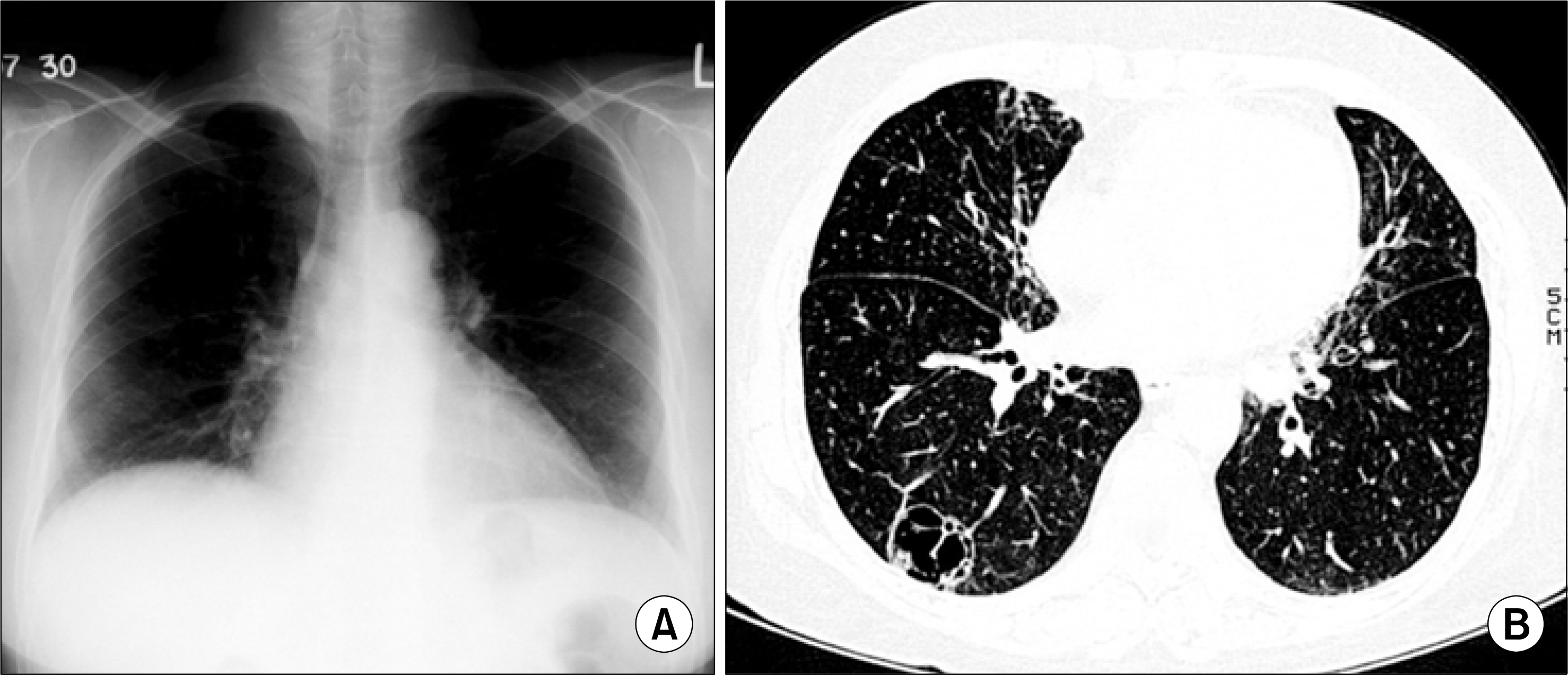 | Fig. 1.Chest radiography (A) and HRCT scan (B) at initial presentation, demonstrating a well-defined mult卜septated cavitary lung lesion in the superior segment of the right lower lobe. |
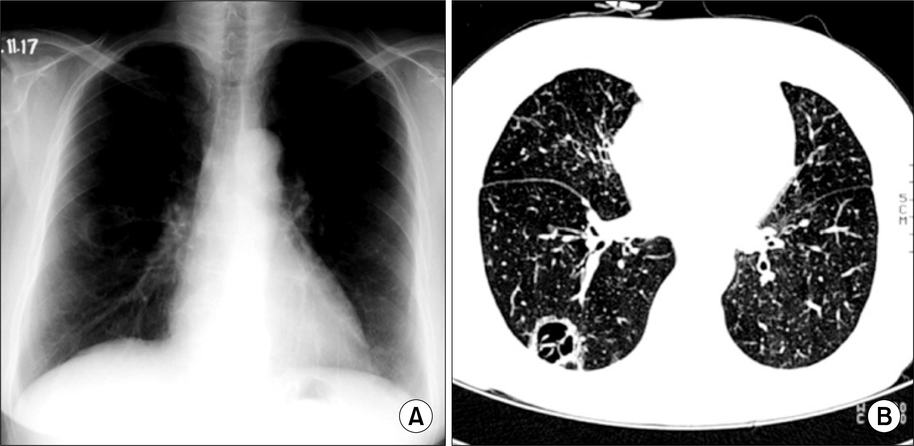 | Fig. 2.Chest radiography (A) and HRCT scan (B) taken 2 years later, demonstrating that the size of lung mass had increased slightly. |
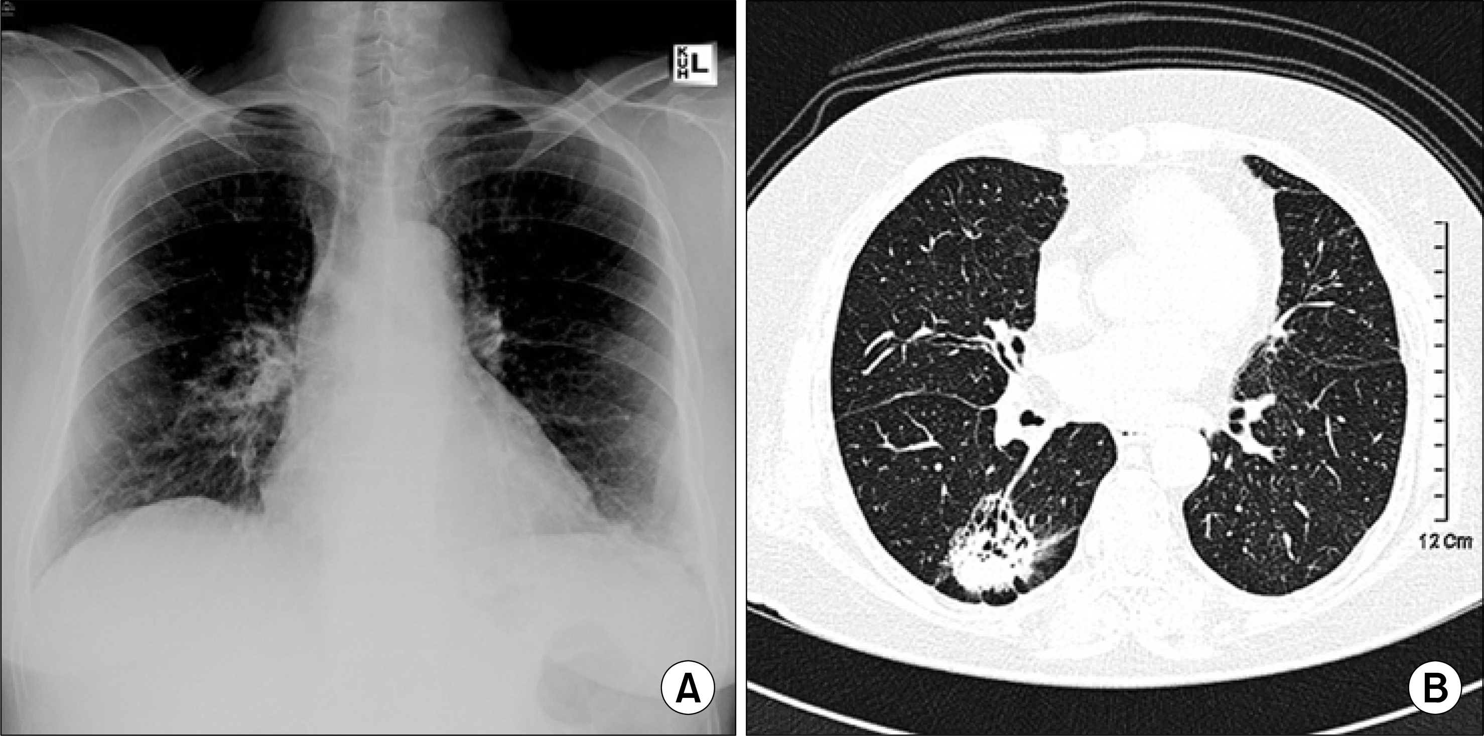 | Fig. 3.Chest radiography (A) and HRCT scan (B) taken 8 years after the initial presentation showing that the cavitary lung mass had increased in size and developed a solid portion. |
 XML Download