

 PDF
PDF ePub
ePub Citation
 Print
Print


Abstract
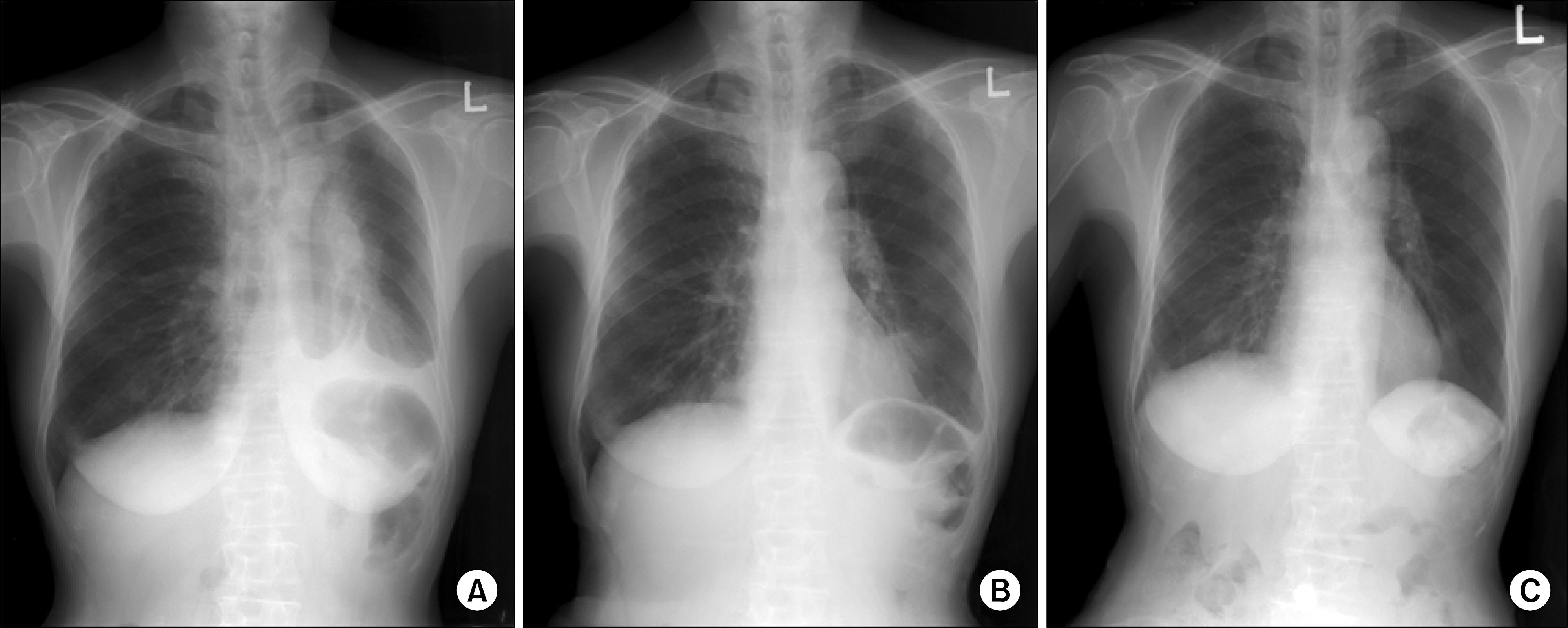
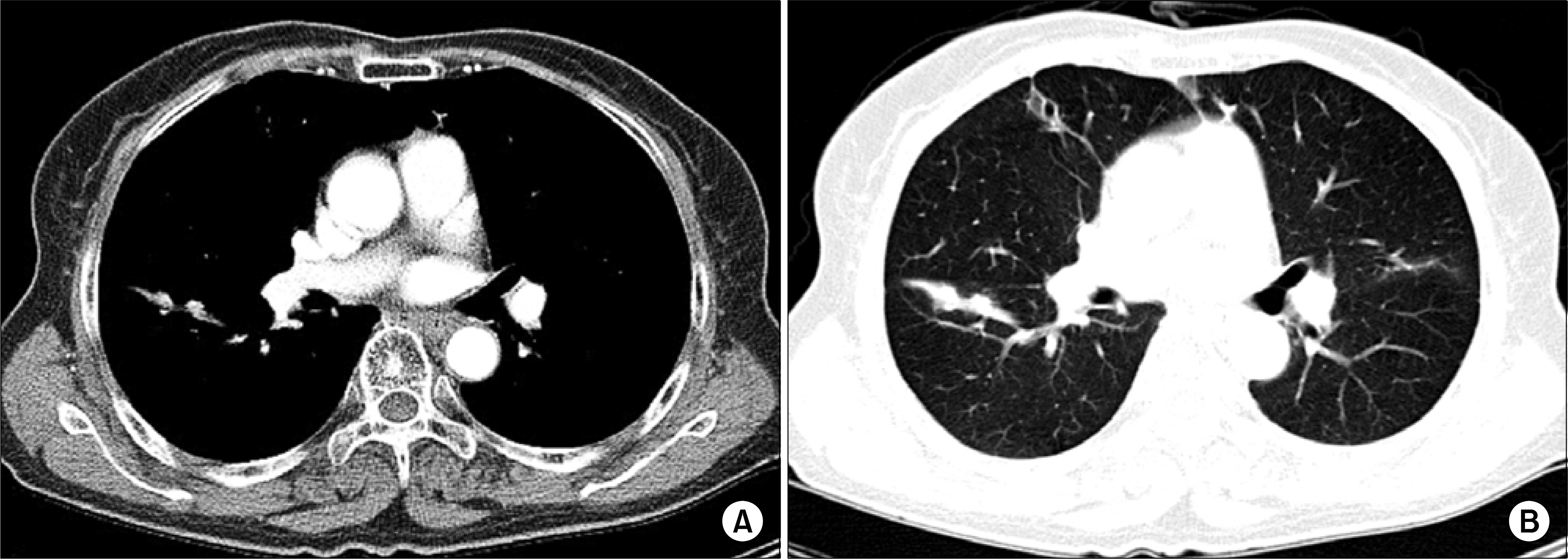
A sixty-five year old female was consulted for radiation therapy due to dyspnea. She was diagnosed as stage IV bronchial-associated lymphoid tissue (BALT) lymphoma at 5 months ago, and then received chemotherapy for 3 months. She have distressed from dyspnea originated from main bronchus obstruction, and should be supplied with oxygen via nasal prong. She underwent 36 Gy radiation therapy to bronchial mass that obstructs left main bronchus in 18 fractions for four weeks. Dyspnea was markedly improved after radiation therapy, and follow-up chest X-ray showed near complete resolution of mass and obstructed lung parenchyma. No recurrence was detected in lung for sixteen months. We experienced that radiation therapy for BALT lymphoma was effective for tumor control. (J Lung Cancer 2006;5(2):114-117)
REFERENCES
1.Harris N., Jaffe E., Stein H, et al. A revised European-American Classification of Lymphoid Neoplasm: a proposal from the International Lymphoma Study Group. Blood. 1994. 84:161–171.
2.Isaacson PG., Wright DH. Malignant lymphoma of mucosa associated lymphoid tissue. A distinctive type of B-cell lymphoma. Cancer. 1983. 52:1410–1416.
3.Zucca E., Roggero E., Pileri S. B-cell lymphoma of MALT type: a review with special emphasis on diagnosis and management problems of low-grade gastric tumors. Br J Hematol. 1998. 100:3–14.
4.Isaacson PG. Mucosa-associated lymphoid tissue lymphoma. Semin Hematol. 1999. 36:139–147.
5.Thieblemont C., Francoise B: Coffier B. Mucosa-associated lymphoid tissue lymphoma. Curr Opin Oncol. 1995. 7:415–420.
6.Thieblemont C., Bastion Y., Berger F, et al. Mucosa-associated lymphoid tissue gastrointestinal and nongastrointestinal lymphoid behavior: analysis of 108 patients. J Clin Oncol. 1997. 15:1624–1630.
7.Zucca E., Roggero E., Bertoni F., Primary extranodal non-Hodgkin's lymphomas. Part 1: gastrointestinal, cutaneous, and genitonurinary lymphomas. Ann Oncol. 1997. 8:727–737.
8.Spencer J., Finn T., Pul ford KA, et al. The human gut contain a novel population of B-lymphocytes resemble marginal zone cells, Clin Exp Immunol. 1985. 62:607–612.
9.Isaacson PG» Wright DH. Extranodal malignant lymphoma arising from mucosa-associated lymphoid tissue. Cancer. 1984. 53:2515–1524.
10.Ferraro P., Trastek VF., Adlakha H, et al. Primary non-Hodgkin's lymphoma of the lung. Ann Thorac Surg. 2000. 69:993–997.

11.Cordier JF., Chailleux E: Lauque D, et al. Primary pulmonary lymphoma. A clinical study of 70 cases in nonimmnocompromised patients. Chest. 1993. 103:201–208.
12.Li G., Hansmann ML., Zwingers T, et al. Primary lymphomas of the lung: morphological, imirmnohistochemical and clinical features. Histopathology. 1990. 16:519–531.
11.Nathwani BN., Anderson JR., Armitage JO, et al. marginal zone B-cell lymphoma: a clinical comparison of nodal and mucosa-associated lymphoid tissue types. J Clin Oncol. 1999. 17:2486–2492.
14.Ahmed S., Kussick SJ., Siddiqui AK, et al. Bronchial-associated lymphoid tissue lymphoma: a clinical study of a rare disease. Eur J Cancer. 2004. 40:1320–1326.

 XML Download