

 PDF
PDF ePub
ePub Citation
 Print
Print


Abstract
Lung cancer with cyst formation is a rare entity. We report a 63-year-old man who underwent surgical treatment of primary lung cancer, which mimics benign solitary cyst. We incidentally found his pulmonary cyst by a low dose chest tomography and followed up for 2 years. Rapid growth of cyst and focal wall thickening evoke us to have a suspicion of its malignancy. Left lower lobectomy via video-assisted thoracoscopic surgery was performed without any preoperative pathologic confirmation. The postoperative pathological finding revealed squamous cell carcinoma with carcinoma in situ on the cyst wall. We emphasize the need for physicians to be aware of the potential of lung cancer in patients with growing pulmonary cyst.
Figures and Tables
Fig. 1
The initial chest computed tomography (CT) scan showed a thin-walled cystic lesion in the left lower lobe. The cystic lesion had grown up during follow-up duration. After two years, the chest CT scan showed newly developed eccentric wall thickening and part solid component on the cyst (A, initial CT; B, after one year; C, after two years).
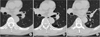
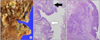
Fig. 2
(A) Gross appearance of resected specimen. Cystic lesion with irregular border was found. (B) Pathologic finding showed a squamous cell carcinoma that focally invaded the lung parenchyma (black arrow). Those were surrounded by carcinoma in situ on the cyst wall (white arrow) (H&E, ×40). (C) Tumor cells showed hyperchromatic nuclei and intercellular bridge, characteristics of squamous cell carcinoma (H&E, ×100).
References
1. Iwata T, Nishiyama N, Nagano K, et al. Squamous cell carcinoma presenting as a solitary growing cyst in lung: a diagnostic pitfall in daily clinical practice. Ann Thorac Cardiovasc Surg. 2009. 15:174–177.
2. Hirai S, Hamanaka Y, Mitsui N, Morifuji K, Sutoh M. Primary lung cancer arising from the wall of a giant bulla. Ann Thorac Cardiovasc Surg. 2005. 11:109–113.
3. Wilson DO, Weissfeld JL, Balkan A, et al. Association of radiographic emphysema and airflow obstruction with lung cancer. Am J Respir Crit Care Med. 2008. 178:738–744.

4. Jakopovic M, Slobodnjak Z, Krizanac S, Samarzija M. Large cell carcinoma arising in bronchogenic cyst. J Thorac Cardiovasc Surg. 2005. 130:610–612.
5. Farooqi AO, Cham M, Zhang L, et al. Lung cancer associated with cystic airspaces. AJR Am J Roentgenol. 2012. 199:781–786.
6. Stoloff IL, Kanofsky P, Magilner L. The risk of lung cancer in males with bullous disease of the lung. Arch Environ Health. 1971. 22:163–167.
7. Takahashi M, Fukuoka J, Nitta N, et al. Imaging of pulmonary emphysema: a pictorial review. Int J Chron Obstruct Pulmon Dis. 2008. 3:193–204.
 XML Download