

 PDF
PDF ePub
ePub Citation
 Print
Print


It has long been recognized that tumors of capillary vessels in the lung are extremely rare. Among this group, pulmonary capillary hemangiomatosis is relatively well known, characterized by a proliferation of pulmonary capillaries in the alveolar septa, perivascular connective tissue, bronchial wall, and pleura (1). It is also known to be a rare cause of pulmonary hypertension and obstruction of pulmonary veins and venules (2).
However, there have been only a few reports of solitary capillary hemangioma (SCH) of the lung (3-6). Although most have reported cases occurring in neonates or children, Fugo et al. reported two cases of adult SCH (5). Awareness of this clinical entity is important because the lesions are difficult to differentiate radiologically from early lung cancer. In the cases in Fugo et al. (5), the lesions were incidentally detected as small nodules with ground glass opacity. The radiologist diagnosed both lesions as early lung cancer, however histologic and immunohistochemical findings were consistent with the diagnosis of SCH of the lung (5).
Herein, we report two further cases of SCH of the lung. Both cases were first detected as a solitary nodule of the lung in chest computed tomography (CT) images. Both lesions were recognized as early lung cancer and surgical resections were performed.
CASE REPORT
Case 1
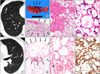
A 55-year-old man visited the thoracic surgery department of the Samsung Medical Center, Seoul, for further evaluation of an abnormal finding on lung examination that had been incidentally detected by routine medical check-up. His physical condition was good, and there were no significant signs or symptoms that were consistent with pulmonary hypertension or veno-occlusive disease, such as hemoptysis or dyspnea on exercise. Other cutaneous or internal organ lesions were not found. Chest contrast computed tomography (CT) showed a growing 9 mm-sized solid nodule in the bottom portion of the right middle lobe (Fig. 1A). The radiologist reported this lesion as representing an area of minimally invasive adenocarcinoma or invasive adenocarcinoma with lepidic tumor growth. No preoperative histological confirmation was performed.
The patient underwent a diagnostic wedge resection of the right middle lobe. The wedge resected lung showed an ill-defined soft hemorrhagic mass. The tumor was not encapencapsulated but was well delineated from the non-neoplastic lung (Fig. 1B, C). The tumor consisted of uniform capillaries with cuboidal or flattened endothelial cells (Fig. 1D, E). The endothelial cells were highlighted by CD31 stain (1:80, Dako, Glostrup, Denmark). There was no other abnormality in the non-neoplastic lung. No significant cytologic atypia was identified. The Ki-67 index was positive in 5% of tumor cells. There was no evidence of recurrence during 12 months of follow-up.
Case 2
A 50-year-old man visited the thoracic surgery department of the Samsung Medical Center, Seoul for detailed evaluation of abnormal findings that had been discovered incidentally by low-dose screening lung CT scan. He had no significant signs or symptoms. There were no other lesions of skin or internal organs. Non-contrast helical chest CT revealed a 10 mm-sized ground-glass opacity nodule in the left upper lobe, and another subpleural small nodule measuring 5 mm in the left upper lobe below the main lesion (Fig. 1F). During follow up, the main lesion slightly increased in size, therefore histologic confirmation was recommended for this lesion. The subpleural small nodule showed no change in size during follow-up. Wedge resections for both nodules were performed for histologic diagnosis.
The 10 mm-sized nodule was histologically confirmed to be adenocarcinoma. The other subpleural mass was well-defined from the adjacent lung parenchyma and pseudo-encapsulated (Fig. 1G). This was a 5 mm-sized round hemangioma composed of dilated capillaries, lined by single layered flattened endothelial cells (Fig. 1H). Endothelial cells were highlighted by CD31 stain (Fig. 1I). There has been no recurrence during six months of follow-up.
DISCUSSION
Vascular tumors of the lung are extremely rare. There have been a limited number of reports of pulmonary capillary hemangiomatosis (1,2), which has been described as comprising multiple nodules in the lung parenchyma or bronchovascular walls, composed of infiltrating thin-walled capillary vessels (1,2). It can be a rare cause of pulmonary hypertension and secondary pulmonary veno-occlusive disease. SCH is even more rare and hemangioma has been seen more commonly in children (3,). There have been only a few case reports of SCH of the lung, most of which have been in very young patients (4-6). Fugo et al. (5) reported two cases of SCH affecting middle-aged adults. Herein, we report two further cases of adult SCH. These are worth reporting because SCH can be a rare cause of a solitary pulmonary nodule. Localized capillary hemangioma manifests as a localized cystic lesion or focal ground-glass opacity nodule in a CT scan (3,5). Therefore it is easily misinterpreted by radiology as cancerous lesion. It is important for pathologists to be aware of this disease entity for accurate diagnosis.
Histologically, capillary hemangioma consists of dilated capcapillaries, lined by single layered flattened endothelial cells. The border of this type of tumor can be either well-circumscribed or relatively ill-defined, as was reported in several previous studies (4,5). The differential diagnosis includes other vascular tumors such as cavernous hemangioma, sclerosing hemangioma or angiosarcoma. Angiosarcoma is easily distinguished from hemangioma due to its cellular atypia. In addition, capillary hemangioma is composed of well-formed capillaries. Cavernous hemangioma is composed of large, dilated vessels filled with blood (6). Galliani et al. (6) reported a case of pulmonary cavernous hemangioma causing massive hemoptysis. Sclerosing hemangiomas of the lung can be confused with capillary hemangiomas due to their prominent angiomatoid features. However, sclerosing hemangiomas are, in fact, a misnomer and current consensus favors these being a benign or very low-grade neoplasm arising from primitive respiratory epithelium. This tumor consists of superficial cuboidal epithelium and round stromal cells with hemorrhagic areas. Tumor cells do not expressive vascular markers and show epithelial differentiation (7).
In conclusion, SCH of the lung is a rare vascular tumor that can be clinically confused with lung cancer. It is composed of dilated capillaries, lined by single layered flattened endothelial cells. It is important to for pathologists to be aware of this entity for differential diagnosis of a solitary pulmonary nodule.
 XML Download