Abstract
Purpose:
β-human chorionic gonadotropin (β-hCG) expressing pulmonary carcinoma is very rare, and little is known about this entity. The aim of this study was to find the characteristic clinicopathologic features of β-hCG expressing pulmonary carcinoma. Materials and Methods: Of all 2790 lobec-tomy specimens of lung excised between January 2006 and December 2010, only six cases of β-hCG expressing pulmonary carcinoma were identified retrospectively. The cases were classified according to the WHO classification, and clinicopathologic features were investigated.
Results:
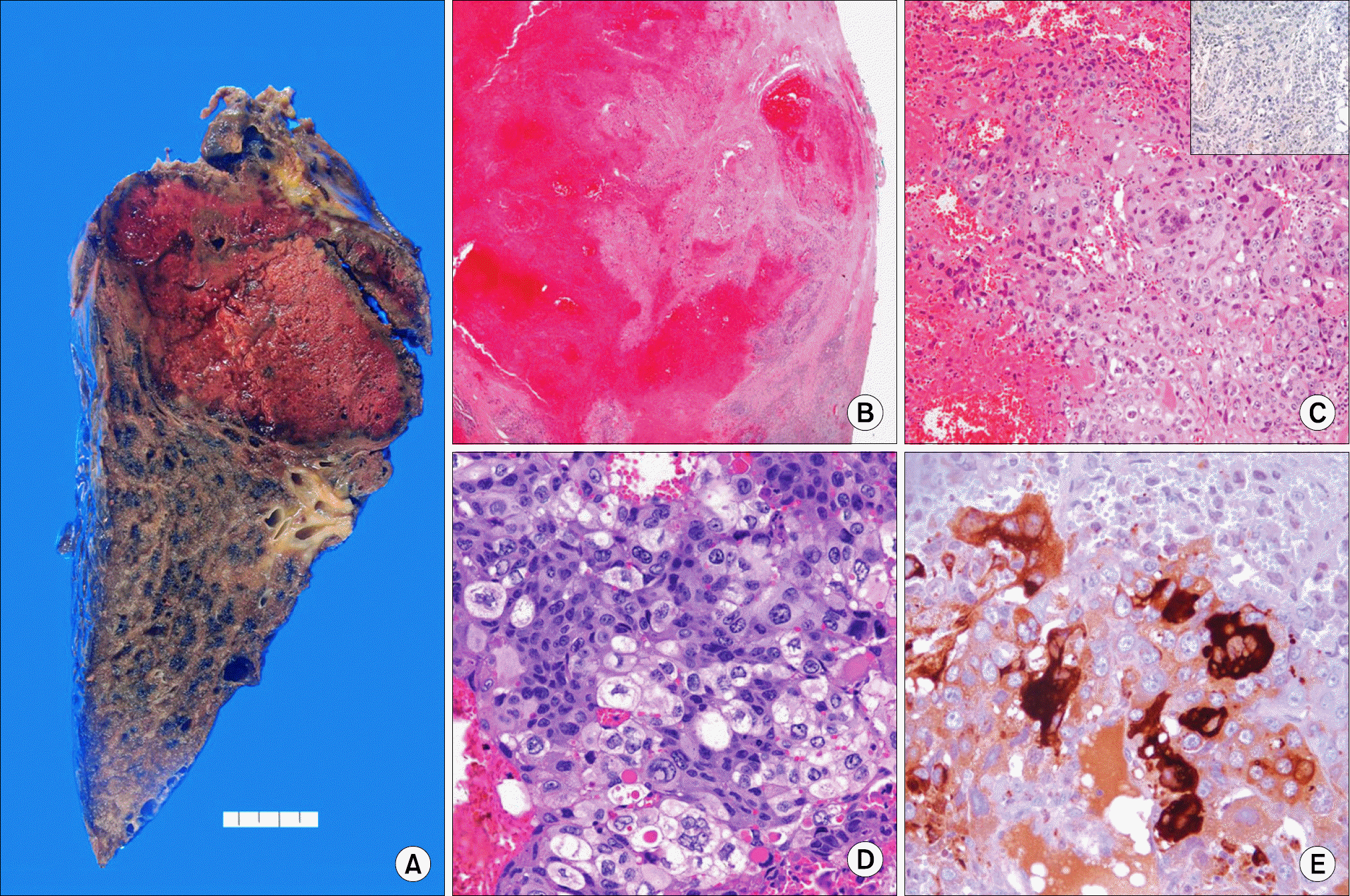
The patients consisted of 4 males and 2 females, and the median age was 64 years. Half of the patients presented with blood tinged sputum or hemoptysis. The median tumor diameter was 4.2 cm. All but one case showed prominent area of hemorrhage and necrosis. All six cases were pleomorphic carcinoma, composed of various types of non-small cell carcinomatous component and giant cell component. All cases showed significant area of β-hCG positivity, and β-hCG was usually expressed in the pleomorphic giant cells.
Go to : 
REFERENCES
1. Fukayama M, Hayashi Y, Koike M, Hajikano H, Endo S, Okumura H. Human chorionic gonadotropin in lung and lung tumors. Immunohistochemical study on unbalanced distribution of subunits. Lab Invest. 1986; 55:433–443.
2. Miyake M, Ito M, Mitsuoka A, et al. Alpha-fetoprotein and human chorionic gonadotropin-producing lung cancer. Cancer. 1987; 59:227–232.

3. Szturmowicz M, Slodkowska J, Zych J, et al. Frequency and clinical significance of beta-subunit human chorionic gonadotropin expression in non-small cell lung cancer patients. Tumour Biol. 1999; 20:99–104.

4. Yokotani T, Koizumi T, Taniguchi R, et al. Expression of alpha and beta genes of human chorionic gonadotropin in lung cancer. Int J Cancer. 1997; 71:539–544.
5. Yoshimoto T, Higashino K, Hada T, et al. A primary lung carcinoma producing alpha-fetoprotein, carcinoembryonic antigen, and human chorionic gonadotropin. Immunohistochemical and biochemical studies. Cancer. 1987; 60:2744–2750.

6. Forst T, Beyer J, Cordes U, et al. Gynaecomastia in a patient with a hCG producing giant cell carcinoma of the lung. Case report. Exp Clin Endocrinol Diabetes. 1995; 103:28–32.
7. Ha SY, Cho HY, Lee JI. Epitheilioid trophoblastic tumor of the lung: a case report. J Lung Cancer. 2009; 8:114–117.

8. Okutur K, Hasbal B, Aydin K, et al. Pleomorphic carcinoma of the lung with high serum beta-human chorionic gonadotropin level and gynecomastia. J Korean Med Sci. 2010; 25:1805–1808.

9. Hirano H, Yoshida T, Sakamoto T, et al. Pulmonary pleomorphic carcinoma producing hCG. Pathol Int. 2007; 57:698–702.

10. Travis WD, Brambilla E, Muller-Hermelink HK, Harris CC, editors. World Health Organization Classification of Tumours. Pathology and genetics of tumours of the lung, pleura, thymus and heart. IARC Press: Lyon. 2004.
11. Krefting IP, Nunez LA, Sherer P, Weinstock A, Kumar A, Travis W. Pleomorphic carcinoma (spindle and giant cell) of the lung. Md Med J. 1994; 43:787–790.
Go to : 
 | Fig. 1.Representative pathologic features of β-hCG expressing pulmonary carcinoma. (A) Gross photography shows a large necrotic mass. (B) Scanning view reveals extensive hemorrhage (hematoxylin-eosin stain, ×1). (C) The tumor was composed of poorly differentiated large tumor cells with pleomorphic giant cells (hematoxylin-eosin stain, ×200). The tumor cells are negative for p63 (inset) (ABC method, ×100). (D) The giant cell component shows marked pleomorphism with occasional multinucleation, which are similar morphology to syncytiotrophoblast (hematoxylin-eosin stain, ×400). (E) Immunohistochemical stains for β-hCG shows intense cytoplasmic positive in pleomorphic giant cells (ABC method, ×400). |
Table 1.
Clinical Findings of 6 Cases of β-hCG Expressing Pulmonary Carcinoma
Table 2.
Pathologic Findings in 6 Cases of β-hCG-Expressing Pulmonary Carcinoma


 PDF
PDF ePub
ePub Citation
 Print
Print


 XML Download