Abstract
A 17-year-old girl presented with polyuria (7 L/day) and polydipsia for one year. Initial urine osmolality was 113mOsm/kg H2O. Following 6 h of fluid restriction, serum plasma osmolality reached 300mOsm/kg H2O, whereas urine osmolality was 108mOsm/kg H2O. Urine osmolality was increased by 427% from 108 to 557mOsm/kg after vasopressin challenge. The patient was diagnosed with central diabetes insipidus, possibly derived from the atypical occupation of a Rathke's cleft cyst at the pituitary stalk following magnetic resonance imaging with enhancement. She was discharged with desmopressin nasal spray (10 µg); urine output was maintained at 2-3 L/day, and urine osmolality was >300 mOsm/kg. Additional pituitary image studies and evaluation of hypopituitarism should be included in the differential diagnosis of patients with central diabetes insipidus.
Clinical disorders characterized by defective urinary concentrations and a variable degree of polyuria include central diabetes insipidus with arginine vasopressin deficiency, and nephrogenic diabetes insipidus caused by the inability of the kidneys to respond appropriately to arginine vasopressin. According to a study of 79 patients with central diabetes insipidus, the most common cause was idiopathic diabetes insipidus, followed by primary or secondary intracranial tumors or infiltrative diseases such as Langerhans cell histiocytosis, neurosurgery, skull fracture, and autoimmune disease12).
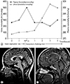
A 17-year-old girl (height, 150 cm; weight, 54 kg; body mass index, 24 kg/m2) presented with polyuria (7 L/day) and polydipsia for one year. She did not have an underlying disease, including diabetes, and denied the use of any medication. Initial urine osmolality was 113mOsm/kg H2O, and specific gravity was <1.005. Her vital signs were normal. Laboratory findings were as follows: white blood cells 5,700/mm3, hemoglobin 13.0 g/dL, and platelets 229 K/mm3; blood urea nitrogen 8.6mg/dL and creatinine 0.5 mg/dL; sodium 141mEq/L, potassium 4.1mEq/L, and chloride 107mEq/L; serum osmolality 282mOsm/kg; and urine sodium 33mEq/L, potassium 11.1mEq/L, and chloride 41mEq/L. Urine analysis revealed no hematuria or proteinuria. Following 6 h of fluid restriction, serum plasma osmolality reached 300mOsm/kg H2O, whereas urine osmolality was 108mOsm/kg H2O. Fig. 1A shows that urine osmolality was increased by 427% from 108 to 557 mOsm/kg after vasopressin challenge. These remarkable changes in urine osmolality can be used to diagnose central diabetes insipidus. Figure (Fig. 1B, C) shows a well-defined hyper signal intensity sellar lesion without an enhanced portion on gadolinium-enhanced fat-suppressed T1-weighted imaging, suggesting Rathke's cleft cyst. Extensive examinations were performed to determine the pathological states of the patient's hypopituitarism(Table 1). Other abnormalities in the release and stimulation of other hormones were not detected. These findings indicate a sole diagnosis of central diabetes insipidus, possibly derived from the atypical occupation of a Rathke's cleft cyst at the pituitary stalk. The patient was discharged with desmopressin nasal spray (10 µg); her urine output was maintained at 2-3 L/day, and urine osmolality was >300 mOsm/kg. She was scheduled for follow-up magnetic resonance imaging (MRI), and pituitary surgery was postponed due to a personal matter.
Rathke's cleft cysts are the most common incidentally discovered benign sellar cysts derived from remnants of Rathke's pouch, the same structure from which craniopharyngiomas arise. In MRI, they typically appear as well-demarcated cystic lesions with homogeneous intensity signals and are sometimes combined with thin cyst wall enhancement3). Among patients with symptomatic Rathke's cleft cysts, 20% showed panhypopituitarism on admission. The serum prolactin levels did not exceed 200 ng/mL, and hypopituitarism was not correlated with cyst size4).
We present a case of central diabetes insipidus accompanied by Rathke's cleft cyst. Additional pituitary image studies and evaluation of hypopituitarism should be included in the differential diagnosis of patients with central diabetes insipidus.
Figures and Tables
Fig. 1
(A) Water deprivation and arginine vasopressin challenge test; (B, C) Sagittal T1-weighted magnetic resonance (MR) image shows well-defined hypersignal intensity sellar lesion (B), without enhancing portion on gadolinium-enhanced fat-suppressed T1-weighted image (C).

Acknowledgments
This research was supported by the Basic Science Research Program through the National Research Foundation of Korea (NRF) funded by the Ministry of Science, ICT & Future Planning (2015R1D1A1A01061037), by Basic Science Research Program through the National Research Foundation of Korea (NRF) funded by the Ministry of Science, ICT and future Planning (2016R1A2B4007870), and by a grant (CRI16013-1) Chonnam National University Hospital Biomedical Research Institute.
Notes
References
1. Kimmel DW, O'Neill BP. Systemic cancer presenting as diabetes insipidus. Clinical and radiographic features of 11 patients with a review of metastatic-induced diabetes insipidus. Cancer. 1983; (12):52:2355–2358.

2. Maghnie M, Cosi G, Genovese E, et al. Central diabetes insipidus in children and young adults. N Engl J Med. 2000; 343(14):998–1007.


 PDF
PDF ePub
ePub Citation
 Print
Print


 XML Download