

 PDF
PDF ePub
ePub Citation
 Print
Print


Ischiofemoral impingement syndrome (IFIS) is a rare clinical entity characterized by chronic groin, buttock or hip pain associated with radiographic evidence of narrowing of the space between the lesser femoral trochanter and the ischial tuberosity.1) The exact prevalence of IFIS is still unknown as this entity is poorly understood and recognized.2) IFIS was first described approximately 40 years ago in patients who presented with persistent groin pain after hip replacement.3) It has since been considered a iatrogenic consequence of hip surgical procedures; however, it was recently described in patients suffering from hip trauma, patients with ischial exostosis, and even as a primary condition in young athletes.1234) The space narrowing is thought to impact the quadratus femoris muscle, altering the morphology and function of this muscle as detected with magnetic resonance imaging (MRI).124) The introduction of MRI into the clinical practice as well as establishment of the radiological definition of the abnormal ischiofemoral distance has led to an increasing interest and recognition of this condition.24)
When diagnosed, treatment may be successful with a multidisciplinary conservative approach; nonetheless, surgery may be necessary in refractory cases.1) To date, only a few reports regarding the surgical treatment of this disease have been published.56789) In the current paper, the authors aim to report successful results of a minimally invasive surgical decompression technique in the treatment of non-remitting cases of IFIS with a review of different arthroscopic treatments and reported outcomes.
CASE REPORT
Case 1
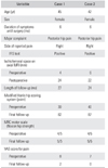
A 45-year-old woman visited University Hospital of Vall d'Hebron with a complaint of progressive, bilateral, posterior buttock pain with distal neuropathic pain radiation. Symptoms were more pronounced on the right limb, and she had no prior traumatic event. On physical examination, the patient had tenderness to palpation of the ischiofemoral space and a positive long-stride walking test. Pain could be reproduced in extension, abduction and external rotation of the hip (Ischiofemoral impingement test).12)
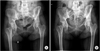
MRI study showed no signs of femoroacetabular pathology, minor L5–S1 disc degeneration, and bilateral reduction of the ischiofemoral distance (4 mm on the right and 5 mm on the left) as well as edema and fibrillar rupture of the right quadratus femoris muscle which compressed and secondarily displaced the right sciatic nerve (Fig. 1).
Conservative treatment yielded no satisfactory results and only partial relief was achieved with computed tomography (CT)-guided local steroid infiltrations. Due to a lack of improvement with the conservative treatment, surgical decompression of the ischiofemoral space through endoscopic total resection of the lesser trochanter was performed.
Case 2
A 42-year-old woman presented with right posterior buttock pain with distal neuropathic pain radiation. On physical examination, the patient had tenderness to palpation of the ischiofemoral space and a positive long-stride walking test. The ischiofemoral impingement test also suggested IFIS. MRI images showed a reduced ischiofemoral distance (6 mm) of the right hip as well as edema of the quadratus femoris muscle. Conservative treatment failed to alleviate pain. After 6 months of conservative treatment, surgical endoscopic decompression through resection of the lesser trochanter was performed.
Surgical Technique
Surgical procedures were performed with the patient in a supine position on a traction table with no traction applied. The hip was externally rotated and flexed to 30° with the intention of relaxing the iliopsoas tendon and bringing forward the lesser trochanter from posterior to anterior position. A 30° standard length arthroscopic probe was used.
Two anterolateral portals were made according to the original technique described by Ilizaliturri and Camacho-Galindo10) for iliopsoas tendon resection in snapping hips. The first portal was an inferior anterolateral portal located 3 cm below and 2 cm anterior to the anterosuperior corner of the greater trochanter. The working portal was 4 cm below the optical portal. A cannulated needle was directed, under fluoroscopic control, through the inferior anterolateral portal until the lesser trochanter was reached, which is easily palpable through this portal. An arthroscopic cannula was advanced over the guide until the lesser trochanter was contacted. Once fluid irrigation was performed, the psoas tendon could be identified as a bright white structure. A new needle was triangulated towards the arthroscope and confirmed with fluoroscopic control. A shaver was used to better visualize the psoas tendon and a hooked radiofrequency probe was employed to release it from the lesser trochanter. Finally, a 4 mm arthroscopic burr was used to resect the lesser trochanter under fluoroscopic control (Fig. 2). Patients were discharged home on the next day following surgery. Full weight bearing was allowed as tolerated and physical therapy was started 1 week after surgery.
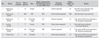
Patients experienced progressive improvement with immediate partial remission of their distal neuropathic radiated pain. Postoperative MRI showed a remarkable improvement of the ischiofemoral distance in both cases, increasing from 4 mm to 24 mm in the first case and from 6 mm to 22 mm in the second case as well as complete remission of the quadratus femoris edema (Fig. 1). Patients' gait also improved progressively, and at the 6-month follow-up, they reported full clinical and functional recovery of the affected limb. The modified Harris hip score improved from a preoperative score of 30 to a postoperative score of 82 in the first case and from 40 to 87 in the second case. Hip flexion strength was recovered in both cases: improvement from a preoperative score of 4/5 to a postoperative score of 5/5 was measured with the Medical Research Council (MRC) motor scale. Moreover, visual analogue pain scale score improved from 8 to 2 in the first patient, and from 7 to 0 in the second patient (Table 1). However, at the 2-year clinical follow-up, the first patient still complained of mild posterior pain in the right buttock. Worsening of her clinical status was attributed to the progression of symptoms on the left side (Fig. 3).
DISCUSSION
The normal ischiofemoral distance that allows the femur to rotate without contacting the ischium is defined as > 2 cm on axial cuts. In the literature, pathological narrowing of this space down to 1.3 cm has been described.24) Such narrowing is thought to affect the quadratus femoris muscle, showing an increased signal on T2 images, with no disruption of muscular fibers. Special caution is advised when interpreting these findings in the absence of suggestive clinical symptoms.1) Patients presented here showed clinical and radiological compatibility with regard to IFIS. They had hip pain with neuropathic radiation in the affected limb, with no clear evidence of a sciatic nerve or lumbar origin. In both cases, MRI showed a decreased ischiofemoral distance, with quadratus femoris edema compressing and displacing the sciatic nerve.
When confirmed, IFIS can be treated using a myriad of conservative techniques that result in variable outcomes. These include nonsteroidal anti-inflammatory medications, guided steroidal local infiltrations, and targeted physical therapy.24) When such treatment fails, surgery is perceived as the only available alternative.156789) Accordingly, our patients had undergone a complete trial of conservative treatments for at least 6 months before surgery was finally indicated. The fact that they responded initially to targeted steroid injections further confirmed our clinical and radiological diagnosis.
Reviewing published literature to date, this paper is one of the firsts to report the positive surgical outcomes of a minimally invasive surgery for IFIS (Table 2).56789) Safran and Ryu5) presented a case of IFIS in a female who had endoscopic resection of the lesser trochanter through an anterior approach with excellent results. Howse et al.6) presented a posterolateral approach for lesser trochanter resection emphasizing that the main advantage of this approach is the preservation of the iliopsoas' attachment. Hatem et al.7) reported an access to the lesser trochanter through a window on the quadratus femoris muscle. In 2015, Jo and O'Donnell8) described a technique of endoscopic resection of the lesser trochanter in a 17-year-old college student diagnosed with IFIS. They performed a partial resection of the lesser trochanter through two anterolateral portals that were established at the level of the lesser trochanter. They reported that the patient's resting and functional pain with adduction and external rotation disappeared within 1 week after surgery, and pain relief was maintained at the 4-month follow-up. The largest series published was by Wilson and Keene9) who reported using an anterior approach on seven patients to release the iliopsoas tendon and resect the lesser trochanter.
Regarding the present report, no intra- or postoperative complications were encountered. Both patients readily agreed to this new technique mostly due to the functional limitation and long-term pain as well as the minimally invasive nature of the procedure.
Despite theoretical morbidities described in previous reports, our functional results showed only mild weakness of the hip flexion strength at the 2-year follow-up. This is in accordance with the results presented by Ilizaliturri and Camacho-Galindo10) who reported preservation of hip strength at 2 and 3 months after complete arthroscopic psoas tendon release. The authors theorized that, possibly, the other flexor muscles compensate for the severed iliopsoas or even the functionality of the psoas muscle may not be completely lost with this technique. Moreover, some regeneration of the psoas tendon has been recently demonstrated after its release with endoscopic procedures.10) Even though our patients experienced a clear initial clinical improvement, the first patient's clinical status worsened after 2 years of follow-up, mainly due to the quick progression of IFIS on the contralateral side, for which she has given her consent for a new endoscopic surgery on the left hip.
IFIS is a rare clinical entity characterized by groin and/or buttock pain, associated with radiological decrease in the distance between the lesser trochanter of the femur and the ischium. MRI imaging is considered the diagnostic study of choice, always correlating to clinical findings. Indications for hip arthroscopy have been growing and therefore arthroscopic techniques may now be used for a number of conditions that were once thought to be extremely difficult to treat with closed techniques. Based on our clinical results and the other reported cases, a minimally invasive surgical decompression technique, such as endoscopic resection of the lesser trochanter, may prove useful for treating resilient IFIS cases. Nonetheless, there is no clear consensus on the best surgical approach for IFIS. We consider that further studies with longer follow-ups are still needed to validate these initial findings.


 XML Download