

 PDF
PDF ePub
ePub Citation
 Print
Print


Continuing progress in the fields of diagnosis and treatment of cancer has led to the increase in the number of patients living with metastatic bone disease (MBD).12) The pelvic bone is the most common site of bone metastases following the axial skeleton.3) MBD of the pelvic bone is usually managed nonoperatively. The indications for surgery include patients with compromised skeletal stability, pain not responding to other modalities of treatment or a solitary or oligometastatic disease.4) Surgery not only relieves pain but also restores structural stability and reduces the disease burden. Surgery on the pelvic bone is a demanding procedure both for the surgeon and the patient.
The outcome of surgical management of MBD of the pelvic bone has been rarely described in the literature contrary to that of the long bones.5) In addition, periacetabular disease remains the cynosure of the available literature on the pelvic bone 67891011) and the studies on the surgical outcomes of MBD of all parts of the pelvic bone involved only small numbers of patients. Hence, this study sought to analyze the complications, local progression and survival after surgery for MBD of the pelvic bone on a larger cohort of patients.
METHODS
The prospectively collected database of Seoul National University Hospital was used to identify 83 patients who underwent surgery for metastases of the pelvic bone between the years 2000 and 2015. The electronic medical records and picture archiving and communication system images of all patients were reviewed. All patients who underwent surgery were included irrespective of the surgical margin and duration of follow-up. Only the index surgery was included. One patient underwent surgery on both sides of the pelvic bone. The study was approved by the Institutional Review Board of Seoul National University Hospital (IRB No. H-1609-077-791).
Patient Demographics and Blood Parameters
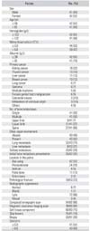
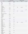
The demographic variables included were age at the time of surgery and gender. There were 41 men and 42 women with a mean age of 55 years (range, 12 to 72 years) (Table 1). For the purpose of analysis, patients were dichotomized based on their age into ≤ 55 years (n = 42, 51%) and > 55 years (n = 41, 50%). Preoperative blood parameters included were haemoglobin level, white blood cell count and albumin level. The mean preoperative haemoglobin level was 122 g/L (range, 85 to 166 g/L), white blood cell count was 5.8 × 10⁹/L (range, 1.8 × 10⁹/L to 11 × 109/L) and albumin level was 39 g/L (range, 26 to 50 g/L). Bone metastasis was the initial presentation of cancer in 25 patients (31%). Thirty-two patients (39%) had bone metastases at presentation. The median time from the diagnosis of primary cancer to bone metastasis was 3 years (range, 4 to 306 months). The diagnosis of pelvic bone metastasis was made based on a biopsy in 23 patients (28%) and on imaging in the rest.
Cancer Type, Burden and Characteristics of Metastatic Lesion
The cancer-related variables included were primary cancer type, number and location of bone metastasis, size of the MBD, presence of skip lesions, soft tissue extension, pathological fracture and presence and location of visceral metastasis. The most common primary cancers were of the kidney (n = 18, 22%) and thyroid (n = 13, 16%). For the purpose of analysis, the primary cancer types were grouped into favourable types (kidney, breast, thyroid or hematolymphoid) and unfavourable types.1213) Pelvic bone metastasis was solitary in 23 patients (28%). Fifty percent (n = 41) of the patients had more than one site of bone metastasis. The spine (66%) was the common location when compared to the extremities (upper limb, 7%; lower limb, 11%) in patients with multiple bone metastases. The most common location in the pelvic bone was the iliac wing (n = 42, 51%). The size was documented as the largest diameter from the pathology report in patients who underwent en bloc resection or from cross-sectional imaging in other patients. For the purpose of analysis, patients were dichotomized based on the tumor size with a cut-off value of 6.8 cm, the mean size of the tumor (range, 2 to 16 cm). Fifteen patients (19%) had skip lesions and 60 patients (60%) had soft tissue component of the metastatic disease. Twenty-three percent of the patients had a pathological fracture (n = 19). Visceral metastases were present in 43 patients (52%), lung being the most common site (n = 32). The radiographic pattern was lytic in 88% (n = 72) and normal in 7% (n = 6).
Treatment
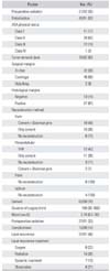
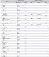
The treatment-related variables included were performance of preoperative embolisation, administration of radiation, American Society of Anesthesiologists (ASA) physical status, surgical margin, type of reconstruction, histological margin, duration of surgery, and blood loss. Preoperative embolisation was done in patients with hypervascular tumors. Embolisation was usually done with gelfoam particles within 24 hours prior to the surgery (Table 2). Preoperative embolisation of the tumor was done in 53% of the patients (n = 43). Twenty-one patients (26%) had received radiation therapy previously. Eleven patients (17%) were class I, 39 (62%) were class II, 12 (19%) were class III and 1 (2%) was class IV, based on the ASA physical status. The decision to perform surgery was taken in a multidisciplinary tumor board on an individual patient basis. Surgery was performed for palliation in a majority of the patients, but also with the intent to reduce tumor burden and cure in a few. No standard criteria were followed, but en bloc resection was generally attempted in patients with metastases from a favourable primary site, solitary metastasis, where feasible, and in whom longer life expectancy was expected. The surgical margins were rated as en bloc in 32 (39%) and curettage in 48 (58%). The mean time from the diagnosis of bone metastasis to the surgery on the pelvic bone was 1 month (range, 0 to 10 months). The method of reconstruction was based on the location of the metastasis. No reconstruction was performed for the metastases involving the pubis or the ischium. Cement with or without Steinman pins was used to reconstruct the ilium. Cemented total hip replacement arthroplasty (THR) with Burch-Schneider Reinforcement cage was used to reconstruct the acetabulum. Only cement was used to reconstruct the pelvic bone in 26 patents (33%), cement and Steinman pins were used in 21 patients (26%) and no reconstruction was performed in 21 patients (26%). Cemented THR was used to reconstruct the hip in 12 patients (15%) which included pasteurised autograft in 2 patients. Histological margins were identified using the pathology report. Negative histological margins were achieved in 19% (n = 13). The mean blood loss was 2.74 L (range, 0.2 to 30 L). The mean duration of surgery was 160 minutes (range, 30 to 560 minutes). In general, postoperative follow-up evaluations were performed at 2 weeks after surgery and then with 3 to 6 months of intervals. The follow-up schedule and modality varied according to each patient's condition. Postoperative radiation was administered to 27 patients (33%). Local recurrences were detected using the available imaging modalities such as X-ray, computed tomography (CT), and magnetic resonance imaging (MRI) scans and positron emission tomography (PET) scan.
Outcome Measures
The outcome measures of this study were complications of surgery, local tumor progression and survival. Complication was defined as any deviation from the ideal postoperative course that was not inherent in the procedure and that did not comprise local progression.14) Local progression was defined as appearance of a new lesion or radiological progression of the existing lesions during follow-up by means of an X-ray, CT scan, MRI scan, PET scan or a bone scan. A patient with no signs of local recurrence at the last imaging was considered censored, meaning that the outcome did not occur by the last moment of follow-up. We only considered the first notice of local recurrence in our time-to-event analyses. Survival was defined as the period between the date of surgery and the date of death.
Statistics
We presented continuous variables as means with standard deviations and categorical variables as frequencies with percentages. We used a chi-square test for categorical variables. We used log-rank analysis to compare local progression and survival between groups. We used Kaplan-Meier plots to demonstrate the outcomes over time and calculate 2-year, 3-year, and 5-year probabilities. A p-value of less than 0.05 was considered to indicate statistical significance. Statistically significant variables on univariate analysis were subjected to multivariate analysis using Cox regression models. Statistical analyses were performed using the IBM SPSS ver. 21.0 (IBM Co., Armonk, NY, USA).
RESULTS
Complications
In all, 13 patients (16%) developed 15 complications. Complication rates were higher in patients who were above the age of 55 years (25% vs. 7.5%, p = 0.034), had albumin levels ≤ 39 g/L (30% vs. 2.5%, p = 0.001) and underwent THR for periacetabular disease than those in whom THR was not done (5/12 [42%] vs. 1/15 [7%], p = 0.030).
The median duration of hospital stay was 14 days (range, 0 to 116 days). Three patients died within 1 month of the surgery; two of them within 24 hours of surgery, one of whom sustained severe blood loss and developed disseminated intravascular coagulation. Six patients developed surgical site infection, which was the most common complication in our series. Five of them developed early surgical site infections, of which 4 needed surgical debridement and one was treated with antibiotics. Surgical debridement was done three times in 1 patient who later developed infectious spondylitis, twice in 1 patient and once in 2 patients. Late surgical site infection was seen in 1 patient who ended up having resection arthroplasty. One patient developed postoperative pneumonia treated with antibiotics. Dislocation of the hip occurred in 2 patients who underwent THR, one of which was managed with closed reduction whereas open reduction was needed in the other patient in whom dislocation was subsequently noted at the last follow-up. Two patients needed postoperative embolisation to control bleeding from the surgical site, one of whom had preoperative embolisation more than 48 hours prior to the surgery. In the other patient, embolization was required due to pseudoaneurysm of the external iliac artery. Electrolyte imbalance and scrotal swelling was noted as a complication and corrected in one patient.
Local Progression
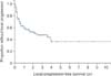
The local progression rate for the series was 46% (n = 37). The median time to local progression was 26 ± 14.3 months (Fig. 1). The median time from local progression to death was 13 months (range, 0 to 81 months). The 2-year and 5-year local progression-free survival were 52.6% ± 6.4% and 36.4% ± 7.6%, respectively. The local progression rate was higher in patients with visceral metastases (57% vs. 33%, p = 0.032). This might be due to the higher rate of en bloc resection with negative margin in patients without visceral metastases. However, neither the surgical margins nor the histological margins were found to have a significant influence on the local progression of the disease.
Local progression was identified with a sophisticated imaging modality such as CT, MRI or a PET scan in the majority, whereas local progression identified on X-ray was confirmed with one of the sophisticated imaging in 2 patients. Biopsy was not performed to confirm local progression. Local progression was seen in the tumor bed or in the tumor bed margin in 27 patients (73%), away from the tumor bed in 6 patients (16%) and in the soft tissue in 4 patients (11%).
Univariate analysis of possible factors associated with local progression revealed that the absence of skip lesions (p = 0.006), absence of visceral metastasis (p = 0.009) and metastases from the breast, thyroid or hematolymphoid malignancies (p = 0.031) were significantly associated with longer local progression-free intervals (Table 3). However, on multivariate analysis, only the presence of skip lesions (p = 0.017) and the presence of visceral metastasis (p = 0.027) were found to be significant factors.
Local progression necessitated treatment in 29 patients. Eight patients underwent repeat surgery and 7 received systemic treatment. In all, 17 patients received radiation therapy including 2 patients who underwent repeat surgery and 1 patient who received systemic treatment. Retreatment rates were significantly higher in patients with single bone metastasis (p = 0.013) and in patients with initial tumor size ≤ 6.8 cm (p = 0.021). The mean survival time from local progression was longer in patients who received retreatment than those who did not receive retreatment (17 months vs. 5 months).
Survival
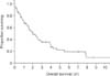
At the time of analysis, 55 patients (66%) had died of disease and 28 were alive. The median survival of all patients was 24 months. The 2-year and 3-year survival rates were 52.5% ± 5.9 % and 35.6% ± 6 %, respectively. For the 55 patients who died of disease, the mean time from surgery to death was 22 months (range, 0 to 95 months) (Fig. 2). The mean follow-up of the 28 survivors was 36 months (range, 2 to 145 months).
On univariate analysis of possible factors associated with survival, favourable primary cancers (of the kidney, breast, thyroid or hematolymphoid) (p = 0.014), absence of visceral metastases (p = 0.008), and albumin levels of more than 39 g/L (p = 0.003), were significantly associated with a better survival (Table 4). On multivariate analysis, all the 3 variables retained statistical significance: favourable primary cancers with a p-value of 0.014, absence of visceral metastasis with a p-value of 0.017 and albumin levels of more than 39 g/L with a p-value of 0.009.
DISCUSSION
The goals of surgical treatment of metastases to the pelvic bone are similar to those for MBD at any other site, which include local tumor control, structural stability and restoration of function.1516) Unlike MBD of long bones that are usually operated by general orthopedists, MBD of the pelvic bone are operated by orthopedic oncologists as the surgery is much more challenging.17) This study aimed to analyze the outcomes of surgery for MBD of the pelvic bone. The authors believe that this is one of the largest series documenting the outcomes after surgery in patients with MBD of the pelvis.
The complication rate of 16% in our series is well within the range of 5% to 50% that has been reported in the literature.51618) As observed in our series, infection is the most common complication after surgical treatment for MDB of the pelvis. This is probably attributable to various factors including, but not limited to, the longer duration of surgery with higher blood loss, lesser immunocompetence due to chemotherapy and disease load. Higher complication rates in patients with lower serum albumin have been reported in other types of surgeries.1920) Increased complications in older patients can be attributed to the poor nutritional status, associated comorbidities and immunosenescence.
The complication rates were higher in patients who underwent THR in our series. The goal of surgery in MBD is to cure sometimes, to relieve often and to comfort always. From this point of view, good functional outcomes could be obtained in patients who underwent THR despite the relatively high complication rates.891011) Patients' increased longevity and potential improvement in quality of life must be justifying the major reconstruction. However, the high rate of complications of THR warrants careful selection of patients with MBD.
The reported local recurrence rates for primary tumors of the pelvic bone are between 0% and 62%.17212223) Although the local progression rate of 46% in our series was similar to the rates reported in the literature, it is considerably high when compared to those seen in the extremities which are between 12% and 17%.182425) The difference in the median survival between the patients undergoing surgery on extremities and on the pelvic bone might reflect the difference in the local progression rate between them. Demanding surgeries on the pelvic bone are carried out in patients with a longer expectancy as compared to those undergoing surgery on the extremities. The reported local progression rate of 16% following surgery on the pelvic bone for metastases in a series of 43 patients in the study by Giurea et al.16) can be attributed to the low rate of median survival of 14 months when compared to the median survival of 24 months in our series. Twenty-one percent (8/37) of the local progression were detected after 14 months in our series. Furthermore, in our series most of the local progression was diagnosed using one of the sophisticated imaging modalities, whereas the surveillance strategy used for local progression was not mentioned in the series reported in the literature.
The factors found to be significantly associated with local progression were the presence of skip lesions and the presence of visceral metastases. The implications of skip lesions in primary bone tumors are well known.2627) To the authors' knowledge, there is no published literature on the implications of skip lesions in the surgical management of MBD. In the case of MBD responding to nonsurgical treatment, skip lesions need not be addressed surgically unless it is deemed necessary to reduce the local progression rate.
The fact that local recurrence rates depend on the adequacy of margins has been well established for primary tumors of the pelvic bone.1721) However, it was not reproducible with metastatic tumours in our series. Although the local progression rates were high in patients who underwent curettage as compared to patients who underwent en bloc resection and in patients who had positive histological margins as compared to negative margins, the differences were not statistically significant. MBD being a systemic disease and the pelvic bone having a rich anastomotic blood supply might explain surgical margins not influencing local progression. Similar results have been reported in the literature.18) However, there are studies reporting contrasting results2528) that might have resulted from the inclusion of patients with metastases to the bones of extremities and pelvic bones or only solitary bone metastasis to the pelvis.
Twenty-nine of the 37 patients who developed local progression received either surgical or nonsurgical treatment. This is in contradistinction to the low treatment rates for local progression in bone metastases to the extremities. The high rate (78%) of treatment for local progression in our series was due to the expectation of prolonged survival among the patients who underwent surgery for their disease.
Overall survival of 56% at 3 years has been reported in a series of 21 patients as compared to 36% in our series. It has been well documented in the literature that patients with MBD with primary cancer in the breast, thyroid, kidney, prostate and of hematolymphoid origin survive long. The results of our series are consistent with those of the available literature. It was also noted that patients with no visceral metastases had a longer survival than those who had visceral metastases. Oncological burden and low serum albumin levels have been well documented in the literature as factors influencing survival in cancer patients. 1329)
Although patients with solitary bone metastases had a longer mean survival than those with multiple metastases (35.1 ± 28.5 months vs. 23.5 ± 27.2 months, p = 0.091), the difference was not statistically significant. There are studies reporting contrasting results in the literature, but they are limited to renal cell carcinoma.2830) In a series of 14 patients who underwent en bloc resection, the median follow-up period was 74.5 months for the surviving patients and 54 months for the nonsurvivors.28) This can be possibly due to the type of primary cancer. In the 14 patients, 6 patients had a primary in the kidney and 2 were from hemangiopericytoma, and all but one had a negative margin. Of the 23 solitary metastases in our series, only 3 patients had a primary in the kidney, and only 5 patients had a negative margin. This might explain the disparity in the results.
Our study did not find any significant difference in survival between patients who underwent curettage and those who underwent en bloc resection. The type of surgical margin had no significant impact even in patients with solitary metastasis. Similar results have been reported in the literature1618) However, contrasting results are also available in the literature.242530) The inclusion of patients with primary cancers from multiple sites in the former studies as compared to only patients with renal cell carcinoma in the latter studies may explain the discrepancy in the results.
Our study was not without its limitations. First, this was a retrospective study and no standard criteria for patient selection were followed. Second, despite the heterogeneity of primary cancers, analyses using rather uniform factors associated with the outcome limit the interpretation of the results of this study. Third, the nonsurgical management was not taken into account although some might have responded to other modalities of treatment such as hormone therapy, chemotherapy, and anti-bone resorbing drugs resulting in the relatively low local progression rate. Fourth, the follow-up periods were varied according to the general condition and oncological burden of the patient. This might have led to the late detection of local progression.
To conclude, surgery remains a viable modality of treatment for MBD of the pelvic bone. Advanced age and low preoperative serum albumin levels are associated with high rates of complications. The presence of skip lesions is associated with local progression. Metastasis from a favourable primary cancer, absence of visceral metastasis and high serum albumin level are associated with a prolonged survival. Surgical margin does not influence local progression or survival in MBD of the pelvic bone. The results of this study need to be confirmed with a larger cohort of homogenous patients.
 XML Download