

 PDF
PDF ePub
ePub Citation
 Print
Print


Pediatric forearm fractures are treated effectively by casting. However, surgical intervention can be required due to failure to obtain or maintain adequate reduction. Currently, intramedullary (IM) nails, K-wires, and plates are used for surgical treatment of pediatric forearm fractures.1,2,3)
Recently, flexible IM nailing has been widely performed for pediatric forearm fractures. Because of advantages such as minimal invasiveness and prevention of pin-related complications, it has changed traditional tenets of pediatric forearm fracture care, gaining more popularity than plating or K-wiring.
Eighty percent of forearm fractures in children occur in the distal one third of the forearm which is further divided into physis, metaphysis, and diaphysis.4) Fractures of the distal radial physis or metaphysis in children are usually treated by conservative methods. In contrast, fractures at the metadiaphyseal junction (MDJ) of the radius is managed differently from fractures of the metaphysis because this region also has diaphyseal characteristics. If the location of a radius fracture is identified as the MDJ, care should be taken to maintain acceptable alignment, especially among adolescents. However, IM nailing at the MDJ has been associated with less stability compared to IM nailing at the diaphysis.
In this study, we investigated the results of flexible IM nailing of radius fractures at the distal MDJ in adolescent patients. Our hypothesis was that there would be no difference between the diaphysis and MDJ regarding restoration of the radial bow, forearm motion, and complication rate. The purpose of this study was to analyze the radiographic and functional outcomes of flexible IM nailing in adolescent patients with forearm fractures at the diaphysis or MDJ.
METHODS
Patient Selection
We retrospectively reviewed the results of 40 patients who underwent IM nailing for the treatment of pediatric forearm fractures in Soonchunhyang University Bucheon Hospital between November 2004 and November 2012. This study was approved by the Institutional Review Board of Soonchunhyang University Bucheon Hospital (SCHBC_IRB 2013-16). Our institution approved the human protocol for this investigation, that all investigations were conducted in conformity with ethical principles of research, and that informed consent for participation in the study was obtained.
The criteria for inclusion in this study were (1) open diaphyseal fracture of both bones of the forearm; (2) ≥ 10° of residual angulation or ≥ 30° of malrotation of the forearm after failed closed reduction; and (3) ≥ 10 years of age. The exclusion criteria were (1) history of previous forearm trauma; (2) concomitant upper extremity fracture; and (3) underlying bone pathology.
Thirty males and 10 females patients were followed for an average of 16 months (range, 12 to 20 months), and their average age was 11 years (range, 10 to 16 years).
The average duration from the onset of trauma to surgery was 3.8 days (range, 1 to 36 days). Four patients had open wounds (Gustillo-Anderson classification grade I; 0.5 cm). One had an associated radial sensory nerve injury. Three cases were converted to IM nailing due to reduction loss after initial attempts involving closed reduction and casting.
Thirty-four patients had fractures of both bones of the forearm. Six patients had a radius fracture alone. Fracture sites were located at the MDJ of the radius in 8 patients (MDJ group; 6 fractures of both forearm bones and 2 radius fractures) while the remaining 32 patients had middle-third fractures (D group; 28 fractures of both forearm bones and 4 radius fractures).
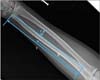
All fractures were classified according to the AO/OTA classification of diaphyseal fractures. Plain radiographic examination to assess restoration of the radial bow was performed in all patients. The magnitude and location of the maximum radial bow were evaluated as a percentage of the total radial length according to the technique suggested by Firl and Wunsch5) (Fig. 1).
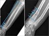
Eight patients who underwent flexible IM nailing for fracture at the distal MDJ were selected according to the following definition of MDJ fracture considering variable age and bony morphology. Fracture at the MDJ was defined as a fracture with (1) the distance between the fracture line and the distal articular surface between 35 mm and 60 mm; (2) the ratio of the length of distal fragment to the total length of radius within 25%; and (3) the ratio of the maximal diameter at 2 cm proximal to the fracture line to that at 2 cm distal to the fracture line within 70% (Fig. 2).
Range of movement (ROM) was assessed using a goniometer and graded according to the criteria proposed by Daruwalla.6)
Surgical Procedure
The operation was performed under general anesthesia using the operative technique described by Lascombes et al.7) Blunt ended titanium elastic nails (Synthes, Paoli, PA, USA) of diameter 2.0 mm were used in all operations. The nail was slightly bent 2 cm from the tip for ease of insertion if passage across the fracture site was difficult.
A transverse skin incision was made 1 cm proximal to the distal radial physis along the wrist crease to leave a less conspicuous scar. The Radial IM nail was introduced in a retrograde fashion just proximal to Lister's tubercle retracting the extensor carpi radialis tendon radialward to avoid radial sensory nerve injury. After radius nailing, stability of the ulnar fracture was checked through full range of pro-supination. If reduction of the ulnar was lost, ulnar nailing was performed. The ulnar IM nail was introduced in an antegrade fashion through a longitudinal incision made 1 cm distal to the olecranon apophysis on the lateral side of the ulnar. After IM nail insertion, limitation of forearm pro-supination was examined intraoperatively to avoid potential rotational malunion. The remaining nail at the entry site was bent and cut 0.5 cm from the bending point for ease of later removal. Skin closure was performed burying the nail tip under the subcutaneous tissue.
Postoperative Management
All patients were immobilized in a long arm splint for 2 postoperative weeks. Then, a short arm cast was applied for additional 4 weeks. Free and full motion of the arm was allowed thereafter.
Statistical Analysis
Continuous data were compared using the Mann-Whitney U-test for angulation and the Kruskall-Wallis test for radial bow with SPSS ver. 14.0 (SPSS Inc., Chicago, IL, USA). Continuous variables included angulation, magnitude, and location of the maximal radial bow. A p-value of less than 0.05 was considered to be statistically significant.
RESULTS
There were 20 patients with simple fractures (22-A3: both bones, 15 patients; 22-A2: radius, 5 patients) and 20 patients in which one or both of the diaphyseal fractures were characterized by a wedge or butterfly fragment (22-B3: both bones, 19 patients; 22-B2: radius, 1 patient). The maximal diameter of the MDJ at the radius fracture site was larger in the MDJ group (average, 12.5 mm; range, 10 to 16 mm) than in the D group (average, 9.5 mm; range, 8 to 11 mm).
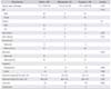
Closed nailing was successful in 32 patients. Open reduction with limited incision was carried out in 8 patients (radius in 4 patients; ulna in 2 patients; and both radius and ulna in 2 patients) due to failure of closed nailing. Out of the 34 patients with both forearm bone fractures, 30 patients had stabilization of both radius and ulna, and only radius was nailed in four patients. In the MDJ group (n = 8; both forearm bones in 6 patients and radius in 2 patients), only radius was nailed in 5 patients and both radius and ulna were nailed in 3 patients. In the D group (n = 32; both forearm bones in 28 patients and radius in 4 patients), only radius was nailed in 5 patients and both radius and ulna were nailed in 27 patients. All patient characteristics are shown in Table 1
Union was achieved in all 40 patients at an average of 8.3 weeks after surgery (MDJ group, 8.8 weeks; D group, 8.3 weeks). All patients had complete restoration of forearm rotation. The results were classified as good in 38 patients and excellent in 2 patients according to Daruwalla criteria (Table 1).
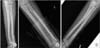
At the latest follow-up, all patients had less than 5° of residual angulation without translation or malrotation (Figs. 3 and 4). The mean preoperative angulation was 6.1° (range, 0.2° to 34.0°) on the anteroposterior radiograph and 12.3° (range, 1.0° to 34.0°) on the lateral radiograph (MDJ group: 3.5° and 16.0°, respectively; D group: 7.0° and 11.0°, respectively). The mean latest follow-up angulation was 1.8° (range, 0.3° to 4.3°) on the anteroposterior radiograph and 3.3° (range, 0.7° to 26.9°) on the lateral radiograph (MDJ group: 1.8° and 2.1°, respectively; D group: 1.9° and 2.8°, respectively) There was no significant difference in the mean angulation between the groups (p = 0.982 and p = 0.393, respectively). The mean magnitude of maximal radial bow was 5.7% ± 1.8% (MDJ group, 5.2% ± 0.8%; D group, 5.9% ± 1.9%). The mean location of maximal radial bow was 58.0% ± 8.8% (MDJ group, 56.4% ± 8.9%; D group, 58.6% ± 8.9%). There was a significance difference in the mean magnitude and location of maximal radial bow compared with the contralateral normal arm (normal: 7.0% ± 1.2% and 50.9% ± 6.0%, respectively; p = 0.002 and p = 0.013, respectively). Although reduction in the magnitude of radial bow and distal translation of the radial bow were observed in both groups, differences in the mean magnitude and location of maximal radial bow between the groups were not statistically significant (p = 0.482 and p = 0.482, respectively). Details of the 8 patients in the MDJ group are shown in Table 2.
Complications
Postoperative complications occurred in 3 patients only in the D group. Two patients developed a symptomatic superficial surgical site infection of the ulnar IM nail at postoperative 6 weeks and 5 months, respectively. Both patients were treated with removal of the ulnar IM nail, but one of them developed delayed union of the radius due to early radial nail removal (postoperative 10 weeks). This patient was treated with an extended period of immobilization (16 weeks) and healed at approximately postoperative 20 weeks. The other complication was refracture. It occurred in a 12-year-old boy and 10-month-old boy with a 22.B3 forearm fracture, whose nail was removed at postoperative 4 months. The child fell while riding a bicycle at 5 months after the index procedure. He sustained fractures of the radius and ulna adjacent to the previous fracture site and was treated with a revision nailing.
The IM nail implant was removed in the operating room in all patients between 4 and 6 months (average, 4.5 months) after the index procedure. None of the patients reported complications related to removal. Activity restriction was not required after nail removal.
DISCUSSION
Among our cohort of 40 patients who underwent IM nailing for adolescent forearm fracture in our hospital, 8 patients had fractures at the MDJ of the radius. All patients obtained satisfactory results without complication.
Unstable forearm fractures can be fixed by several methods. When surgery is indicated, percutaneous pining has been commonly used for metaphyseal fractures and IM nailing or plating for diaphyseal fractures.
The average maximal diameter at the radius fracture site was about 3 mm larger in the MDJ group (12.5 mm) than in the D group (9.5 mm), which seemed to jeopardize immediate stability of distal fragment supported by the IM nail. In this study, it was noted that the degree of step-off at the radius fracture line frequently increased after radius nail insertion in the MDJ group. It could be explained that the larger canal diameter in the MDJ group resulted in less restrain of nails against re-displacement than in the D group. In 1 patient, we inserted additional K-wire through the fracture line to reduce the step-off, which was later removed at postoperative 4 weeks. However, in the remaining 7 patients in the MDJ group, no additional K-wire was inserted in spite of visible step-off after nail insertion. We believed that the IM nail alone would be sufficient to resist re-angulation or maintain the radial bow. Our data supported the assumption that alignment or radial bow maintained by IM nails would not be significantly different between the MDJ group and the D group.
Distal translation of the radial bow induced by IM nailing in the forearm bone fractures did not correlate with limitation of forearm rotation in a study by Shah et al.8) Our date also showed complete restoration of forearm pro-supination even though there were differences in the average magnitude and location of the maximal radial bow compared with the normal contralateral arms.
We determined that the minimal distance between the fracture line and the distal articular surface should be > 3.5 cm for IM nailing. If a fracture site was located closer to the physis (< 3.5 cm), it was considered unsuitable for IM nailing. Too short distal working length provided by a physeal sparing IM nail would lead to less favorable results than crossed K-wire pinning. Although transphyseal pinning and intramedullary fixation across the physis into the epiphysis have been reported to exhibit low rates of complications, efforts should be made not to violate the intact physis.9)
The union time in the MDJ group (8.8 weeks) was not short compared to that in the D group (8.3 weeks), even though none of the patients in the MDJ group required open reduction. In general, the union time at the metaphysis takes less than 5 weeks in the adolescent. Thus, this finding may suggest that a delay in union time of the MDJ could be caused by several factors, although we could not determine whether it was due to the diaphyseal characteristics of the MDJ or insufficient immediate stability of the distal fragment supported by IM nails.
A biomechanical study noted that IM nails had more recoil and did not induce a new fracture line in a canine model of pediatric forearm fractures compared to Kwire. IM nails were clinically superior to crossed K-wires for pediatric forearm fractures despite biomechanically less rigid fixation.10)
Percutaneous K-wire cross pinning in the MDJ of the radius can be challenging because of the relatively smaller diameter than the metaphysis. Our experiences have suggested that K-wire fixation in the MDJ of the radius is prone to loss of reduction. The insertional difficulties due to small diameter or less precise wire placement may result in loss of reduction after crossed K-wire pinning. Another drawback of K-wiring is percutaneous pin-related problems, resulting in late displacement after removal at 5 to 6 postoperative weeks.
For this reason, percutaneous K-wire pinning in the MDJ of the radius may not be appropriate especially in the noncompliant adolescents because they require both well molded cast immobilization and long period of pin site care postoperatively to avoid late displacement.
Although the results of single bone fixation in pediatric diaphyseal fractures of both forearm bones have been reported comparable to those of both bone fixation, it should be noted that fixing the radius rather than the ulna provides better outcomes.11,12) In this study, we assessed intraoperatively fracture stability and alignment of the unfixed ulna after radius nailing. When reduction of the unfixed ulna found lost in fluoroscopic evaluation after full range of pro-supination, we determined to nail the ulna to prevent re-displacement. Of the 34 both forearm bone fracture patients, radius nailing only without ulnar nailing was performed in 4 cases. In the D group, only one out of the 28 both forearm bone fractures did not require ulnar nailing. On the contrary, in the MDJ group, 3 out of the 6 both forearm bone fractures did not require ulnar nailing. Although more proximal forearm deformity has been known to impart greater restriction in forearm rotation, it has not been determined whether fracture location is related to stability of both forearm bone fractures because no study has addressed this issue to date. Our small series has demonstrated that fracture location affects the treatment of the remaining unfixed ulna after radius nailing in adolescent patients with both forearm bone fractures.
Forearm fractures in the MDJ of the radius could be treated effectively with IM nailing without any complications in our small series. All three complications occurred only in the D group, even though it was not possible to determine whether this was related to the fracture location or injury severity or both. Refracture occurred in 1 patient while elective nail removal was performed 4 months after the index procedure. Refracture risk has been reported to be 4%–7% in the first 12 months after a forearm fracture. It has been suggested that the forearm fracture line still visible at the time of cast removal is a risk sign.4) So we think that IM nails should be kept in place if delayed union was suspected especially in the diaphyseal forearm fractures.
There were a number of limitations to the current study. First, the study involved a relatively small number of patients and had a retrospective design. Second, we included patients with radius fracture alone; however, the aim of the current investigation was to assess the outcomes of IM nailing at the MDJ of the radius. Third, intraoperative stability evaluation was dependent on the author's experience. Finally, universal application of our definition of the MDJ relying on radiologic arbitrary distance measurement can be controversial although we considered age variance in adolescents.
In conclusion, IM nail fixation in adolescent forearm fractures provided satisfactory results, even though the fracture was located at the MDJ of the radius. Although careful intraoperative assessment of the remaining unfixed ulna after radius nailing is required, IM nailing of the radius alone can be effective to maintain adequate stability of both forearm bone fractures at the MDJ in adolescents.


 XML Download