

 PDF
PDF ePub
ePub Citation
 Print
Print


Tibial tuberosity-trochlear groove (TT-TG) distance is a measure of lateralization of the tibial tuberosity in relation to femoral trochlea. Many studies have confirmed that lateralization of the tuberosity is linked to patellar instability, thus TT-TG is an important parameter for evaluation of patellofemoral disorders.1234) However, the degree of lateralization that may contribute to actual dislocation varies among studies.234) Alemparte et al.5) studied healthy volunteers and found that normal values for TT-TG were 13.6 ± 8.8 mm, indicative of large variation. Dejour et al.2) reported that the TT-TG in the control group was 12.7 ± 3.4 mm and > 20 mm in pathological cases. Thus, there is reportedly a high degree of variability in the normal TT-TG values and furthermore, some methods of measurement are complicated with significant intra- and interobserver measurement errors.67)
Anterior knee pain is a common finding in young adults with no definite etiology or diagnostic test.8) Patellofemoral instability is often considered an important contributing factor and X-ray and computed tomography (CT) scans are used to understand the aberrations in the normal anatomy contributing to instability.8) However, a variety of methods for performing these scans and a wide normal range of various studied parameters are reported, which makes the diagnosis of patello femoral syndrome a challenge.
The TT-TG distance represents the radiographic measurement of the quadriceps vector, which represents a lateral force displacement on the patella during knee motion. It is > 20 mm in patients with recurrent patellar dislocations, as compared with 13 mm in control subjects.2) Thus, a lateralized tibial tubercle may be a relevant anatomic indicator for a distal realignment procedure. However, TT-TG distance parameter values at different developmental stages are not defined in the current literature. Additionally, the reference of TT-TG is based upon the value obtained in European or American populations. Since the pathological significance of the TT-TG distance depends on the force vector of the extensor mechanism resulting from individual knee size, different somatometric values are expected to be related to the TT-TG distance in Koreans. Therefore, the purpose of this study was to evaluate the TT-TG distance in Korean patients with normal knee joint function using rotational profile CT scans.
METHODS
Patients
We recruited 100 completely asymptomatic patients of both genders (50 each) to obtain unilateral standard lower extremity rotational CT scans for evaluation of hip joint pain and anteversion angle of the affected acetabulum. Patients with no clinical signs or symptoms in the knee joint for 1 year prior were included in the study. Exclusion criteria were poor quality CT scan data that was unreadable for evaluating anatomic landmarks and newly detected deformities at the level of the knee joint or below the knee joint. Based on the 2 exclusion criteria, six patients and nine patients were excluded, respectively.
TT-TG Measurement
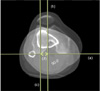
TT-TG was measured using a Light Speed Plus (General Electric Medical Systems, Milwaukee, WI, USA) with a scan time of 0.5 second and 2.5 mm slice thickness. The X-ray tube voltage was 120 kV with a scan exposure of 250 mA. Each subject was examined in a supine position during CT examination. The TT-TG distance was assessed according to Schoettle et al.9) The first transverse craniocaudal image that depicted a complete cartilaginous trochlea was used to determine the deepest point within the trochlear groove. A line was drawn through the deepest point of the trochlear groove, perpendicular to the posterior condylar tangent. A second line was drawn in parallel to the trochlear line through the most anterior portion of the tibial tubercle. The distance between the 2 lines represented the TT-TG distance (Fig. 1).
The study was performed in 2 stages. Stage 1 of the study involved the analysis of interobserver and intraobserver reliability of the CT scan. Three observers were involved in the study, including one consultant and two trainee surgeons. Radiographs of the 85 patients were evaluated by all three observers independently, on 3 separate occasions at least 2 weeks apart. Stage 2 of the study assessed the validity of the measurement by comparing each data set. The results obtained by the senior consultant were used for validation purpose and compared with those obtained by other observers.
RESULTS
General Characteristics
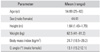
The study included 44 male and 41 female patients with an average age of 54.89 years (range, 25 to 82 years). Table 1 showed the mean weight, height, and body mass index of the patients. The mean Q angle was 13.1° with a maximum angle of 22°.
Inter- and intraobserver agreement for TT-TG readings was excellent, with statistical significance (p < 0.001 for both). The mean kappa value for interobserver reliability was 0.61 (range, 0.59 to 0.63) and the mean kappa value for intraobserver reliability was 0.74 (range, 0.39 to 0.86; intraclass correlation coefficient, 0.912).
CT Scans
Overall median TT-TG distance was 11.24 mm, and ranged from 2.1 to 17.35 mm. The median value for male patients was 10.19 mm and the median value for female patients was 10.36 mm, with no statistical significance (p = 0.20). The mean TT-TG distance in the entire cohort was 10.24 ± 0.8 mm. The mean TT-TG distance was 10.36 ± 0.5 mm for female subjects and 10.19 ± 0.5 mm for male patients. Increased TT-TG distance of > 20 mm is reportedly associated with pathologic patellar instability.2)
DISCUSSION
In the study by Dejour et al.,2) the mean TT-TG distance for the control group was 12.7 ± 3.4 mm and the mean TT-TG distance for the patellar instability group was 19.8 ± 1.6 mm in addition, 20 mm was determined as the cutoff value for patellar instability. In our study, we established the cutoff value of TT-TG distance according to their result. Few studies have reported the TT-TG distance, including under pathologic conditions, in Koreans. Hence, the cutoff value used in the current study was based on studies in Western populations despite some bias. Normal range of the TT-TG distance value varies among studies from 9.4 ± 0.6 mm to 13.6 ± 8.8 mm;45678) and most of these studies were performed in Europeans or North Americans. The conclusion of these studies is that patients with patellar instability have higher TT-TG distances.1210)
Reports of the normal ranges of the Q angle are varied with slight differences between male and female.11) Our findings were in agreement with this observation. However, in our study, despite the differences in Q angle between female and male patients, the TT-TG of female and male patients were not significantly correlated. Biedert and Warnke12) reported similar results in their study of 56 knees with patello-femoral pain.
We determined the TT-TG distance of asymptomatic knees in a Korean population based on the recommendations by Wright and Feinstein13) and achieved low variability in the study data. All patients were evaluated under similar conditions with similar test protocols. All tests were performed with a relaxed quadriceps muscle in the supine position. Tests were performed by the same radiologist using the same equipment and technique. Also, all three trained operators performed the measurements in all cases by consensus.
Reports on normal TT-TG values in the Korean population are rare, hence our study serves as the basis for future work. The mean TT-TG distance for the normal population was 10.24 ± 0.8 mm, with similar average values between female or male patients. Furthermore, rotational profile CT scan is a reliable and reproducible method for evaluating TT-TG. The TT-TG value provides a guideline for the use of a distal realignment procedure.
The limitation of our study was that our study population was mainly aged over 20 years with very few young patients. CT scan data of the younger age group could not be obtained due to the special feature of the clinic that provides health care to primarily adults. This could have biased the results and CT scans from a younger population may provide a different range of values. Determination of the pathologic TT-TG value in Korean patients is an ongoing process. Future prospective studies in a more homogeneous group are required to further establish the normal range of TT-TG values.
In conclusion, the TT-TG distance in normal Korean people is approximately 10.24 mm with no statistically significant difference in the median TT-TG distance between genders.
 XML Download