

 PDF
PDF ePub
ePub Citation
 Print
Print


Supracondylar fracture of the humerus is a common childhood injury. The morbidity of this injury is related to a lesser degree to the fracture itself but results more often from associated injuries. The incidence of a supracondylar fracture with an ipsilateral forearm bone fracture, known as floating arm injury, is a very rare paediatric injury and has been reported to occur in 2% to 13% of children. This combined injury has been termed floating elbow in view of the fact that the elbow is completely dissociated from the rest of the upper limb. There are relatively few case reports in the literature on an ipsilateral supracondylar fracture associated with a proximal humeral fracture, the so called floating arm injury. We describe the case of a 9-year-old boy who had a combination of the above-mentioned injuries in the ipsilateral limb. To the best of our knowledge, this is the only case report of a combined floating elbow and floating arm injury in the same limb.
CASE REPORT
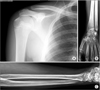
A 9-year-oldboy was brought to the emergency ward of Government Hospital for Bone and Joint Surgery in Srinagar 7 hours after the trauma of a fall from a tree (10 m high) onto an outstretched hand with the forearm pronated and the wrist hyperextended. The boy complained of severe pain in the right upper limb. The right elbow, proximal part of the right arm, and the right distal forearm were swollen and tender with a palpable crepitus. There was a 3-cm laceration over the anterior aspect of the right elbow. The hand was well-perfused (red and warm). Distal neurovascular status was normal. Pulses could be felt at radial and ulnar nerves. Movement of fingers and thumb was precise. Roentgenograms demonstrated a displaced fracture at the proximal humeral metaphyseal-diaphyseal junction, with an extension (Gartland type III) supracondylar humeral fracture with a Salter-Harris type II physeal injury of the distal forearm (Fig. 1). All injuries were found in the right upper limb. His upper extremity was splinted using a Cramer wire splint extending right from the tip of the shoulder to the fingertips, and a cuff and collar bandage was applied.
Under general anaesthesia, debridement of the Gustilo-Anderson type II open supracondylar fracture of the humerus was done, and it was copiously irrigated with normal saline. Open reduction of the fracture was done. Reduction was stabilized using 2 lateral Kirschner wires. Next, under fluoroscopic control, closed reduction of the proximal humeral fracture was done using a temporary Kirschner wire. The Kirschner wire was driven from lateral to medial in the humeral shaft and used as a joystick. Reduction was stabilized using 2 Kirschner wires. Lastly, closed reduction of the distal forearm injury was done, and the radial physeal injury was stabilized using 2 lateral Kirschner wires. All the Kirschner wires were left protruding from the skin and covered with a sterile dressing. After completion of the surgical procedure, a long plaster of Paris splint extending right from the tip of the shoulder to the fingertips was applied, and an arm pouch was given.
The wound over the elbow healed uneventfully and delayed primary closure was done. The Kirschner wires were removed 4 weeks after radiological confirmation of healing or reunion. It took almost 6 weeks for reunion to be observed (Fig. 2). A splint was provided to support the hand and forearm movement, which was followed by range of motion exercises that began1 week after the use of the splint support. A 6-month follow-up was scheduled. The patient achieved a full wrist, elbow extension/flexion of 0°–130° and pronation/supination, and normal shoulder range of motion when measured with a hand-held goniometer, and had no functional deficits (Fig. 3).
DISCUSSION
Various descriptive terms have been used to describe a combination of upper limb injuries at different sites. The "floating elbow" describes a combination of ipsilateral forearm and supracondylar fractures.12) The term "floating arm" refers to a rare injury, describing a combination of ipsilateral supracondylar and proximal humeral fractures.345) To the best of our knowledge, this is the first case report in English literature describing a combination of an ipsilateral floating arm and a floating elbow, which resulted from a fall from height. The intensity of trauma was so severe as to result in 3 level upper limb injuries in the patient. Various authors have recommended that the supracondylar fracture should be fixed before other injuries because of the much greater risk of complications associated with this fracture.45) In our case, we used the method described by Parmaksizoglu et al.6) of using a temporary Kirschner wire as a reducing joystick to aid in closed reduction of the proximal humeral fracture. The distal radial fracture was reduced in a closed manner with traction at the fingers and counter-traction at the level of the forearm. The swelling and the less than adequate traction and countertraction precluded perfect reduction of the distal radial physeal injury; however, this was considered acceptable due to the remaining growth and distal nature of the physeal injury.
In conclusion, this is the first report of a floating upper limb, a combination of a floating elbow and a floating arm. Due to the severe nature of the injury with the attendant swelling, closed reduction is difficult to achieve. The use of a temporary Kirschner wire in the humeral shaft helps in closed reduction of the proximal humeral injury.

 XML Download