

 PDF
PDF ePub
ePub Citation
 Print
Print


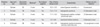
Osteochondral lesion of talus (OLT) is a broad term used to describe an injury or abnormality of the talar cartilage and adjacent bone, often symptomatic enough to warrant treatment. Treatment for OLT has substantially increased over the last decades. Antegrade transmalleolar drilling is a popular option due to its relative ease and satisfactory results. However, bone drilling may present problems related to cartilagenous injury of distal tibia which might cause cyst formation by check-valve mechanism, and heat production that could cause irreversible osteonecrosis leading to morbidity. We report five cases of patient who had cystic formation of medial malleolus after drilling (Table 1).
All patients had antegrade medial transmalleolar drilling combined with anterior ankle arthroscopy for drill guidance. Tourniquet was used in all procedures. In the succeeding months, repeat imaging showed bone cyst formation on the medial malleolus accompanied by osteolysis corresponding to the drill pathway. We did not perform any surgical procedures for these cases because their symptoms were not so severe and fortunately the lesion did not increase in size during the follow-up period.
CASE REPORTS
Case 1
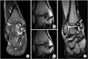
A 20-year-old man presented with lateral complex instability. After seeking consultation, initial magnetic resonance imaging (MRI) showed 10.1 mm × 12.3 mm talar osteochondral lesion (Fig. 1A). The patient underwent distal tibia drilling using 1.6 mm Kirschner (K)-wire with surgical time lasting for 62 minutes. At 8 months postoperative follow-up, repeated MRI revealed the tract traversed by bone drill and evidence of bone edema on medial malleolus (Fig. 1B and C). At 3 years follow-up, MRI showed presence of cystic lesion on medial malleolar axilla (Fig. 1D).
Case 2
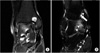
A 31-year-old man came in with symptoms of impingement syndrome. Initial MRI showed 12.4 mm × 11.0 mm talar osteochondral lesion. Distal tibial drilling was done using 1.6 mm K-wire with duration of surgery lasting for 56 minutes. On follow-up, MRI demonstrated floating necrotic bone fragment within a cystic lesion over the medial malleolar axilla (Fig. 2A).
Case 3
A 24-year-old man sought consultation due to multiple periarticular spurs and talocalcaneal coalition. There was an 11.7 mm × 14.3 mm talar osteochondral lesion seen on initial MRI. The patient underwent distal tibial drilling using K-wire with surgery lasting for 72 minutes. On follow-up, MRI revealed cystic lesion over medial malleolar axilla.
Case 4
A 43-year-old man complained of insertional Achilles tendinitis symptoms. Initial MRI revealed an 8.7 mm × 6.8 mm talar osteochondral lesion. Distal tibial drilling was performed using 1.6 mm K-wire lasting for 49 minutes. At 10 months postoperative follow-up, MRI showed a tract tracing bone drilling pathway and evidence of cystic lesion over the medial malleolus (Fig. 2B).
Case 5
A 31-year-old woman consulted due to impingement syndrome. Initial MRI showed a 9.6 mm × 10.8 mm talar osteochondral lesion. Drilling of the distal tibia was done using K-wire with surgery lasting for 66 minutes. On follow-up MRI, cystic lesion over the medial malleolus was found without relation to the patient's symptom.
DISCUSSION
The treatment of OLT mainly involves arthroscopic excision, curettage and bone marrow stimulation, autologous chondrocyte implantation and osteochondral autograft transfer system. In case a lesion is hard to reach because of its location on the talar dome, the lesion can be drilled through the malleolus.
In this case report, authors present iatrogenic cyst formation of distal tibia after drilling which might have been caused by many possible etiologic factors.
Simonian et al.1) reported cystic formation and tunnel enlargement around tibial tunnel after anterior cruciate ligament reconstruction, which can be successfully treated by curettage and bone graft if the lesion is large enough to intervene. And they assumed that the reason for cyst formation might be due to osteolysis after joint fluid leakage. There are few reports on iatrogenic origin bone cyst formation, postoperative bone cyst formation of tibia are extremely rare. The pathogenesis of the iatrogenic bone cystic formation still remains a matter of conjecture and several theories have been suggested. Microtrauma is the most frequently discussed etiologic factor in the formation of iatrogenic bone cyst formation. The blood clot liquefies and adjacent bone is destroyed by enzymatic activity.2)
Other possible etiologic factor can be cyst formation due to joint fluid leakage with synovial origin. Crane and Scarano3) reported two cases of synovial cyst in distal tibia and carpal bone which they mentioned the cause might have been the leakage of joint fluid and synovial proliferation in to the cyst. Histologically, the wall of cyst was composed of dense fibrous and loose fibroblastic mucinous tissue, resembling synovial membrane, and there was no giant cell reaction, new bone formation, inflammatory response, of pigment and fat deposition. In this case series, transmalleolar drilling might have made pathway of joint fluid into distal tibia, but confirmation through biopsy was not possible because all of five patients underwent conservative therapy.
Last possible consideration can be osteonecrosis due to thermal damage during drilling. In this situation, catabolic effect is driving force for the bone cyst formation. Two possible causes described above are uncontrollable factors by the practitioner, but thermal necrosis is much more influenced by practitioner's surgical technique. In our study, drilling was performed according to the procedure of Kumai et al.4) where a K-wire or drill bit is inserted about 3 cm proximal to the tip of the medial malleolus and directed across the medial malleolus into the lesion through intact cartilage. The drill hole was made at an interval of about 2 to 3 mm at a depth of about 1 cm. Anterior ankle arthroscopy was also performed on all patients to provide drill guidance for the chondral lesion. However, for patients no. 2 and no. 5, the size of the K-wire inserted could not be verified since they were performed at another institution.
During drilling, the resistance of compact cortical bone causes increase in bone temperature, which might result in thermal bone necrosis.5) Determining specific threshold for thermal damage in living bone tissue is rather a complex problem. The cellular death caused by heat is immediately evident with temperatures above 70℃.6) Others found that 50℃ caused irreversible cortical bone necrosis. Lundskog7) performed thorough biomechanical, histochemical and morphologic studies on thermal damage to rabbit tissue, and demonstrated that the threshold for irreversible enzymatic disturbance to cortical bone is 50℃ during 30 seconds. Bonfield and Li8) reported irreversible bond weakening of the bone–collagen hydroxyapatite complex at 50℃. On the other hand, Eriksson and Albrektsson9) found that the minimum critical temperature for delayed death of osteocytes were around 47℃ till 3 weeks or more after the injury. The temperature exposure to 47℃ for a minute causes bone resorption and also disturbs the middle- and long-term anchorage of implants. They constructed optical thermal chambers for in situ microscopy that makes it possible to follow the "true" tissue reactions after a defined heat trauma by repeated light microscopic observations.78)
Bonfield and Li8) reported that external irrigation is the single and most important factor in decreasing the increase in bone temperature during drilling while considering all other factors. In all our patients, the absence of cystic lesion over the talar dome on follow-up imaging may be due to the cooling effect of the irrigating fluid routinely used in arthroscopic procedures.
All five patients in our series underwent a trial of conservative treatment wherein each was advised to avoid prolonged weight-bearing over the affected ankle and to refrain from sports activities. It was emphasized that close and regular follow-up must be maintained to monitor progression of the lesion. Fortunately, in all cases, there was no progression of the lesion and even resolution of the lesion in one case.
D'Hooghe et al.10) reported a case of a combined osteochondral kissing lesion involving the talar dome and tibia after transmalleolar drilling. The tibial cartilage may also be injured when the K-wire is inserted into the lesion.1) The authors believe that this may explain the presence of a necrotic bony fragment within the cystic lesion in patient no. 2. It has been shown that once the cartilage is injured, and it is difficult to regenerate completely.
Aside from thermal damage, another problem that can arise in bone drilling is the difficulty in maintaining a freehand control of the drill even when using a drill guide in attaining geometrical accuracy in hole size and location. When the drill bit begins to enter the bone surface, it tends to "walk" or slip on the bone surface.11)
Antegrade transmalleolar drilling remains a reasonable treatment method for OLT. However, the surgeon needs to be aware of the possibility of heat-induced osteonecrosis, talotibial kissing lesion, or iatrogenic cyst formation as it may lead to morbidity if not detected early and managed accordingly. Therefore, we recommend other treatment methods such as arthroscopic microfracture, with minimal iatrogenic risk, as more viable treatment options for these lesions of the talar dome.
 XML Download