

 PDF
PDF ePub
ePub Citation
 Print
Print


Tuberculosis rarely involves the lower cervical spine, which has a high association with neurological complications.1) Its presentation varies in children and adults. The adult type is a localized disease with the involvement of a vertebral body and less pus formation as compared to the pediatric version.1) Spondylolisthesis is rarely seen in the cervical spine as compared to the lumbar spine and is most commonly degenerative.2) Although pathological spondylolisthesis due to various causes has been described in the cervical spine, tuberculous spondylolisthesis has not been described.2) Spondylolisthesis in case of tuberculosis has been described in lumbar or lumbosacral spine.34567) Anterolisthesis the common presentation.7) There is only one report of posterolisthesis in a tuberculous lumbar spine.7) There is controversy as to whether spondylolisthesis coexists or precedes the tuberculosis of spine and the exact mechanism of this has been inadequately described in the literature.3)
We report a rare adult type of paradiscal lower cervical tuberculous infection with posterolisthesis in a 67-year-old female which was managed surgically with a three-year follow-up period. This case highlights the varied and complex presentation of cervical tuberculosis and gives insight into its pathogenesis, diagnosis, and management.
CASE REPORT
A 67-year-old female patient presented with neck pain and stiffness for the past 4 months. The neck pain radiated to both upper limbs and was associated with tingling and numbness. The symptoms had increased in intensity over the past month.
The patient had constitutional symptoms, including loss of appetite, loss of weight (5 kg over a month), evening rise of temperature, and night cries. She was disabled and bedridden due to her severe imbalance while walking. She did not have any history of trauma or coexisting medical illnesses.
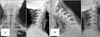
On physical examination, there was severe tenderness over the lower cervical spine. Neck movements were painful and grossly restricted. There was no sensory or motor weakness. Reflexes were brisk. Bladder and bowel function was normal. There were no other respiratory or systemic complaints. X-rays of cervical spine showed a kyphotic deformity, destruction of C6 vertebral body with only a thin part of inferior end plate of C6 remaining, and the associated posterolisthesis of C5 over C6 (Fig. 1).
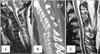
Blood reports showed lymphocytosis with raised erythrocyte sedimentation rate and C-reactive protein values. A magnetic resonance imaging (MRI) showed infective spondylitis of C5/C6 with a pre/paraspinal abscess from C2 to T2 which indented the pharyngeal wall (Fig. 2A). A computed tomography scan showed posterolisthesis of C5 over C6 with bony spinal canal stenosis, anterior wedging of C6, erosion of the superior end plate of C5, and prevertebral soft tissue from C3 to C6 (Fig. 2B).
A diagnosis of infective spondylodiscitis of C5 and C6 secondary to tuberculosis was made. Although the patient was intact neurologically, there was extensive bone destruction and high grade spondylolisthesis; thus, an operative line of management was preferred.
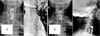
Since operative management was likely and the MRI was highly suggestive of tuberculous spondylodiscitis, a preoperative biopsy was not considered. The patient was scheduled for anterior decompression and stabilization. The surgery was performed under general anesthesia in the supine position through the right anterior approach. C5 and C6 corpectomy using an anterior cage and cervical plating from C4 to C7 was performed (Fig. 3). Tissues were taken for biopsy and sent for histopathology examination. Postoperative antitubercular treatment was started. The histopathological examination confirmed tuberculosis.
Postoperatively, there was no neurological deficit and patient was given a cervical collar. The anti-tuberculous treatment was continued for 18 months. After 2 years, an MRI was performed, which showed that the disease had been healed (Fig. 2C). A radiograph showed solid fusion (Fig. 3). After 3 years, the patient was symptomatically better and did not have any recurrence of the disease.
DISCUSSION
The spine is the most common site of skeletal tuberculosis. The thoracolumbar spine is the most commonly affected.3) The cervical spine is a rare site for tuberculous infections. Clinically, pain is the most common symptom in cervical spine tuberculosis, with varying degrees of kyphosis and instability.1) Constitutional symptoms are non-specific and do not lead to the diagnosis.8)
Tuberculosis of the lower cervical spine presents differently in children and adults. In children, it is of an aggressive nature with diffuse involvement and abscess formation. Healing results in significant kyphosis.19) In adults, it is more localized and involves a single vertebral body with less pus formation. The resulting kyphosis in adults is not as severe because the articular surfaces are spared.19)
Spondylolisthesis is a rare presentation in of any cervical spine pathology. It is most commonly due to trauma and degeneration.2) Although pathological spondylolisthesis in the cervical spine have been reported in cases of aneurysmal bone cyst, neurofibromatosis, and skeletal fluorosis, 2) spondylolisthesis as a presentation of tuberculous has not been reported.37) In our case, there was a pathological posterolisthesis subsequent to the tuberculosis. Table 1 shows the reports of concomitant tuberculosis with spondylolisthesis described in the literature. In the case described by Ratliff,5) anterolisthesis was present before the vertebral tuberculous affection. Nissen-Lie (as described by Ratliff5)) in his report, postulated that, since the disease involved posterior elements of vertebra, it lead to the development of a stress lesion, causing anterolisthesis. The details of cases by Newman and Stone4) and Tuli6) are obscure. Chadha et al.3) described three cases, in which one case had preexistent anterolisthesis and other two had tuberculosis of the posterior elements causing the anterolisthesis. Kirkman and Sridhar7) described a case of grade 4 posterolisthesis due to a vertebral tuberculous affection in a 12-year-old girl. Our case did not have any prior complaints of neck pain or radiculitis, so we feel that the destruction of the vertebral body due to the tuberculous process may have led to the instability and subsequent posterolisthesis.
Treatment of cervical spine tuberculosis should be undertaken early to prevent neurological deficits and deformities.10) Anterior surgery remains the mainstay treatment of cervical tuberculosis with instability, allowing for the debridement and evacuation of an abscess with stabilization of the anterior collapsed segment.810) Posterior surgery is rarely used in lower cervical tuberculosis unless there is a posterior abscess.9) Our case showed destruction of the vertebral body with posterolisthesis and a prevertebral abscess, so we performed an anterior decompression and stabilization. The fixation was adequate and additional posterior fixation was not required.
Thus, we conclude that tuberculosis of the cervical spine can present as spondylolisthesis. This is rarely reported in literature and should be borne in mind. The exact mechanism of this infection is inadequately understood and reported. Early and prompt diagnosis is necessary to initiate the appropriate treatment and obtain a satisfactory outcome.

 XML Download