

 PDF
PDF ePub
ePub Citation
 Print
Print


Intertrochanteric fractures are among the most common types of hip fracture in elderly. However, mortality rate of these patients are high and almost half of these patients cannot return to their previous level of activities.1234) Immediate mobilization and early weight bearing of these patients is utmost important to protect them from possible complications of long lasting immobilization such as atelectasia, deep venous thrombosis, pulmonary embolism and bed sores. Partial weight bearing is usually not enough for immediate mobilization and full weight bearing is mandatory due to frail conditions of such elderly patients. Moreover, it is very important to avoid secondary operations to the maximum extent in this patient group.
Various surgical treatment methods and implants have been employed for the treatment of unstable intertrochanteric hip fractures such as fixed angle plates, dynamic hip screws, proximal femoral nails, and prosthetic replacement. When fracture fixation is chosen, the primary stability of the fracture and fixation system is crucial for early mobilization. However, in case of unstable intertrochanteric fracture, particularly in patients with marked osteoporosis, primary stability of the fixation may not allow full weight bearing and secondary operations due to implant failure may occur.56) In these patients, calcar replacement hemiarthroplasty is usually advocated. Although calcar replacement hemiarthroplasty allows immediate full weight bearing, the stability of the prosthesis is at risk due to removal of calcar region, as the calcar region is very important for long time stability and survival of prostheses.
In the present study, we have proposed that preservation of the calcar region and using an uncemented collared femoral stem will provide immediate full weight bearing as well as a considerably long life for the prosthesis. The union of the calcar region and osteointegration to the prostheses will provide further stability. The purpose of this prospective cohort study was to report the results of patients with unstable intertrochanteric fracture treated with uncemented collared bipolar hip prosthesis, preserving the calcar region.
METHODS
This prospective cohort study involved 64 consecutive patients with unstable intertrochanteric fractures treated with calcar preserving uncemented collared bipolar hemiarthroplasty between May 2011 and December 2012 in Private Ortopedia Hospital. This study was carried out in accordance with the ethical standards laid down in the 1964 Declaration of Helsinki and its later amendments. Institutional Review Board approved the study protocol and all patients and/or primary caregivers gave informed consent prior to their inclusion in the study.
All consecutive elderly patients aged 75 years and over with osteoporotic (Singh index ≤ grade 3) unstable intertrochanteric fractures (AO/OTA type 31-A2 and 31-A3) were included in the study. Patients with immobility before the injury, pathological fractures, polytraumatic patients, patients with simultaneous fractures of the ipsilateral extremity and finally patients, who refused participation were excluded from the study.
No patients died in the hospital after surgery. Three patients died from cardiopulmonary disease, two patients died from renal failure, one patient died in a road traffic accident within a year after surgery and three patients died before the minimum follow-up period with unrelated reasons of the fracture treatment and one patient could not be contacted, thus leaving a total of 54 patients for the final analysis. Etiology of all fractures was low-energy trauma such as simple falls at home. All operations were performed by a single experienced surgeon with an interval of 0 to 3 days after initial injury in accordance with the concomitant comorbidities.
Surgical Technique
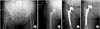
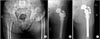
All patients were operated under spinal anesthesia in lateral decubitus position using a posterior approach. The femoral neck was cut with an oscillating saw similar to the preparation of femur in total hip arthroplasty and the femoral head was removed leaving the rest of the femoral neck and femoral calcar. Based on the assessment of fracture pattern and type, if the stability between the implant and femur could be achieved by compaction (mostly AO/OTA type 31-A2 fracture and/or trochanter minor fractured with a little spike), an extra fixation of the neck and trochanter minor (such as trochanteric grip or cable) was discarded, then the prosthesis was tapped into place following standard reaming (Fig. 1). If it was thought that stability may not be achieved by the compaction technique of the trochanter minor fracture fragments (mostly AO/OTA type 31-A3 fracture and/or trochanter minor fractured with a large part towards distal) or if it was decided after compaction that stability had not been achieved, then cable was used. If there was a concomitant fracture of the trochanter major, then stability was achieved by binding both the trochanter major and minor (Fig. 2).
The femoral stem was inserted in a distal press-fit manner into the femoral canal. After the stability assessment and insertion of the appropriate femoral head, the hip joint was reduced. A suction drain was applied and the surgical wound was closed. In all cases, we have used Echelon Primary Hip System (Smith and Nephew Inc., Memphis, TN, USA).
On the first postoperative day all drains were removed, and ambulation with full weight bearing with aid of a walker or crutches was started and encouraged. Patients with no wound complications and those who could take a few steps were discharged subsequently. Follow-up evaluation was performed on third day, 45th day, third month, sixth month, and every six month postoperatively. Anteroposterior and lateral plain hip radiographs were taken after surgery and at each follow-up. Radiographs were examined for evidence of nonunion, component position (alignment), loosening and subsidence. Union was defined as bony bridging across the fracture line on at least three cortices. More than 3° varus and valgus malalignment and more than 5 mm longitudinal subsidence of the femoral stem were considered significant.7)
Clinical evaluations were performed using Palmer and Parker mobility score and pain was evaluated with visual analogue scale (VAS) at each follow-up.8) The duration of operation (minutes), amount of blood transfusion (number of units), and duration of hospitalization (days) were recorded. Any complication during the surgery and follow-up period were also recorded. Repeated measures were used for statistical analyses which were performed using SPSS ver. 16. (SPSS Inc., Chicago, IL, USA). Significance level was accepted as p < 0.05.
RESULTS
The study finally included 15 male and 39 female patients with a mean age of 81.3 years (range, 75 to 93 years). The mean duration of operation was 86.6 minutes (range, 60 to 125 minutes). The mean transfused blood units were 1.2 units (420 mL; range, 0 to 3 units). The average duration of hospital stay was 5.3 days (range, 3 to 7 days). And mean follow-period was 31 months (range, 24 to 52 months). Bone mineral density (BMD) was available for only 32 patients, and mean T score was -3.9 (range, -2.5 to -4.6). Singh index was ≤ grade 3 in all patients.
Clinical Results
The mean mobility score obtained from patients and their relatives before fracture was 6.20 (standard deviation [SD], 1.37). Postoperative first day mobilization was attempted for all patients with the aid of a walker or crutches. Although mobilization was mostly achieved 40/54, some could only stand still, 14/54 therefore patients were re-evaluated on postoperative third day. Mobility assessments were also repeated on all follow-up periods. The results of the statistical analysis revealed mobility scores to have increased at each follow-up after three days (Greenhouse-Geisser: F = 27.236, df = 2.554, p = 0.000) (Table 1).
When the visual analog scale was evaluated preoperatively and postoperatively, the mean scores were seen to decrease over time. The mean scores were statistically significant between all follow-up periods except between 12 months and 24 months (Greenhouse-Geisser: F = 136.999, df = 3, p = 0.000) (Table 1).
Radiological Results
Radiological interpretation revealed bony union in all cases. With regard to the changes in the alignment of the implant, there was no case with more than 3° varus or valgus malalignment. The degree of subsidence of the femoral stem was 1, 2, 3, and 4 mm in 9, 6, 6, and 2 patients, respectively. There was no more than 5 mm subsidence or osteolysis in any of the cases.
Complications
One patient had deep vein thrombosis (treated with anticoagulation), one patient had early postoperative delirium, two patients had superficial wound complications with drainage and delayed healing (treated with antibiotherapy). None of the patients had deep infection. There was one posterior dislocation seven days after the surgery. The patient was treated with closed reduction and physiotherapy. This complication did not influence the patient's mobility score. In one patient, cable penetrated the trochanter major at third month of operation where the cable was used to stabilize the trochanteric fracture. There was no need for an additional operation for this patient because she was able to walk without any pain or symptom.
DISCUSSION
There is still no consensus on the best treatment method for intertrochanteric fractures and fixation materials. For several decades, the treatment of choice for unstable intertrochanteric fractures in elderly patients has been open reduction and internal fixation. Sliding hip screws are widely used,91011) but it has been reported that treatment with dynamic hip screws has resulted in implant problems at a rate of between 1% and 20%. Problems related to fracture type, BMD, reduction quality, implant shape, and localization of lag screw in the femoral head have been reported.12) In addition, osteoporotic elderly patients, in particular, have been reported to have a high prevalence of unsatisfactory functional results, with unacceptable shortening and external rotation deformity of the limb, high cut-out, and varus displacement rates with extramedullary devices.1314)
The use of intramedullary devices has become more widespread with few researchers claiming that intramedullary devices have shown fewer complications and re-operations.1516)
The Cochrane Collaboration performed a systematic review of trochanteric fractures with 43 trials.17) Twenty-two trials involving 3,746 patients compared the gamma nail with any number of sliding hip screw designs and gamma nail was found to be associated with an increased risk of operative and later fractures of the femur and an increased reoperation rate. There were no major differences between implants in terms of the rates of infection mortality, or medical complications. Five trials involving 623 patients compared the intramedullary hip screw (IMHS) with sliding hip screw. Fracture fixation complications were more common in the IMHS group and results for postoperative complications, mortality and functional outcomes were similar in both groups.
Early mobilization is of particular importance for these patients as it might decrease the risk of mortality and morbidity, although older patients are unable to walk soundly enough and are only capable of partial weight-bearing in the postoperative period following internal fixation methods.1819) Therefore, many researchers have shown interest in arthroplasty in trochanteric fractures. Haentjens et al.20) compared the clinical results of internal fixation and bipolar arthroplasty for unstable trochanteric fractures and reported 75% satisfactory results and fewer postoperative complications in the latter group. They insisted that early weight-bearing was the major factor responsible for decreasing postoperative complications. Elderly patients, who are often unable to cooperate with partial weight-bearing (due to neurovascular problems, gonarthrosis, spinal stenosis and other walking difficulties) required after an internal fixation are able to accept full weight-bearing easily. Lu-Yao et al.4) reported that 27% of hospitalized patients with a hip fracture die within 12 months of occurrence of fracture. When we consider this concept with respect to time frame, it becomes obvious that additional six months with full weight bearing means one fourth of the rest of an elderly patient's life.
Many surgeons prefer arthroplasty for the treatment of unstable trochanteric fractures in the elderly in order to decrease complications. Choy et al.7) used arthroplasty and reported mean 80.6 ± 9.3 Harris hip score at the end of two years follow-up period. Andress et al.21) showed adequate osteointegration of the implant, supporting the conclusion that an uncemented prosthesis can be used successfully to treat complex, unstable trochanteric fractures.
Although the clinical outcomes were similar for patients having internal fixation and hemiarthroplasty, the latter had a lower postoperative complication rate and the patients were capable of earlier weight-bearing.20) Kayali et al.5) reported a significant difference in full weight-bearing time between the two groups. Though more costly, cone hemiarthroplasty is an appropriate treatment option for patients with unstable intertrochanteric fractures, which can achieve earlier mobilization. Furthermore, systemic complications due to prolonged immobilization can also be prevented.
There are a few published studies in treatment of unstable intertrochanteric fractures with total hip arthroplasty. Successful treatments were reported especially concurrent to osteodegeneration.222324) However, it has been noticed that total hip arthroplasty involves more operation time, a large amount of blood loss, more risky for dislocation, and also much expensive when compared to bipolar hemiarthroplasty. It is proposed that the use of bipolar arthroplasty instead of total hip replacements can reduce these complications to an acceptable rate.2526)
Our method revealed similar mean transfused blood units (1.2 unit [420 mL] vs. 1.3 units,5) 350 mL,26) 460 mL,22) and 1,050 mL25)) and similar surgery time (86.6 minutes vs. 90 minutes,5) 71 minutes,26) 110 minutes,22) and 151 minutes25)) as has been reported in literature. The mean hospital stay for our patients was shorter than (5.3 days vs. 13 days,5) 10.9 days,26) 9.5 days,22) and 19 days25)) that reported in literature. The early postoperative mobility scores of our patients were high and the postoperative 24th month scores were similar to the preoperative scores. When the VASs were compared with preoperative scores, mean full reduction was observed with this method of treatment. Thus, the 24-months follow-up revealed no loosening of the cables, no subsidence, and no femoral fractures, indicating that the early results from this method may be valuable.
Retaining of lesser trochanter and reconstruction of femoral calcar are important for improving periprosthetic biomechanics and reducing local complications.27) As the placement of the prosthesis is established distally (distal fitting femoral stem), there is no weight-bearing in the calcar region in the early period when full weight-bearing is allowed. The calcar region carries little weight thus allowing the possibility of a complication-free recovery. In the long-term, union in the calcar region and osteointegration of the prosthesis increases the survival term of the prosthesis. Moreover, the torque to the iliopsoas muscles which inserts to trochanter minor allows the continuing muscle function to strengthen the muscles around the hip.
The most frequent late complications following fixation of intertrochanteric fractures are malunion, nonunion, pseudoarthrosis, osteonecrosis of the femoral head, joint damage (associated with fixation slippage or chondrolysis), soft tissue irritation from the implant, bursitis, impingement, infection, trochanteric problems, and chondrolysis. In some series, particularly in patients with unstable fractures, revision or reoperation rate has been published between 5.5% and 22.5% with internal fixation devices.2528)
Majority of above mentioned problems can be avoided with calcar preservation hemiarthroplasty. Hence, it is proposed that this technique can be considered as a definitive surgery in terms of reducing the risk of secondary operations which will further increase the mortality and morbidity and reduce the quality of life. We believe that hemiarthroplsty resolves both mechanical and biological problems in unstable trochanteric fractures at the same time.
In conclusion, calcar preservation arthroplasty is a good treatment option for elderly patients with unstable intertrochanteric fractures, severe osteoporosis, frail constitution and who have high risk of secondary operations. Although the number of cases is low in this study, the desired results were obtained in the early and middle postoperative period. Long-term follow-up studies are required to confirm these results.

 XML Download