

 PDF
PDF ePub
ePub Citation
 Print
Print


Synovial chondromatosis is a rare disorder that is characterized by cartilaginous and osseous metaplasia of the joint synovium. This condition is almost universally monoarticular, and it usually affects large joints such as the knee, hip, elbow and shoulder. It is exceptionally rare in the wrist and hand. There are only a few reported cases affecting the distal radioulnar joint1) and involving the tenosynovium of the digits and the wrist.2) Here, we present a case of synovial chondromatosis of the pisotriquetral joint in which radiographs and magnetic resonance imaging (MRI) did not demonstrate evidence of calcified bodies.
CASE REPORT
The patient was a 57-year-old, right handed female with a five-month history of pain that worsened on ulnar deviation, and swelling and erythematous change at left wrist were present. There was no history of preceding trauma, and she had not participated in any kind of sports. She had no significant medical history of note or symptoms in any other joints.
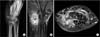
On physical examination, the patient had swelling and tenderness over the left distal radioulnar joint and flexor carpi ulnaris (FCU) tendon, approximately 3 cm proximal from the pisiform of one of its insertion sites. The active range of motion of the left wrist included 50° extension, 50° flexion, 5° ulnar deviation, and 20° radial deviation. The active range of motion of the normal right wrist included 70° extension, 80° flexion, 20° ulnar deviation and 30° radial deviation. The patient complained of pain especially at the pisiform of one of the insertion sites of the FCU in resistive motion during ulnar deviation, wrist flexion and in passive motion during wrist extension, and radial deviation. In addition, she had an aching pain around the distal radioulnar joint in active wrist flexion and ulnar deviation. A neurovascular exam including electromyography, nerve conduction velocity and Tinel's sign at Guyon's canal was normal. Plain radiographs indicated diffuse soft-tissue swelling and widening in the pisotriquetral joint without any other calcified bodies except for the carpal bone (Fig. 1A). MRI demonstrated multiple masses around the pisiform, triquetral bone and perilesional soft tissue swelling (Fig. 1B and 1C).
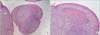
At surgery, the masses were exposed through a volar approach in the Guyon's canal, and the ulnar nerve was not compressed by the mass of synovial chondromatosis (Fig. 2A). Taking care with the ulnar nerve and the artery injury, the pisiform was excised with the mass for the treatment of FCU tendinitis, and to ensure complete excision of the tumor. Most of the chondral bodies were free in the pisotriquetral joint (Fig. 2B). However, parts of the chondral bodies were attached to the synovium in the pisotriquetral joint. For the treatment, synovium and all chondral bodies were excised (Fig. 2C). Histopathological examination revealed synovial chondromatosis with the beginnings of woven bone formation but no evidence of malignancy (Fig. 3).
The patient was discharged and then followed as an outpatient. She started with an active and passive range of motion exercises after the first postoperative week. She regained full range of wrist motion after the fourth postoperative week. The final examination at six months after the surgery showed that the patient had no pain, no residual motor deficits and no radiographical evidence of soft tissue swelling or widening of the pisotriquetral joint that would indicate recurrent disease.
DISCUSSION
Synovial chondromatosis is a rare condition, and its etiology is largely unknown. Benign metaplasia of the synovial membrane results in the formation of multiple intraarticular cartilaginous bodies.
The knee is most commonly affected joint, followed by the elbow and hip. Involvement of the hand and wrist is extremely rare;234) however, Roulot and Le Viet2) have also reported primary synovial chondromatosis of the hand and wrist. Chondromatosis have been classified into two categories within the hand and wrist. Intraarticular synovial chondromatosis is the most common variant, the other being tenosynovial chondromatosis that develops in the synovial lining of the tendon and is consequently the most common in the hands and feet. In this article, the primary sites within the hand and wrist have a variety of locations such as the distal radioulnar joint, the trapeziometacarpal joint, the midcarpal joint, the metacarpophalangeal joint, the interphalangeal joint in intraarticular synovial chondromatosis and the flexor tendon, the extensor tendon in tenosynovial chondromatosis. Ossific bodies were seen on plain radiographs in 11 out of 20 patients, including 5 with intraarticular disease and 6 with tenosynovial disease. A cortical erosion opposite the lesion was visible in 6 out of 20 patients including 2 with tenosynovial disease. There is a wide variation in the age-range with a peak in the 20s and 30s; however, this disease may occur at any age.245)
In the early stage of synovial chondromatosis, plain radiographs may be normal. In the advanced stage, plain radiographs may show diffuse swelling, erosion of the underlying bone or radiopaque densities, depending on the degree of calcification or ossification. If the cartilaginous bodies were not calcified, it would create a problem in the diagnosis of the lesion. This occurs in about one-third of cases.367) Associated radiological features are the erosion of bone, occasional osteoarthritis, and regional osteoporosis. For cases in which radiolucent bodies are present, computed tomography (CT) and MRI scans may be able to identify the lesions. CT scans can detect erosions that may not be seen radiographically.8) MRI scans can demonstrate non-calcified and non-ossified chondral bodies, and identify the size and extent of the lesion. Three different MRI patterns were identified in synovial chondromatosis.7) Pattern A was characterized by a lobulated homogeneous intraarticular signal that was isointense or slightly hyperintense on a T1-weighted image, and hyperintense on a T2-weighted image. Plain films in patients with pattern A showed no calcification. Pattern B is the most common pattern. It was similar to pattern A; in addition, it was characterized by focal areas of signal void on all imaging sequences. These areas corresponded to foci of calcification on plain film. Pattern C had similar features to those of patterns A and B, but included foci of peripheral low signal surrounding central areas characteristic of fat. Plain film and CT scans in these patients revealed multiple foci of calcification and ossification.
The differential diagnoses include rheumatoid disease, osteochondritis dissecans, crystal arthropathy, tuberculous arthritis, trauma-related osteochondral proliferations, periosteal chondroma, synovial sarcoma, psoriatic arthropathy, pigmented villonodular synovitis and degenerative joint disease.
Milgram9) described three stages of the disease. Phase 1 consists of active synovial disease with no free or loose bodies, phase 2 has both active synovial disease and loose bodies in the synovial tissue and in the joint cavity or bursa, and phase 3 consists of multiple osteochondral loose bodies without active synovial disease.
Synovial chondromatosis typically affects a single large joint. The appearance of this lesion in the wrist is exceptionally rare, with some cases in the distal radioulnar joint appearing in the literature. However, cases involving the pisotriquetral joint is extremely rare in the literature. As a result of the rarity of synovial chondromatosis in the pisotriquetral joint, other diseases including infection, tenosynovitis, autoimmune disease, triangular fibrocartilage pathology are suspected at first. The comparatively high prevalence of other conditions in this region, combined with a low index of suspicion of synovial chondromatosis, may result in a failure to diagnose the condition and to provide the appropriate treatment.
Due to the fact that no calcification or ossification of the cartilage occurs in roughly one-third of cases, making a radiographical diagnosis of synovial chondromatosis is difficult. In these cases, MRI would appear to be an ideal method for the evaluation of synovial chondromatosis.
The presence of intraarticular bodies can lead to degenerative arthritis with associated pain and loss of function, and spontaneous regression is rare. Also, there have been a very small number of reported cases of sarcomatous transformation of synovial chondromatosis.10) For these reasons, standard treatment was synovectomy and removal of loose bodies by open surgery or arthroscopically.1)

 XML Download