

 PDF
PDF ePub
ePub Citation
 Print
Print


Butterfly vertebra is a rare congenital defect of vertebral body formation characterized by anterior and median aplasia.1) The vertebra has a cleft through its body and a funnel shape at the ends that gives the appearance of a butterfly on the anteroposterior (AP) view of a plain radiograph. It may be associated with other congenital syndromes23456) and vertebral anomalies;7) however, there are few cases reporting the presence of butterfly vertebra as an incidental finding.8) The butterfly vertebra often causes spinal deformities including kyphosis,9) but rarely causes neurological symptoms. The orthopedic surgeon has to identify this benign spinal anomaly, which may be confused with a pathologic fracture, infection, or associated vertebral anomalies and syndromes.
In the present report, we describe a 40-year-old woman that presented to our emergency department after a simple fall with low back pain, left sciatica, and a L4 butterfly vertebra.
CASE REPORT
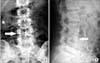
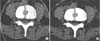
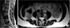
A 40-year-old female patient presented to our emergency department after a simple fall. She reported pain upon palpation of the lower lumbar spine and radiating pain to her left buttock and lower leg. The patient reported a history of low back pain and left sciatica, with exacerbation of the symptoms after the fall. Clinically, no neurologic deficit was present. The straight leg raising test was positive on the left side. Simple radiological control of the lumbar spine revealed a sagittal cleft of the body of L4 vertebra on the AP view (Fig. 1A) and a wedge-shaped deformity of the same vertebra on the lateral radiograph (Fig. 1B). Additional hematologic evaluation that included total leukocyte count, erythrocyte sedimentation rate, serum calcium, serum alkaline phosphatase, and serum protein electrophoresis was performed, in order to rule out a pathologic fracture or infection. All values were within normal limits. Computed tomography (CT) imaging of the lumbar spine showed a large, symmetrical cleft-like defect involving the entire vertebral body of L4 with no evidence of canal compression (Fig. 2A). The spinal arch of L4 was intact and no paravertebral soft tissue swelling was evident (Fig. 2B). A posterolateral prolapse of the L5-S1 intervertebral disc was evident, which correlated with the patient's symptoms. The diagnosis of L4 butterfly vertebra was then established. A thorough clinical examination of the patient, the patient's medical history, and her family's medical history did not reveal any information associated with the incidental finding of the butterfly vertebra. Subsequently, a simple radiological evaluation of the entire spine was performed, in addition to ultrasonography of the heart, urogenital, and hepatobiliary systems, so as to exclude associated congenital abnormalities. A magnetic resonance imaging (MRI) of the lumbar spine reconfirmed the sagittal defect of the L4 vertebral body, occupied by intervertebral disc tissue that included nucleus pulposus material (Fig. 3A). The intervertebral discs L3-L4 and L4-L5 communicated with a bar of disc material which prolapsed in the cleft of the L4 body. Incidentally, a hemangioma was found to be present in the body of L4 vertebra (Fig. 3B).
The patient was treated for her low back pain with analgesics and physiotherapy. The benign nature of the butterfly vertebra was explained to her and she was informed that no further treatment was required. Treatment of the L5-S1 disc prolapse was deferred for a later stage.
DISCUSSION
Butterfly vertebra is usually an asymptomatic and isolated finding. However, it may be associated with other congenital anomalies such as Mullerian hypoplasia/aplasia,2) Alagille syndrome,3) Jarcho-Levin syndrome,4) Crouzon syndrome,5) and Pfeiffer syndrome.6)
This defect is thought to occur between the third and sixth week of embryonic development. The vertebral body is formed by the fusion of two lateral sclerotomes that derive from the somites. Failure of the fusion of the two sclerotomes results in the formation of butterfly vertebra.1) Butterfly vertebrae is seen predominantly in the lumbar spine.1)
In this case, the patient did not present with features of any known associated syndromes. Further evaluation with CT and MRI for other vertebral anomalies such as supernumerary lumbar vertebrae, spina bifida, diastematomyelia, and kyphoscoliosis, which could be associated with butterfly vertebra7) were all negative.
In simple AP radiographs, the butterfly shape of the body of the vertebra is easily identifiable, while the pedicles may look divergent. In the lateral X-ray view, the butterfly vertebra usually presents with as a wedge-shaped deformity, which may be confused for a compression fracture. That is the reason why the AP radiographic view is particularly useful in establishing the diagnosis. In case of doubt, or in the setting of previous trauma, a CT scan is indicated.10) An MRI may show possible degenerative changes of the overlying and underlying intervertebral discs, as well as the presence of disc material in the body defect of the butterfly vertebra. This can be explained by the ectopic development of disc tissue caused by the deficiency of sclerotomic substance during gestation.1) The degenerative changes of the intervertebral discs above and below the butterfly vertebra are suggestive of possible altered biomechanics in the spine. To our knowledge, there are no data regarding the long-term effects of this condition on the stability of the spinal elements.
The congenital malformation of the butterfly vertebra is usually isolated. However, a thorough clinical examination of the patient, a detailed family history and, if necessary, additional imaging and laboratory evaluation may be needed in order to exclude pathologic fracture, infection, and congenital anomalies which are associated with this malformation. Furthermore, in the emergency setting, awareness of this entity is needed so that a correct diagnosis can be established efficiently.
 XML Download