

 PDF
PDF ePub
ePub Citation
 Print
Print


Hip arthroplasty in the young has remained a challenging subject for the orthopaedic surgeon. The need for greater implant longevity and tolerance to more active lifestyles provide the bulk of the challenges when selecting prostheses. The realisation that early primary surgery increases the probability of the requirement for revision has led to the development of bone preserving resurfacing arthroplasty instead of conventional total hip replacement. However, resurfacing relies on the bone quality of the proximal femur. Poor femoral head bone quality is a relative contraindication to resurfacing arthroplasty. Hence, one must be selective when choosing patients undergoing conventional resurfacing arthroplasty. Patients who would normally be considered for resurfacing but have been otherwise excluded due to poor femoral head quality now have an option other than total hip replacement. The Birmingham Mid-Head Resection (BMHR) device is a metal on metal bearing implant with an un-cemented short stem. When using the BMHR, the deficient femoral head is excised at its base rather than being resurfaced so that little of the head but all of the neck remains. The femoral neck is prepared to receive the proximal load-bearing cone of the BMHR stem. This is different to when conventional resurfacing is performed, when most of the femoral head is preserved and a resurfacing 'cap' is placed over the top.
This implant is a novel solution to addressing this challenging group of patients. Currently, there is very little clinical literature on this implant in press. So far, only 1.2 to 5.3 years survival results have been published.12) This case report is the first to describe the complication of periprosthetic fracture with this implant in vivo. Hence, this report is important as it gives an insight as to what to expect with if this complication occurs and how it can be managed successfully.
CASE REPORT
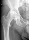
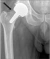
We report a case of periprosthetic fracture of a 57-year-old male patient with BMHR. The patient, having previously had a left total hip replacement for osteoarthritis, subsequently underwent right hip BMHR for osteoarthritis (Fig. 1). The surgery was uneventful and notching had not been noted at the time of surgery. The patient recovered successfully and was discharged home. Postoperative radiographs had shown a small notch in the superior femoral neck. This has been highlighted with an arrow in Fig. 2. This patient, who was a retired delivery driver, was walking pain free and unaided postoperatively.
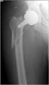
Twelve months later, he presented to casualty via ambulance with a clear history of a mechanical trip, right hip pain and inability to weight bear. This was not a high-energy mechanism and simply involved a fall from standing height with the patient landing directly onto the right hip. Subsequent radiographs revealed an unusual periprosthetic fracture pattern around his right BMHR (Fig. 3).
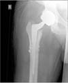
The fracture started high on the superior lateral side of the femoral neck and spiralled distally to the subtrochanteric region. The implant had bonded well to the stem but the fracture rendered the stem not sufficiently 'well-fixed' to be retained. He subsequently underwent revision surgery with a long uncemented stem. Cables were used to support the proximal fracture (Fig. 4) and the patient was eventually discharged home. A long stem, which was distally fitting, was used so as not to rely on the fractured proximal metaphysis.
DISCUSSION
We describe a case of periprosthetic fracture in a patient with a BMHR implant. This has never been described in the literature previously. In our case, the fracture was complex, spiralling from the femoral neck to the subtrochanteric region. This is unlike the usual fracture pattern seen in conventional hip resurfacing arthroplasty and that seen in cadaveric BMHR studies.
Cadaveric studies have suggested that standard resurfacing arthroplasty fracture rates are reduced with slight valgus placement of the femoral component compared to anatomic position.34) As resurfacing implants are biomechanically similar to the BMHR, one may assume that valgus placement of the BMHR might also be protective against fracture. Again, until recently there was no corroborating evidence to suggest that this was also the case. A recent cadaveric study using the BMHR has actually found that valgus placement does not strengthen and varus placement does not weaken femora.5) However, valgus implant placement had a relatively protective effect on full cortical thickness superior neck notch. This is important as aiming to place the implant in valgus may result in placing it too valgus, which may result in notching and fracture as in our reported case. This latest cadaveric study seems to imply that the BMHR may be a more forgiving implant to use in terms of its positioning compared to its resurfacing cousins. However, this evidence needs to be strengthened with other such trials. The BMHR is an uncemented metaphyseal fixed implant, which is different to the epiphyseally cemented Birmingham Hip Resurfacing (BHR). The shape of the BMHR femoral stem is conical and is said to allow for improved physiologic loading similar to that of the intact femur.6)
Femoral neck notching has been shown to be a significant contributor to subsequent fracture of resurfacing implants.27) Cadaveric "no notch BMHR femora" have been found to have similar loads failure points to "no notch BHR femora" (5,002 N vs. 5,302 N). Thus, both implants have similar failure points when there are no deficiencies in the femoral neck cortex. However, when 2 mm notches were tested in BMHR femora, there was not a significant reduction in the load failure point compared with "no notched femora." This was unlike 2 mm notched BHR femora, which failed with significantly reduced loads. In fact load failure rates were comparable (4,060 N vs. 4,043 N) with 5 mm notched BMHR femora and only 2 mm notched BHR femora.5) Thus, it appears that femoral notching can still increase the risk of fracture but requires more significant notching or stronger loading forces than with standard hip resurfacing implants before failure occurs. This correlates with our case where our patient also had a notched neck of femur. Our patient walked well for many months before developing a fracture and this was due to a fall on to the affected side.
Olsen et al.8) found that cadaveric femora with a BMHR implant tested to failure required significantly less axial force than those with BHR implants (average of 1,578 ± 865 N less force, P < 0.001). Olsen also describes two distinct fracture patterns: 19 of the 32 fractures of the transcervical vertical shear type and the remaining 13 femora sustained subcapital fractures.
The fracture patterns in our case seem to be unusual compared with what has been documented in the literature. In the case we present, the fracture appears to have emanated from the notched superior neck cortex and spiralled distally to the subtrochanteric level. The valgus placement of the implant and radiographs suggest that reaming had thinned the medial calcar, potentially weakening the femur further. The fracture, however, has not spread in the usual transcervical or subcapital pattern. This difference may represent the difference between laboratory tested axially loaded cadaveric femora and real life when a fall is more likely to result in rotation forces caused by the femoral head-neck anteversion. Time will tell when further case reports, biomechanical and follow-up studies emerge if spiral subtrochanteric fractures are more common than suggested by current biomechanical studies. This will be of great clinical significance as revision surgery will be more technically challenging and may result in longer operating times, greater blood loss, longer hospital stays and poorer patient satisfaction outcomes although it should be emphasised that it is still too early to tell.
 XML Download