

 PDF
PDF ePub
ePub Citation
 Print
Print


A requirement in military parachuting is a system enabling soldiers to jump from an aircraft and land on the ground in a fit state to fight. The parachute system should enable the aircraft to fly as low as possible and the soldiers should exit the aircraft as fast as possible to minimize the time the aircraft spends over the drop zone. Military occupational medicine requires mandatory analysis of injuries resulting from military parachuting and appropriate treatment. Parachute injuries can occur between leaving the aircraft and removal of the harness, and can be grouped as problems with exit, descent, and landing. The former 2 problems can cause fatal losses; the main cause of military hospitalization is the latter problem. Approximately 70% of the parachute injuries are due to improper landing techniques.1,2) The ankles are injured in approximately one-third of parachutists.1,3,4,5) The rate for fractures in parachute injuries is 0.5-2.0 per 1,000 jumps, half of which are ankle fractures.1,4,5,6,7,8)
The purpose of this study was to analyze the etiology of ankle injuries of paratroopers in special operation units making descents for air invasion through evaluation of the frequency of fractures and fracture patterns. We also evaluated the treatment options and clinical outcomes.
METHODS
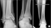
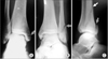
Medical records and radiographic outcome were collected retrospectively from 56 patients with parachuting-related ankle fractures sustained between January 2005 and April 2010. These ankle fractures occurred in members of the Korean Special Forces army brigade. The mean age was 23.57 years (range, 20 to 44 years). Twenty-eight patients each had ankle injuries on the right and left side, respectively. Radiographic fracture patterns were used to classify the injuries by the Lauge-Hansen classification,9) Weber classification,10) and the positions of the fracture fragments. The Lauge-Hansen classification describes the position and motion of the foot with respect to the leg; specifically, these injury patterns are supination-external rotation (SER), supination-adduction (SA), pronation-abduction (PA), and pronation-external rotation (PER). The Weber system uses the position of the level of the fibular fracture in relation to the height at the ankle joint. Type A fractures are below the ankle joint, type B fractures are at the level of the joint, and type C fractures are above the joint level (Fig. 1). The ankle fractures were also identified by the positions of the fracture fragments: medial, lateral, and posterior malleolar fractures; simple and complex fractures; and deltoid rupture and syndesmotic diastasis (Fig. 2). The American Orthopedic Foot and Ankle Society (AOFAS) ankle-hindfoot score,11) Olerud and Molander score,12) patient satisfaction, and return to full activity were recorded at the final follow-up evaluation and by telephone interview. The patients were asked if they were satisfied or dissatisfied with the outcome of their surgery (excellent, good, fair, and poor). The patients were also interviewed regarding the degree of return to physical activity, as follows: full activity with descent, competitive sports without descent, jogging, walking, limited walking due to pain, crutch walking, and requiring additional operative management. The fixation devices were removed in 19 of 56 cases.
We also evaluated the efficacy of each treatment option. Statistical significance was defined at the 5% level (p < 0.05) using an independent t-test on SPSS ver. 16.0 (SPSS Inc., Chicago, IL, USA).
RESULTS
The mean follow-up period was 23.8 months, and all patients achieved complete bony union with the exception of 2 cases of medial malleolar non-union.
Twenty-two patients had simple fractures and 34 patients (60.71%) had complex fractures. The average number of fracture sites per patient was 1.75. The total number of injuries was 116, including a syndesmosis and a deltoid injury; the average number of injury sites per patient was 2.07.
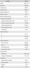
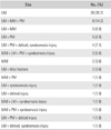
Based on the Lauge-Hansen classification, SER injuries were found in 20 cases, PER injuries in 11 cases, and SA injuries in 22 cases. Two cases had distal tibial fractures and medial malleolar fractures. Weber type A, B, and C fractures were found in 4, 38, and 14 cases, respectively (Table 1).
Fifty-two cases (92.86%) had fibular fractures, 23 cases (41.07%) had posterior malleolar fractures, and 21 cases (37.5%) had medial malleolar fractures. Deltoid ligament ruptures were diagnosed in 7 cases (12.50%) with medial mortise widening and operative findings. Syndesmotic injuries were diagnosed in 11 cases (19.64%) with mortise ankle anteroposterior views. The syndesmotic injuries were treated surgically, as follows: 30 cases with plate fixation; 15 cases with screws; 8 cases with plate and tight rope fixation; and 2 cases with intramedullary nail fixation. One of the cases was treated conservatively with cast immobilization. Greater than 20% of cases with posterior tibial articular involvement were treated with open reduction and posterior screw fixation, and intraoperatively a > 2 mm displacement after fibular fixation was also treated with open reduction and posterior-to-anterior screw fixation through a posterolateral approach (Table 2).
The postoperative mean AOFAS score was 85.4 points (standard deviation [SD], 8.9; range, 67 to 100 points) at the last follow-up evaluation. The Olerud and Molander score was 77.5 points (SD, 12.6; range, 40 to 100 points) at the last follow-up evaluation. The self-assessment questionnaires yielded an 80.36% (45 patients) excellent and good satisfaction rating; 24, 21, 9, and 2 patients rated their satisfaction as excellent, good, fair, and poor, respectively. The patients were also interviewed on the degree of return to physical activity. Eighteen patients (32.14%) returned to full activity with descent, 7 patients (12.50%) returned to competitive sports without descent 16 patients (28.57%) returned to jogging, 8 patients (14.29%) returned to walking, 1 patient (1.79%) required crutch walking, and 1 patient (1.79%) required additional revision surgery for nonunion. The mean AOFAS score of ankle fractures with and without posterior malleolar fractures was 82.7 and 88.9, respectively (p = 0.336). The mean Olerud and Molander score of ankle fractures with and without posterior malleolar fractures was 71.6 and 78.6, respectively (p = 0.563). Postoperative complications included 2 medial malleolar nonunions, 2 irritations by screw protrusion and 1 deep infection.
DISCUSSION
Impact with the ground causes majority of military parachuting injuries. Several methods have been designed to prevent injuries in parachute descents. During the early days of parachute training, a forward landing roll was used. The British and United States Army developed an alternative landing roll, a sideways roll, which caused significantly fewer injuries. The sideways roll, referred to as the 5 landing position, in which the parachutist lands and conducts a sideways roll onto the forefoot, outer side of the leg, thigh, and buttocks onto the opposite shoulder. The parachutist also has to adopt a relaxed position, with the legs held forward, the knees slightly bent, and the toes lifted so that the feet land flat on the ground.8,13)
Most of the fractures in this study involved one side. Most patients did not recall the accident events. However, a small portion of patients recalled the cause of injury due to the improper single leg landing. If the landing is made with the feet apart, one foot is likely to strike the ground before the other foot and can be injured more easily. Yeow et al.14) reported the hip and knee are predominantly involved in energy dissipation during double-leg landing; the hip and ankle were the dominant energy dissipaters during single-leg landing in frontal plane. Total energy dissipation of double-leg landing and single-leg landing in the sagittal plane are 29.7% and 45.7%, respectively. Ellitsgaard and Warburg15) described the movements causing ankle fracture. The improper landing attitude most commonly means 'legs apart' and the resulting injury can occur as SER and PER fracture. The rear foot receives an eversion force upon contact with the ground. Small bumping or obstacles on the ground surface can force the foot to the pronation or supination position. The authors recommended the improvement of landing and steering techniques and the support of semi-calf boots. Amamilo et al.5) also reported 24.4% of ankle injuries were due to improper action of the paratroopers with the feet apart. According to the paratrooper's textbook, the parachute landing fall recommends both feet and knee pressed tightly together. In this position, the legs constitute a flexible shock break, which is much safer than landing with the legs apart. Once the feet contact the ground, the knees are pressed together and the hips twist and bend to provide direction to landing.16) External rotation injuries were the most common injury pattern based on the Lauge-Hansen classification and accounted for 55.36% of ankle injuries in the current study. Thus, one of the main mechanisms of paratrooper ankle fracture injuries was landing with the feet apart and external rotation forces.
Ekeland1) reported that 71% of all injuries occurred during improper landing; uneven terrain and wind accounted for and 8% and 6% of injuries, respectively. Second fracture pattern is SA injuries (39.3%) belong to Lauge-Hansen classification. SA pattern is caused by axial loading and inversion stress. SA injuries are usually induced by oscillation (unintentional swing of the parachutist under the canopy). Even though the paratroopers typically gather their feet together, the impact on the lateral margin of the forefoot will force the foot into supination due to increased landing velocity with oscillation, improper steering, or high wind speed.15)
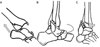
The ratio of operative procedures by mechanism of injury are SER injuries in 60% and PER injuries in 38% of civilian ankle fracture cases.17) Posterior malleolar fractures occur in 8%-21.1% of lower-leg fractures.18,19,20,21) Parachuting ankle injuries included 41.07% (23 cases) accompanied with posterior malleolar fractures. Ruedi et al.22) first described in detail the combination of an axially-applied injury in combination with foot position. Posterior malleolar fracture occurs if the vertical compression force is applied with the foot in plantar flexion. This mechanism of posterior malleolar fractures ranged from falls down slopes or steps. However, isolated posterior malleolar fractures were not included in this study, so pure axial loading with foot plantar flexion position were not suggested as the main mechanism of posterior malleolar fractures in paratroopers. The fracture patterns, including posterior malloear fractures, were 13 SER, 8 PER, and 2 SA. Excessive rotational forces were more appropriate for the posterior malleolar fracture mechanism. The sequence of morphologic changes giving rise to posterior malleolar fractures involves failure of the lateral side with the foot in supination resulting in violent talar external rotation, especially combined with an axial load. The first injury is an oblique fracture of the fibula, and further talar rotation results in a fracture of the articular lip of the tibia (Fig. 3). High-energy axial loading with rotational force leads to posterior malleolar fractures in parachute jumping.
A higher risk of injury was associated with higher wind speed, uneven terrain in the drop zone, greater body weight, night jumps, jumps wearing additional equipment, and lower parachute skill. Several authors have reported the use of an ankle brace to effectively reduce the incidence of ankle injury hospitalization among Army Airborne School students.2,23) The outside-the-boot parachute ankle brace could prevent extreme ankle inversion and eversion, while allowing plantar flexion and dorsiflexion. The authors also reported that during ankle brace use periods, the occurrence of other types of traumatic injuries did not increase. Expenditures were $30,000 per year for braces, but the saved cost was $835,000 per year for hospital care and rehabilitation.23,24) Therefore, our armed forces also need to adopt the ankle bracing protocol for paratroopers.
Postoperative AOFAS and Olerud and Molander scores were not significantly decreased in ankle fractures combined with posterior malleolar fractures. Jaskulka et al.19) reported a definite rise in the incidence of osteoarthrosis when a fracture of the posterior tibial margin was present, even in cases with a small marginal fragment of the lip of the tibia. However, many authors agree that internal fixation of posterior malleolar fractures yields better clinical results.18,19,25) Lee et al.26) reported results of open reduction and internal fixation in patients with ankle fractures and concomitant posterior malleolar fractures had equally favorable outcomes to patients with ankle fractures without concomitant posterior malleolar fractures. Our patients were injured by high-energy trauma and underwent suitable stable fixation of the articular involved posterior malleolar fragment. Thus, these patients were not affected by the posterior malleolar fracture.
Several limitations of this study were that the surgeries were performed by several army surgeons; further, the retrospective design of our study limited our ability to consider certain variables; we were not able to assess compound factors, such as chondral damage, syndemostic fixation, and an accurate size of the posterior malleolar fragment. We have no comparative group like as recreational sports injury or traffic accident (non-parachute injury group) and relatively small size of patient numbers and short-term follow-up. We collected the data with AOFAS scores retrospectively. AOFAS scores are no longer recommended for use to validate outcomes with ankle injury. Hence, we added the Olerud and Molander score.
Of the paratrooper ankle fractures, 41.07% were accompanied by posterior malleolar fractures, thus paratroopers ankle fractures usually resulted in operative treatment. The most common injury mechanism was an external rotation injury that was rated by 55.36%. The ankle brace should be an important prevention method of ankle fractures.
 XML Download